Medical editor: Strokina O.A. - therapist, functional diagnostics doctor. November, 2021.
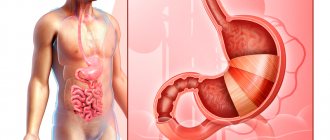
Ultrasound examination of the intestines (ultrasonography) is a diagnostic procedure that allows you to examine the intestines along its entire length and identify the pathologies present in it with sufficient accuracy.
Modern ultrasound equipment makes it possible to examine areas of the intestinal tract that are inaccessible to other diagnostic methods. Standard procedures (colonoscopy, anoscopy, sigmoidoscopy) cause the patient significant psychological discomfort and pain, have many contraindications, and ultrasound is a completely painless and safe medical procedure.
Indications for intestinal ultrasound
The content of the article
An intestinal ultrasound is prescribed by a therapist, gastroenterologist or proctologist. You can go through the procedure yourself. More often, intestinal ultrasound is included in a comprehensive ultrasound of the abdominal cavity, since gastrointestinal diseases are always interconnected and to identify the main cause of the disease, all organs need to be examined.
An ultrasound examination of the intestine is indicated for:
- stool disorders - constipation, diarrhea, unstable stools;
- presence of blood in feces;
- flatulence;
- abdominal pain of unknown origin;
- disorders of the digestive system;
- suspicion of the presence of a neoplasm in the intestine;
- shift of an organ detected by x-ray;
- chronic colitis;
- changes in the shape of the rectum detected during rectoscopy or colonoscopy;
- oncological diseases;
- endometriosis in women and prostate cancer in men (to exclude intestinal damage);
- intussusception;
- anemia, fever of unknown origin;
- monitoring the condition of organs in the postoperative period (to exclude relapse of the oncological process).
Intestinal diseases occur not only in adults, but also in children, including infants. An examination of the children's intestines is carried out if pathology is suspected:
- Hirschsprung's disease;
- irritable bowel syndrome;
- dolichosigma (elongation of the sigmoid colon);
- Crohn's disease;
- nonspecific ulcerative colitis;
- abnormal structure and localization of the intestine;
- fecal stones;
- change in intestinal motility.
Intestinal ultrasound can be used as a primary or additional diagnostic method to clarify the results of previously conducted studies.
What pathologies does ultrasound examination of the intestine reveal?
The high information content of ultrasound makes it possible to diagnose many diseases that cannot be detected by other research methods.
The doctor can easily detect the following pathologies:
- fluid in the abdominal cavity (but ultrasound cannot reveal its nature);
- inflammation, cysts and tumor formations;
- enteritis is an inflammatory process in the small intestine, in which its basic functions are disrupted;
- abdominal abscesses - limited ulcers in the pyogenic capsule;
- Hirschsprung's disease is a congenital abnormal development of the large intestine;
- intestinal deformation, ulceration of the mucous membrane;
- diverticulitis - inflammation of blindly ending protrusions of the intestinal wall;
- abdominal hematomas;
- acute mesenteric thrombosis - disturbance of blood flow in the vessels of the mesentery;
- nonspecific ulcerative colitis – chronic inflammation of the colon mucosa;
- chronic spastic colitis (irritable bowel syndrome) – disruption of intestinal motor function due to dysregulation;
- intestinal bleeding;
- abnormal localization of the intestine;
- Crohn's disease is a chronic inflammatory disease of the gastrointestinal tract;
- appendicitis in acute and chronic form;
- ischemic intestinal disease - blockage or narrowing of blood vessels;
- intussusception – intestinal obstruction “gut in gut”;
- oncological diseases.
Types of intestinal examination
There are several types of ultrasound examination of the intestine using ultrasound:
- Transabdominal ultrasound
- through the anterior wall of the abdomen. The study can be carried out standardly or using contrast (ultrasound irrigoscopy). The disadvantage of the method is poor information content due to limited organ visualization capabilities; - Endorectal ultrasound
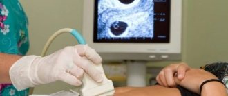
- by inserting a sensor into the rectum. To improve visualization, the procedure may be performed with contrast (a sterile liquid is injected through a transducer catheter). This is the most informative method, capable of quickly and accurately identifying the pathological focus, but due to the inconvenience of its implementation, it is prescribed only to clarify the pathology if it is not visible during ultrasound of the abdominal cavity, performed using the abdominal method. - Transvaginal ultrasound
- by inserting a cavity sensor into the vagina. This method is used in women in very rare cases, as an additional method. It allows you to examine the wall between the intestines and the vagina and other important areas.
Each type of ultrasound has relative contraindications and disadvantages, therefore, to obtain the most complete picture of the condition of the intestines, the doctor is not limited to one research method. For children, only the external method is used.
Features of devices for diagnosing diseases of the small intestine and colon
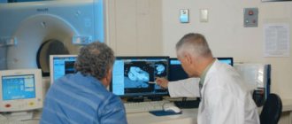
In our clinic, examinations are carried out using stationary ultrasound equipment of expert and high class. It allows you to get a good quality image. The results of the study can confirm or refute the suspected diagnosis.
Ultrasound diagnostics of the intestine is performed in B-mode (from the English brightness - brightness). On the screen, the doctor sees a black and white two-dimensional image in one plane. For examinations in our clinic, convex ultrasound sensors are used, which operate at a frequency of 2-7.5 MHz. They allow penetration to a depth of 25 cm. The doctor will determine the exact operating frequency of the device depending on your build.
This is interesting! The first ultrasound of the intestines was performed by the English scientist John Wilde in 1949. He used this diagnostic method to determine the thickness of the intestinal wall. For this development, the scientist was called the “father of medical ultrasound.”
To examine the rectum, a rectal ultrasound sensor is used, which has a small thickness. The sterility of the device and painless insertion helps to preserve the special condom.
How to prepare for an ultrasound bowel examination procedure
The rules for preparing for each type of ultrasound differ, so you must first clarify with your doctor which method the procedure will be performed.
Preparing for a transabdominal ultrasound
When examining with an abdominal probe, you should:
- Avoid eating and drinking 6 to 8 hours before the procedure. Children under 1 year of age should take a break from eating for 2–4 hours (ultrasound is performed immediately before feeding), children 1–3 years old should not eat for 4 hours, and over 3 years old should not eat for 5–6 hours;
- For 3–4 days, follow a slag-free diet, excluding from the diet foods that increase flatulence (fresh vegetables and fruits, dairy and yeast products, carbonated drinks, etc.);
- Drink 1 - 1.5 liters of non-carbonated liquid within an hour and do not empty your bladder. If for some reason it is not possible to fill the bladder naturally, then it is filled through a catheter;
- 3 days before the procedure, start taking medications that relieve gases in the intestines (espumisan, activated carbon, smecta, etc.) and food enzymes (mezim, Creon, etc.);
- If ultrasound irrigoscopy is also planned, then the evening before the ultrasound it is necessary to thoroughly cleanse the intestines with 1 - 2 enemas (volume up to 2 liters). It is not recommended to replace them with fortrans, microenemas, glycerin suppositories or laxatives, since these drugs will not be able to fully cleanse the intestines.
Preparing for an endorectal ultrasound
For such a study, an empty bladder and cleansed intestines are required. The procedure is carried out on an empty stomach, but it is not necessary to observe a 6-hour break in meals.
It is necessary to cleanse the intestines the night before using one of the following methods:
- taking an osmotic laxative – fortrans (dissolve 3 sachets in 3 liters of water and drink within an hour);
- setting 1 – 2 cleansing enemas (up to 2 l);
- taking a herbal laxative (picolax, senade, etc.).
Immediately before the ultrasound, you need to empty your bladder.
Preparing for a transvaginal ultrasound
This type does not require special preparation. Immediately before the procedure, you only need to empty your bladder. It is also recommended to take medications that eliminate flatulence (activated carbon, espumizan, smecta, etc.) 2–3 days before the ultrasound.
Preparation
Normally, the gastrointestinal tract is filled with feces and air inclusions. This state of the organ makes it difficult to obtain informative images reflecting the condition of the walls of the gastrointestinal tract, which reduces the diagnostic value of the study. Before conducting any examination of the gastrointestinal tract, preliminary preparation is necessary.
General principles of preparation:
- For several days before diagnosis, it is necessary to follow a diet aimed at reducing the load on the gastrointestinal tract and reducing gas formation;
- limit as much as possible the consumption of legumes, cabbage, onions, mushrooms, dairy products, eggs, sweets, carbonated drinks;
- exclude alcohol;
- drink at least 2 liters of fluid per day;
- It is recommended to include lean meat, chicken, cereals, and vegetable broths in the diet;
- meals are taken in small portions 5-6 times a day;
- You must come for the study at least 6 hours after your last meal;
- if diet is not enough to reduce the amount of gases in the gastrointestinal tract, you should take activated charcoal and espumizan on the day before the examination;
- if constipation occurs, you should use laxatives;
- On the eve of the procedure, it is necessary to cleanse the large intestine with an enema.

Neglecting the preparation rules leads to insufficient cleansing of the stomach, which is a relative contraindication for the procedure. If an examination is carried out in such a condition, then there is a high probability of missing a small pathological formation, and the diagnostic value of the study will be reduced.
When visiting a doctor, you must take a referral for testing and medical documentation related to your current state of health - an advisory opinion from the attending physician with a preliminary diagnosis, laboratory test results. If an intestinal ultrasound or colonoscopy is repeated, you must have the results of previous examinations with you. The maximum amount of information about the patient’s health status helps the doctor make an effective diagnosis.
How is an intestinal ultrasound performed?
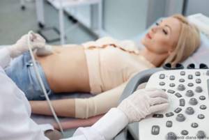
Transabdominal method.
The patient lies with his back on the couch and bends his knees to relax the abdominal cavity. The doctor applies a sound-conducting gel to the abdomen and scans using an ultrasound probe. First, he examines the sigmoid colon, then moves the sensor from the underlying sections to the overlying ones. The intestine is examined in longitudinal, transverse and oblique sections with measured pressure applied to the abdominal cavity by a sensor. If the results are unclear, the doctor may ask the patient to change position, inflate the stomach, or take a deep breath.
If ultrasound irrigoscopy is required, the patient should expose the lower part of the body and lie on his side with his back to the doctor. About 2 liters of sterile saline solution is injected through a catheter inserted into the intestine. This is done so that under the pressure of the liquid the intestinal walls straighten and make it possible to thoroughly scan all sections. After filling the intestines, the study continues.
Endorectal method.
The patient bares the lower part of the body and lies on his right side, bending his legs and pulling them towards him. The doctor carefully inserts a cavity sensor into the anus to a depth of 10–12 cm, onto which he has previously placed a condom and lubricated it with lubricant.
Transvaginal method.
The patient is naked below the waist, lies with her back on the couch, bending her knees. The doctor puts a condom on the transducer, lubricates it with lubricant and carefully inserts it into the woman’s vagina, after which he examines the condition of the intestines.
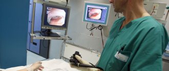
How is a colonoscopy performed?
The success of the procedure depends on the quality of the patient’s preparation and the accurate implementation of the doctor’s instructions. The examination is carried out using local or general anesthesia.
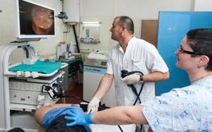
The patient undresses, lies on the table on his left side and pulls his legs to his chest. The tip of the colonoscope is inserted into the anus, to which a flexible tube is attached. The advancement of the tube into the large intestine occurs gradually, with the injection of a small amount of air, which ensures a moderate expansion of the walls of the gastrointestinal tract and easier passage of the tube.
Through the tube, a video camera and instruments can be brought to the walls of the tract to perform operations, stop bleeding, and take a biopsy. During the procedure, the mucous membrane is examined. The detected polyp can be removed using a surgical instrument inserted through a tube. The presence of a neoplasm or ulcer means taking the material with special forceps for examination in the laboratory.
The duration of the examination depends on the volume of manipulations, on average – 20-60 minutes. After the procedure, the patient may experience discomfort - accumulation of gas, the urge to defecate. The condition normalizes on its own within a few hours; to speed up the process, you can take espumizan or activated carbon.
Ultrasound of the fetal intestine
When examining the fetal intestines, the doctor can determine its echogenicity. The detection of increased echo density indicates the accumulation of feces in the intestines. This is not a direct pathological sign, since it can be observed in both healthy babies and patients with chromosomal diseases. In combination with a discrepancy between the size of the fetus and the gestational age, hyperechogenicity of the intestine indicates a delay in the development of the child.
If such a phenomenon has been identified, the doctor will refer the woman to an appointment with a geneticist, and will also prescribe a blood test for antibodies to TORCH infection. A repeat ultrasound is performed a month later.
General preparations
|
| Preparation for intestinal examination is an important and integral part of the ultrasound procedure. |

The main purpose of an intestinal ultrasound is to conduct a thorough examination of the wall of a hollow organ filled with air, food debris and feces. The contents of the intestine represent the main barrier to the passage of ultrasonic waves, which, when colliding with an obstacle, are immediately reflected, and further study of the condition of the wall becomes impossible. Therefore, before the examination it is necessary to carry out some preparatory measures.
Recommendations for preparing for an ultrasound scan can be general or specific. For general preparatory activities
relate:
- When undergoing an ultrasound for free, under the compulsory medical insurance policy, you must have a referral from the attending physician and a valid compulsory medical insurance policy registered in the attached clinic.
- When undergoing an ultrasound for free under a voluntary health insurance policy, you must have another referral from the insurance company and a valid VHI policy registered with this insurance company.
- When undergoing an ultrasound scan for paid medical services, there must be an agreement with the clinic and a receipt for payment for the procedure.
- On the appointed day, the patient must come for examination on an empty stomach, in comfortable clothes, with a sheet to put on the couch, wipes to remove gel from the skin and shoe covers, or replacement shoes (if the clinic does not provide this).
Interpretation of data: normal intestinal ultrasound indicators
The sonologist scans the intestines in transverse and longitudinal sections and evaluates the following indicators:
- intestinal size and shape;
- localization of the organ in relation to the uterus, bladder, prostate and seminal vesicles;
- thickness, structure, layering and echo density of walls;
- length of different parts of the intestine;
- condition of surrounding tissues;
- size and structure of regional lymph nodes;
- the presence of an abnormal structure, formations, inflammation, etc.
A “normal” rectum has a round, slightly elongated shape, an even contour and a rim extending from the muscle layer. Decoding is carried out by a proctologist depending on the type of study being performed.
Standard indicators for external ultrasound examination
| Index | Meaning |
| Terminal length | 5 cm |
| Middle section length | 6 – 10 cm |
| Length of the mid-ampullary section | 11 – 15 cm |
| Wall thickness | up to 9 mm |
| Gut lumen | It is revealed that there are no deformations |
| Number of wall layers | 2 |
| Regional lymph nodes | Not rendered |
The methodology for performing endorectal ultrasound is different, so it determines different standards
| Index | Meaning |
| Number of wall layers | 5 |
| Layers with increased echo density | 1, 3, 5 |
| Layers with reduced echo density | 2, 4 |
| Internal and external contours | Smooth |
| Regional lymph nodes | Pararectal, up to 5 mm |
Pathological indicators detected by intestinal ultrasound
| Pathology | Signs on ultrasound |
| Enteritis | dilated intestinal loops; increased contraction of the walls; the presence of contents with different echo densities in the lumen |
| Hirschsprung's disease | an additional loop is detected, the size of the colon or part of it is increased |
| Intestinal obstruction (mechanical) | loops are expanded 2 - 3 times; thin, hyperechoic walls; circular folds of the mucous membrane; homogeneous liquid echogenic chyme in the intestinal lumen; pendulum-like movement of the contents; collapsed loops of the small intestine; bidirectional contraction of the walls; stratification of the contents into anechoic sediment and liquid (in later stages); hypoechoic formation that does not provide a shadow (if the pathology is caused by a foreign body) or with a distal shadow (from a gallstone) |
| Intestinal obstruction (paralytic) | the intestinal lumen is dilated; no contractions of the wall occur; the loops are tightly compressed together; echogenic content |
| Acute mesenteric thrombosis | there are no contractions of the affected area; the area is hypoechoic; no layering |
| Nonspecific ulcerative colitis | uneven internal contours due to ulceration of the mucosa; the affected area does not shrink; wall thickness increased; hypoechogenicity and inelasticity; haustrae are not visualized; lack of layering |
| Chronic spastic colitis | the intestinal wall is thickened; muscle bands are visualized as hyperechoic stripes |
| Ischemic colitis | the wall is unevenly thickened and hypoechoic; lack of layering |
| Crohn's disease | uneven hypoechoic wall thickenings; accumulation of fluid in the lumen; peristalsis is reduced or absent; haustrae and wall layers are not defined; reduced elasticity; hyperechoic growths and polyps |
| Acute appendicitis | congestion in the pelvic area; thickening of the bladder wall; double contour at the point of contact with pus; anechoic formations in the peritoneum; accumulation of fluid in intestinal loops and the appearance of abscesses (in case of peritonitis) |
| Cancer | circular increase in wall thickness; stenosis; layers are not detected |
Colonoscopy results
A healthy large intestine has a smooth, shiny, pink mucous membrane, without areas of hyperemia or edema. Cicatricial changes, diverticula, pathological narrowing or expansion of the intestinal lumen, ulcers, and polyps are normally absent.
What some diseases look like:
- with nonspecific ulcerative colitis, the mucous membrane becomes bright red, swelling, granularity, and areas of hemorrhage appear;
- intestinal diverticula look like branches of the gastrointestinal tract with an entrance diameter of up to 2 mm, at the site of the lesion the tone of the intestinal wall is increased, the folds are thickened;
- cancer looks like a heterogeneous neoplasm of irregular shape with a granular surface, surrounded by an area of edema, and bleeds when touched.
The results of examining the intestinal walls with a video camera can be obtained shortly after the procedure. Based on the results of the conclusion, the attending physician makes a diagnosis or refers the patient for further diagnostics. If a biopsy was performed, the result of the examination of the fragment will be issued within two weeks. Microscopic examination allows you to make the most accurate diagnosis.
Contraindications
There are only relative contraindications for intestinal ultrasound:
- infectious and inflammatory bowel diseases in the acute stage;
- abdominal skin damage;
- severe mental disorder of the patient (inadequacy);
- rectal stenosis.
It is not recommended to perform the procedure if a study using a contrast agent or colonoscopy was performed shortly before it.
Features of ultrasound examination of the liver and other obstructions during pregnancy
For women who have a history of chronic diseases of OBP, it is advisable to undergo an ultrasound scan in the early stages of pregnancy - preferably before 16 weeks. After all, the longer the period, the more difficult it is to examine certain abdominal organs on an ultrasound. This is due to the fact that after 15 weeks of pregnancy the uterus extends beyond the bony ring of the pelvis and gradually fills the abdominal cavity. By 37 weeks, the fundus of the uterus almost reaches the chest, rising to a height of 32–37 cm from its original position.
In fact, after 36 weeks, an ultrasound of the liver or other OBP does not make sense, as it will be uninformative. Factors such as multiple pregnancies, polyhydramnios, or a large fetus can complicate the ultrasound diagnostician’s task even at earlier stages.
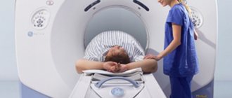
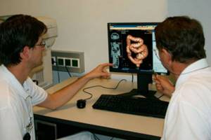
Research Alternative
Along with ultrasound, there are other methods of examining the intestines, such as colonoscopy, endoscopy, MRI and irrigoscopy. Ultrasound research is less informative than other methods. However, he has no equal in diagnosing cancer of a given location at any stage of its development. In addition, this is the cheapest and most harmless method.
Ultrasound of the intestines is an absolutely safe and painless method. It does not expose the body to radiation, does not cause complications and has no age restrictions. The procedure can be performed multiple times even during pregnancy. This is the most gentle diagnostic method for children, elderly and debilitated patients.
An ultrasound of the intestine is performed for a relatively short time - about 10 - 20 minutes. Patients note the presence of discomfort only during the insertion of the sensor, after which the discomfort goes away, which cannot be said about colonoscopy.