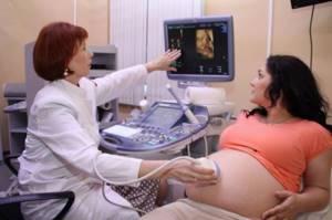
Modern ultrasound examination during pregnancy is a non-invasive way to learn about the condition of the fetus and associated tissues without surgical intervention. 3D ultrasound is the best way to get complete information about the baby’s condition without harming the mother and child.
THE COST OF AN ULTRASOUND DURING PREGNANCY IN OUR CLINIC IS FROM 1000 rubles. CONSULTATION WITH A GYNECOLOGIST ON THE RESULTS OF AN ULTRASOUND OR ANALYSIS—RUB 500.
CLICK TO MAKE AN APPOINTMENT, TEST OR ULTRASOUND
Using 3D ultrasound, diagnostics are made in three dimensions - height, width and depth. With such a study, not a flat picture is displayed on the monitor screen, but a multidimensional image. This allows you to get a three-dimensional image and see the appearance of the child even before his birth. The equipment allows you to see in detail how the embryo develops and whether the pregnancy has frozen.

The Diana Clinic (St. Petersburg) has a modern 3D device, and the results are interpreted by a specialist of the highest category. We invite expectant mothers to undergo a safe and inexpensive diagnosis with us, without a queue and at any convenient time. You will receive the results in your hands. Disc with recording - as a gift!
3D ultrasound: is it necessary?
The content of the article
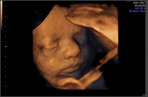
With the development of medicine, it has become possible to see your baby not only after birth, but also in the womb. Young parents use 3D ultrasound to record a video with their unborn baby in the lead role. Moreover, on the screen you can see all the baby’s movements: how he sucks his fingers, moves, opens his mouth, squints his eyes.
Some time ago, three-dimensional ultrasound was considered something exotic. Now thousands of couples want to capture unforgettable moments of a baby’s intrauterine life for a family video archive. 3D diagnostics has become a popular service from the paid list of modern clinics. Its cost is not so high, so any family can afford this pleasure.

But 3-D ultrasound is not just a movie for memory! During pregnancy, ultrasound is the most important diagnostic procedure. It is considered mandatory and is carried out to determine the child’s condition. For the mother, this is the first acquaintance with her future baby. During the entire pregnancy, ultrasound is performed 3 times: in the third, fifth and eighth months. In some cases, if a natural delivery is suspected, a fourth ultrasound is performed towards the end of pregnancy.
Ultrasound diagnostics are carried out to determine the age and position of the fetus, the presence of birth defects, diagnosis of multiple pregnancy and singleton pregnancy. Ultrasound does not pose any danger to either the mother or the baby. This is confirmed by many years of practice in using this diagnostic method in obstetrics.
Still, it is better to use an ultrasound after a doctor’s prescription, although without a referral; at the request of the pregnant woman herself, the diagnosis will, of course, be carried out. It is advisable to follow the recommended timing, then you can obtain reliable information about the condition and development of the fetus.
Science has NOT proven that...
1. Ultrasound is generally unpleasant for the fetus. How to check whether he is pleased or not remains a question.
2. Ultrasound leads to fetal growth retardation. Typically, ultrasound is performed more often when fetal growth is restricted in order to monitor weight gain. And it turns out like in a joke: all dead people ate cucumbers, which means cucumbers lead to death.
3. Ultrasound leads to the development of brain cysts. Fetal brain cysts form during and during growth and usually go away on their own. Ultrasound does not affect their growth and development in any way.
4. Ultrasound leads to the formation of moles in a child. Apparently, adherents of this theory believe that moles appeared in humans along with ultrasound... Apparently, before the advent of ultrasound, no one had moles.
Key principles for the safe use of ultrasound
1. Ultrasound examination should be used only for the purpose of establishing a medical diagnosis.
2. Ultrasonic equipment should only be used by personnel who are fully familiar with its safe and proper use.
This requires:
- understanding the possible thermal and mechanical biological effects of ultrasound;
- full awareness of equipment settings and understanding of their impact on the ultrasound output power level.
3. The examination time should be as short as necessary to establish a diagnosis.
4. Ultrasound output power should be kept as low as possible to obtain useful diagnostic information.
5. Ultrasound scanning during pregnancy should not be performed when the purpose is only a “souvenir” video or photograph.

How does 3D ultrasound work?
The method is based on diagnosing the body using ultrasound. Ultrasound is sound waves up to 20 kHz frequency. The human ear does not perceive such low-frequency sounds, but this does not mean that they do not exist. In nature, ultrasound can be found in the noise of the sea surf or the communication of dolphins and bats. Simply put, ultrasound is of natural origin and is not produced artificially.
Almost all technical equipment used in the diagnostic process operates in pulse mode. This reduces the duration of ultrasound exposure. In addition, no more than half of all radiation reaches the fetus being examined. The other part is absorbed and reflected by the underlying tissues of the mother.
The diagnosis itself is not considered an absolutely safe procedure and occurs as follows. The device that the doctor uses to move it over the belly of a pregnant woman is called an ultrasound emitter or scanner. It receives signals coming from the fetus and transmits the received data to the control panel, which converts sound vibrations into an image, which appears on the device monitor.
This is the essence of ultrasound diagnostics, be it 3D ultrasound or a conventional procedure. But there are many more questions about three-dimensional research.
WE DO ALL TYPES OF ULTRASOUNDS FOR PREGNANTS / We do all kinds of ultrasound - 2D, 3D, 4D diagnostics with Doppler
Our clinic has an ultrasound scanner SonoAce X8 - a universal ultrasound scanner with Doppler, from Samsung Medison - one of the best expert devices used in European medicine. We use one of the best ultrasound scanners with the SonoAce X8 doppler from the company SAMSUNG MEDISON. The device for the diagnostic combines the maximum set of functions and a wide range of diagnostic capabilities. SonoAce represents all kinds of sensitive Doppler, has an excellent degree of visualization, the possibility of panoramic scanning and the construction of images in real time in 2D, 3D and 4D formats. The kit includes all necessary ultrasound sensors of any type – transvaginal, transabdominal and transrectal.
Advantages and disadvantages of 3D ultrasound
First, let's look at all the negative aspects of this procedure:
- Availability of time restrictions
. 3D ultrasound is especially informative at a later stage, when you can clearly see the face, body structure, emotions and movements of the fetus. - The baby's face is not always accessible
. For example, if the fetus has its back to the sensor, then it is unlikely that its face will be visible. But this is exactly what future parents want to see. This factor has nothing to do with the quality of the examination, but mothers are offended in this case. - Duration of the procedure
. A regular ultrasound is quick and takes no more than 15 minutes. 3D ultrasound may take 2-3 times longer.
3D ultrasound has many more advantages, including:
- In terms of the intensity and power of the sound wave, 3D ultrasound is no different from conventional ultrasound. This means that the examination is harmless, contrary to all the myths spread by employees of clinics that do not have good new equipment.
- Obtaining additional data. This is especially important and makes it possible to detect various developmental defects at an early stage. A three-dimensional ultrasound examination will allow parents to verify the normal condition of the baby and the absence of external defects. Often, 3D ultrasound is prescribed precisely because of suspicion of possible abnormalities in fetal development.
- The picture obtained during three-dimensional diagnostics allows us to examine the baby’s facial expressions and his mood. You can understand how he feels at the moment. Everyone knows that the harmonious development of the fetus is influenced by positive emotions. What if the child’s face is sad or distorted from pain? The reason probably lies in a lack of oxygen or abnormalities in the development of internal organs.
- The three-dimensionality of the image reveals to the specialist those structures that are not visible on conventional ultrasound. For other doctors, the three-dimensional image makes it much easier to understand the condition of the fetus.
- For timely diagnosis of anomalies in the structure of a child’s torso, a three-dimensional projection is very important. This information is necessary when determining defects in the development of the spine, limbs and face. The sex of the unborn child is much more easily determined by a three-dimensional study.
- After a 3D ultrasound, future parents will have a video and digital photo as a souvenir, which they will review more than once. Any mother dreams of seeing her baby, examining him, and deciding who he looks like. Of course, 3D ultrasound can help with this.
Do pregnant women need a referral for a 3D ultrasound?
Usually this procedure is recommended by the gynecologist himself due to the suspicion that the child has certain abnormalities and defects. If there is nothing like that, then you must first decide for what purpose such research will be carried out. There is no need to do an ultrasound often just to find out if everything is okay with the baby. There are other diagnostic methods for this.
The difference between conventional and three-dimensional diagnostic methods is that 3 D ultrasound provides a more complete picture of the intrauterine state of the fetus.
Safety of the procedure
Many women are afraid to go for an ultrasound during pregnancy, and this is understandable. Since childhood, we have become accustomed to the fact that we can only get a “photograph” of internal organs using a dangerous x-ray with all its radiation. It is not surprising that many mothers think that the picture they see on the screen is the result, as it were, of their body being scanned. In fact, ultrasound is performed using sound waves that are “bounced” off objects and returned to the sensors.
The operating principle of an ultrasound machine can be compared to the echo that we hear in the forest. That is, ultrasound has nothing in common with x-rays.
Accordingly, the ultrasound procedure is no more dangerous than, for example, a regular blood test.
When is a 3D ultrasound performed: exact timing
According to the order of the Ministry of Health, during a normal pregnancy, three screening ultrasound examinations must be performed. If pregnancy proceeds with complications, the obstetrician-gynecologist prescribes additional ultrasound examinations.
- The first mandatory ultrasound should be done between 10 and 14 weeks.
During this period, it is best to diagnose significant developmental disorders of the fetus. For example, a 10-week examination will show disorders of the cerebral hemispheres and skull, if any. It is at this time that chromosomal pathology can be seen, manifested in changes in the collar space. If menstruation was irregular, then starting from this ultrasound, you can correctly calculate the due date. - The second mandatory ultrasound is done in the second trimester at a period of 20 to 24 weeks
. At this time, fetal anatomy can best be assessed. The main part of the organs of the unborn child has already been formed, so with the help of this diagnosis it is possible to accurately determine developmental defects. Thanks to the information received, doctors determine how to further manage the pregnancy. If a serious defect incompatible with life is identified, it is not too late to terminate the pregnancy. - The third 3D ultrasound examination is done at 32, 33 or 34 weeks
. At this time, the defects that appear the latest are visible. At this stage, fetal growth retardation syndrome is diagnosed.
Purpose of scanning depending on the period
During the prenatal period, ultrasound monitoring is carried out as planned. The timing is determined by the obstetrician-gynecologist observing the woman, guided by medical standards:
- From 5 to 8 weeks, you can establish the presence of pregnancy, make sure that the fertilized egg is firmly attached to the wall of the uterus, and the embryo is viable.
- From 10 to 14 weeks, ultrasound allows you to determine the exact date of conception, indicate the expected date of birth and obtain data on the compliance of the child’s development with norms.
- From 16 to 23 weeks, a specialist can rule out the presence of developmental defects in the embryo, monitor the formation of vital organs, and check whether the baby’s size corresponds to the established deadlines.
- From 30 to 32 weeks the procedure is carried out in combination with Doppler ultrasound. This allows us to draw a conclusion about the general development of the fetus and its motor activity.
- Monitoring before birth is necessary to detect presentation, control the health of the embryo and its body weight.
3D ultrasound in the first trimester of pregnancy
In the first trimester, the baby's main organs and systems are formed. A woman, having visited a 3D ultrasound, makes sure that the pregnancy is progressing well and finds out its exact date. The screen shows where the embryo is attached, whether there are any malformations or placenta previa. The number of fruits is determined. This allows you to choose the right pregnancy management tactics.
Indications for 3D ultrasound in the 1st trimester
All women should undergo such an examination, but in some cases it is vital. These are women at risk:
- over 35 years old;
- with the threat of interruption;
- those who had miscarriages, frozen pregnancies and children with chromosomal diseases;
- working in hazardous industries;
- have had infections and taken unwanted medications;
- those suffering from chronic diseases of the kidneys, liver, heart;
- those in a consanguineous marriage.
The study is carried out at 11-13 weeks, when the length of the embryo reaches 45 mm. The doctor evaluates the development parameters of the embryo and compares them with normal ones. The baby's growth, development of the bones of the limbs and the symmetry of the brain hemispheres are determined. The condition of the placental vessels, the tone of the uterus and the condition of its pharynx are assessed. The sufficiency of amniotic fluid is necessarily assessed - the amniotic index.
What the doctor sees and measures
This is a modern examination that allows you to examine the embryo in detail:
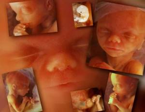
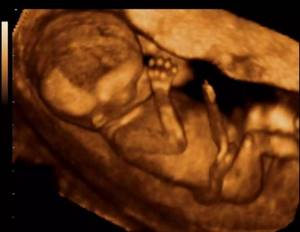
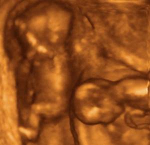
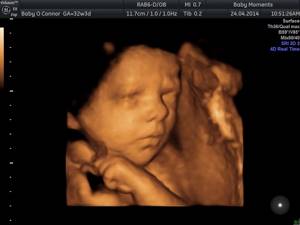
- With a 3D ultrasound, a three-dimensional image of the unborn child is displayed on the screen. The result is a picture similar to a three-dimensional photograph. The body and facial features of the embryo are visible. Parents can also see what the baby looks like.
- On a 3-D ultrasound of the fetus at 12 weeks of pregnancy, the length of the embryo is measured - the so-called coccygeal-parietal size (CTD).
- The device clearly shows how the fetal neural tube develops. This allows us to exclude serious pathologies - underdevelopment of the brain, its absence (anencephaly), dropsy and cysts in the head. The doctor will measure the size of the baby’s head - head circumference, fronto-occipital and biparietal dimensions.
- Assessment of fetal development allows us to identify severe hereditary diseases - Edwards, Down, Patau, Cornelia de Lange, Smith-Opitz syndromes. To do this, the thickness of the collar zone is measured - the distance between the skin and tissues of the neck.
You can see in 3D how your child's face develops. With severe hereditary diseases, facial features are “blurred” and the nasal bone is not visible. If hereditary anomalies are suspected, the woman is referred for other tests and examinations. By going through this simple and inexpensive procedure, you can be sure that the baby does not have any serious pathologies yet. If something goes wrong, it will not be too late to decide not to get pregnant.
Patient reviews
The 3D scanning procedure is considered quite popular and more and more women are interested in the best time to do a 3D ultrasound during pregnancy. Reviews from pregnant women indicate that the procedure guarantees future parents a lot of positive emotions. Especially if the study is carried out at 24 weeks. At this time, the baby is already quite developed and you can examine his facial features, fingers and toes in detail. The expectant mothers were especially impressed by the baby’s serene smile.
For women who care about the health of their child, it is important not only to get a photo as a souvenir, but also to make sure that the baby has no pathologies. Experienced specialists, having studied the data obtained, immediately name the approximate due date, explain how the fetus is developing, and whether there is growth retardation. Patients note that if ultrasound is performed free of charge, the emphasis is on the peculiarities of the formation of organs and systems. Women who underwent the procedure in a private clinic were able not only to obtain information about the health of the baby, but also to observe him. And photographs recorded on digital media can be shown to all relatives and friends.
3D ultrasound does not pose a health risk. But you should not exceed the number of procedures recommended by your doctor.
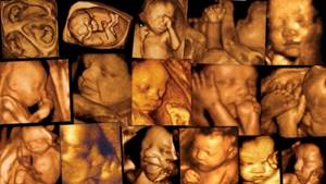
Photo of 3D ultrasound during pregnancy
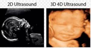
difference between 2D and 3D
3D photo of a baby at different stages of pregnancy
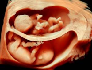
3D ultrasound multiple pregnancy
3D ultrasound 5 weeks
Medical indications
Patients often ask a pregnancy specialist at what time it is better to do a 3D ultrasound of the fetus. The optimal time for the procedure is selected taking into account medical indications:
- the risk of developmental pathologies in the embryo;
- genetic predisposition to dangerous diseases;
- detection of fetal defects;
- surrogate pregnancy;
- undergoing IVF procedure;
- multiple pregnancy;
- determining the sex of the child.
Many institutions allow 3D diagnostics without a medical referral so that parents can see their baby for the first time. It is believed that this allows future fathers to better bond with the baby. It is important to independently ensure that the frequency of studies does not exceed the norm.
Ultrasound in 3D format in the 2nd trimester
This examination is done at 18-21 weeks. It allows you to find out how the pregnancy is progressing and whether there are any complications. Thanks to 3D technologies, you can now view your baby from different angles and take photos and videos for the family album.
What is determined by ultrasound in the second trimester?
Although even at the first ultrasound it was determined whether the child had developmental abnormalities, problems could appear later. For example, if the mother suffered from an infectious disease or took medications that were undesirable during this period. Some defects are poorly diagnosed at the beginning of pregnancy, being determined only at the second ultrasound examination. At the second screening, the sex of the child is determined. 3D technologies make it possible to find out who will be born, even if the baby has his back turned. An ultrasound examination determines whether the position of the placenta has changed or whether its detachment has occurred. They determine the amount of amniotic fluid, identifying polyhydramnios or oligohydramnios, diagnose problems with the vessels of the umbilical cord, assess the viability of the cervix, and exclude isthmic-cervical insufficiency. 3D ultrasound of the second semester is the most important study, so do not ignore it! The doctor performing the ultrasound:
- measures the volume of the fetal head and torso, drawing conclusions about their proportionality;
- assesses the symmetry of the cerebral hemispheres and determines the degree of development of brain structures;
- examines the baby’s spine and elements of the skeletal system;
- evaluates the child's face in frontal and lateral projection. An increased distance between the eye sockets indicates Down syndrome;
- reveals anomalies in the development of the jaws and hard palate - at this stage it is already possible to diagnose “cleft palate”, “cleft lip”, and irregular tooth formation;
- determines the correct development of the cardiovascular system;
- assesses the condition of internal organs
If pathologies are detected, the woman is prescribed additional examinations (blood test, genetic consultation).
Preparing for the examination
Ultrasound diagnostics in the second trimester is carried out without preparation, since the intestine is pushed away by the growing uterus to a sufficient distance and it does not cover the child. The place of the bladder (it is displaced during pregnancy) is taken by amniotic (amniotic) fluid.
How is the examination carried out?
The examination follows the classic procedure - without pain or discomfort. The woman lies on her back, her stomach is lubricated with a special gel that improves the passage of ultrasound. The ultrasound results are displayed on the screen. Sometimes parents are afraid that the child is thin. This is fine. By the time he gives birth, he will gain weight and become plumper, and folds will appear on his legs and arms. The main thing is that the baby is healthy!
Screening ultrasound 3D (4D) in the third trimester of pregnancy - Doppler
The word “screening” translated into Russian means “sifting.” The examination is carried out at 30-34 weeks. It allows you to see the development of the fetus, the condition of the placenta and umbilical cord. Doppler testing allows you to assess the condition of blood vessels and identify placental insufficiency, accompanied by a lack of oxygen in the baby’s blood.
How is 3D (4D) screening ultrasound performed in the third trimester?
During the examination, the fetus, placenta, membranes and umbilical cord can be examined. The doctor sees how correctly the baby’s organs have developed, whether there is enough amniotic fluid, and whether there are any signs of a threat of premature birth. The resulting image shows the presentation of the baby and its position in the uterus. The umbilical cord and other problems are discovered that complicate the birth process. This diagnosis allows you to decide on the tactics of obstetric care. Since the child is practically formed during this period, the doctor evaluates:
- the state of the cardiovascular system - whether there are any heart or vascular defects - this is an ultrasound of the fetal heart;
- brain development - whether its parts have formed correctly;
- fetal biometry (measurements according to protocols), height and weight of the child;
- absence of “early aging” of the placenta, Doppler of the uterine arteries and umbilical cord vessels;
- ultrasound of all internal organs of the baby;
- factors that interfere with natural childbirth.
With 3D ultrasound, a three-dimensional image appears that resembles a stereo image. The fruit on it can be seen from three sides. With 4D ultrasound, a video is taken of what the child looks like. The results are recorded on disk and given to future parents.
Why is doplegramography (USD) performed in the third trimester?
This examination resembles an ultrasound, but during it the condition of the blood vessels is assessed:
- uterine arteries;
- umbilical cord vessels;
- middle cerebral artery and fetal aorta.
A color picture of the state of the vessels appears on the screen: blue indicates the vessels coming from the sensor, red - to the sensor. The darker the shade, the lower the blood flow rate. Most often, this diagnosis is carried out together with ultrasound by the same specialist. If technical capabilities allow, the same apparatus is used. The examination allows you to assess whether the fetus is suffering from a lack of oxygen and whether emergency delivery is required. We are conducting an examination with a new Doppler device. Read about it here.
How is the examination carried out in the third trimester?
No preparation is required for the 3D ultrasound procedure. The examination is carried out regardless of food intake and bladder fullness. The woman is placed on her back, her stomach is lubricated with a special gel that improves the conductivity of ultrasound. The doctor moves a special sensor along the abdomen. The examination result is visible on the screen. Ultrasound and Doppler Doppler examinations are harmless to mother and baby and can be performed as many times as the doctor prescribes. Ultrasound does not cause radiation or fetal abnormalities.
How much does 3D ultrasound cost in St. Petersburg?
In our clinic, the price of ultrasound during pregnancy with 3D visualization depends on timing and other factors. The table shows the base price, excluding discounts. To get a discount and find out the exact cost of a pregnancy examination, call the clinic.
| Fetal examination from 11 weeks. 15 weeks | 2000 |
| Fetal examination in multiple pregnancies from 11 weeks. for 15 weeks | 2900 |
| Fetal examination 15 weeks. for 40 weeks | 3100 |
| Fetal examination in multiple pregnancies from 15 weeks. for 40 weeks | 4000 |
| Fetal examination from 15 weeks. for 40 weeks + dopplerometry | 3600 |
| Fetal examination in multiple pregnancies from 15 weeks. for 40 weeks + dopplerometry | 4600 |
| Screening ultrasound examination in the third trimester during multiple pregnancy (30.0 – 34.0 weeks) | from 4000 |
| Fetometry (ultrasound dynamics of fetal growth) | 1600 |
| Fetometry during multiple pregnancy (ultrasound dynamics of fetal growth) | 2500 |
| Doppler testing during pregnancy (study of blood flow in the middle cerebral artery, fetal umbilical cord arteries, uterine arteries) | 1000 |
| Doppler testing during multiple pregnancy (study of blood flow in the middle cerebral artery, fetal umbilical cord arteries, uterine arteries) | 2000 |
| Cervicometry | 1000 |
| Determination of the amount of amniotic fluid, assessment of the placenta, regardless of the stage of pregnancy | 1700 |
Carrying out diagnostics
Once the patient has decided on the date for 3D ultrasound during pregnancy, she needs to sign up for diagnostics. The procedure is carried out in the same way as a two-dimensional scan - using a standard sensor. To facilitate the sliding of the device, the woman’s stomach is lubricated with a special gel. Thanks to the special principle of moving the device, the computer can build a three-dimensional image based on the received data. The scan takes 5–10 minutes. It takes another 30–40 minutes to process the result.
The study will be informative only if the woman managed to find out at what time it is better to do a 3D ultrasound. It is also worth understanding that the baby can turn the back of his head towards the sensor.
Why do modern mothers increasingly choose 3D diagnostics?
Conventional two-dimensional diagnostics does not lose its relevance, especially during a short period of pregnancy (up to 12 weeks). In some cases, the gynecologist himself may prescribe a 3D diagnostic procedure to exclude the possibility of ectopic pregnancy, genetic diseases, pathologies at the chromosomal level, and defects in the circulatory system. hypoxia, umbilical cord entanglement.
After the procedure, the pregnant woman will receive comprehensive information about the condition of the fetus, its position and the exact duration of pregnancy. 3D ultrasound is especially necessary for women whose professional activities are closely related to hazardous work.
Three-dimensional diagnostics is becoming increasingly popular. However, some women consider this procedure dangerous to the baby's health. In fact, everything is not like that. Ultrasound, which is used in the ultrasound process, has nothing to do with harmful ultraviolet rays. Ultrasonic vibrations do not affect human genetics and do not lead to pathologies. The baby in the womb does not feel any impact due to the small amount of ultrasound that penetrates the body of the expectant mother.
For women who experience complications after IVF, miscarriages or health problems, a three-dimensional study is prescribed. Such cases require constant monitoring of the fetal condition. Some women undergo this procedure not on the recommendation of the supervising doctor, but at their own request. And it is possible. After all, the main thing during pregnancy is positive emotions. And what else can bring more joy than the contemplation of a little man who lives inside, moves, moves his lips and smiles.
<
p style=”text-align: justify;”>
CLICK TO MAKE AN APPOINTMENT, TEST OR ULTRASOUND
If you find an error, please select a piece of text and press Ctrl+Enter
Does the procedure harm the baby?

Ultrasound radiation can penetrate the human body. For this reason, many expectant mothers worry that the baby will be harmed. It must be taken into account that data on the destructive effects of ultrasound were obtained as a result of the use of amplified sound waves. This study does not use such power.
Experts say that an ultrasound performed in accordance with all the rules does not harm either the mother or the embryo. According to WHO, it is considered safe to undergo four ultrasound examinations, and at a period of less than 10 weeks the procedure should not be performed unless absolutely necessary. In many modern clinics, 3D ultrasound is not performed without a corresponding referral. Experts refer to the conclusion provided by the American FDA, which does not allow the possibility of additional ultrasound sessions solely for the peace of mind of parents. In this case, you need to contact a doctor managing the pregnancy, who can recommend at what time it is better to do a 3D ultrasound and write out a referral.