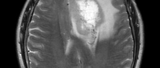
MRI diagnostics is based on the interaction of hydrogen protons present in the body and an artificially created magnetic field. The result of the study is layer-by-layer images in three projections - axial, sagittal and coronal, on the basis of which it is possible to create a three-dimensional model of the structures in question. The high accuracy of the method allows you to detect the smallest pathological changes.

Newborn baby
Magnetic resonance imaging is used at any age to examine various parts of the body. MRI of newborns is done to diagnose diseases of the brain, spine, limbs, abdominal cavity, pelvis, etc. Scanning is necessary for babies with birth injuries, developmental delays, suspected pathologies of the central nervous system, including those caused by infection, problems with nutrition, digestion of food, the presence of symptoms such as sleep disturbances, constant crying for no apparent reason. Lack of timely diagnosis can result in serious, including life-threatening, complications.
Indications for MRI of the brain in children
An examination is prescribed if a child has the following problems:
- deterioration of hearing and vision;
- unexplained changes in behavior;
- dizziness and headaches;
- convulsions and periodic fainting;
- head injury with concussion;
- speech development delay.
Frequent headaches or dizziness as usual
The cause of pain may be disturbances in the blood supply to the brain and inner ear. For correct diagnosis, it is necessary to undergo a modern, highly informative study.
Fainting occurs periodically without visible external influences
Fainting in children that occurs repeatedly may indicate serious illness. If such signs occur, an MRI of the head and brain with contrast is prescribed; a special substance is injected into a vein for better visualization.
Convulsive syndrome occurs periodically
This is a nonspecific reaction of the body to irritants, which manifests itself in involuntary muscle contractions. To determine the causes of seizures, specialists are consulted and an MRI of the child’s head is performed.
Markedly progressive decrease in vision and/or hearing
The study provides an image of the structures located inside the skull. The presence of layer-by-layer images is an opportunity to conduct an accurate diagnosis.
MRI of a newborn under anesthesia
For high accuracy of results, the subject must lie still. Therefore, MRI of an infant is most often performed under general anesthesia. On the eve of the procedure, the anesthesiologist talks with the baby's parents. The specialist is interested in indications for tomography, a history of allergic reactions to medications, concomitant diseases, and behavioral characteristics of the baby.
The size difference between newborn and adult patients requires special anesthetic technique. Children of this age have small lower and upper respiratory tracts, blood vessels, and a small body surface area. Certain anesthetic nuances are associated with the miniature size of the patient.
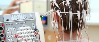
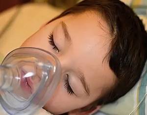
For MRI of the brain and other organs, infants generally use inhalation anesthesia: the drug is administered using a mask. During the diagnostic procedure, doctors monitor:
- arterial pressure;
- heart rate;
- blood oxygen level;
- work of the heart muscle;
- body temperature.
Mask anesthesia
At the slightest deviations in indicators, specialists take measures to eliminate violations. At the end of the examination, the little patient is observed until he recovers from anesthesia.
What will general anesthesia do? The brain goes into a state of rest: while the drug is in effect, reflexes fade, the nerve centers responsible for the ability to feel pain stop functioning, and muscle tone decreases.
For the procedure to proceed without complications, simple preparation is required:
- on the eve of the MRI, you need to put the child to bed two hours later than usual and wake him up one hour earlier;
- anesthesia is used on an empty stomach - when putting the patient into a medicated sleep, it is necessary to prevent aspiration (entry of the contents of the digestive organ into the respiratory tract) - that is why, immediately before an MRI, you cannot eat under general anesthesia:
- infants feeding on mother's milk are fed 4 hours before scanning, infants using artificial formula - six;
- for a child over the age of one year, the fasting pause lasts at least a quarter of a day;
- a few hours before the diagnostic session, the baby is no longer given water;
- Before the procedure, the baby needs to empty the intestines.
Detailed information about why anesthesia is needed and how to properly prepare for the procedure can be obtained from your attending physician or anesthesiologist.
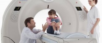
Features of the procedure in children
Parents are interested in how MRIs of the brain are done for children. The main problem is the difficulty of getting the child to remain still while the tomograph takes pictures. You are required to remain motionless for 15-20 minutes. The device is noisy and can frighten the baby. However, patient immobility is a mandatory requirement, so tomography in some cases is performed under anesthesia. Indications for the use of anesthesia are age under 5 years, fear of confined spaces, panic fear of examinations, hyperactivity, and the presence of mental illness. To conduct examinations in infants, special incubators are used, which are used in conjunction with a magnetic tomograph. Anesthetic agents are propofol-based drugs, which are characterized by:
- short action;
- low level of toxicity;
- minimizing side effects;
- falling asleep quickly;
- minimal impact on the central nervous system;
- no effect on the respiratory centers;
- rapid elimination from the body.
When using anesthesia that does not affect the central nervous system, the baby's infancy is not a contraindication. Doctors at specialized diagnostic centers have extensive experience in conducting examinations of children of different ages. Parents ask what an MRI of the child’s brain shows. The study allows you to visually analyze the state of the brain and identify pathological changes that appear due to difficult pregnancy and childbirth. Using the method, problems in brain development caused by intrauterine development disorders, previous diseases, bruises and injuries are identified.
MRI for newborns, how is it done?
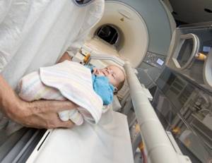
Preparing your baby for scanning
Before an MRI is performed, items with metal elements are removed from the baby. The little patient is then placed on a mobile tomograph table. The torso and head of infants are fixed with special devices to prevent movement. If contrast is required, a catheter is inserted into the vein to inject a dye. Before going into medicated sleep, sensors are attached to the newborn's body to monitor vital signs.
Diagnostics takes from 15 minutes to half an hour. First, a scan is done without an amplifier. When the required number of images is obtained, a drug is administered to improve visualization and the manipulations are repeated.
There are usually no complications after MRI in children. If the procedure was performed under anesthesia, after emerging from drug-induced sleep, individual reactions may appear in the form of weakness, nausea, etc. Side effects go away on their own after some time.
In many clinics, one of the parents can be in the room where MRIs are performed on infants. It is worth clarifying whether your presence at the examination is acceptable when making an appointment for the scan.
How is the research performed?
Doctors create the most comfortable atmosphere for the child so as not to frighten him. It is necessary to ensure the patient's immobility; if this is not possible, anesthesia is used for children. Older children undergo the test in the presence of their parents, who can help reassure them. The child is placed on a special table, which slides into the apparatus. The use of an open type tomograph allows the baby to see his parents and feel comfortable. The equipment is equipped with a feedback system for communication, listening to fairy tales and music. You can watch a video of how an MRI of the brain is done for children
.
MRI for children: without fear and anesthesia
Experts have long known: MRI studies performed on children differ significantly from this procedure involving adult patients. It is not easy for children to spend a certain amount of time in a tomograph without moving; they are wary – and many with fear – of unfamiliar surroundings, people in white coats, and equipment that is incomprehensible to them. But “Clinic Expert” Orenburg accepts more and more children from year to year, parents bring them here quite willingly. Why? We are talking about this with the director of the clinic, Yuri Andreevich Podlevskikh.
– Yuri Andreevich, is it difficult to work with children?
– Yes, the procedure is quite difficult in many cases, even adults find it difficult to undergo it, but MRI and children are a separate matter. We conduct research on them from infancy to adolescence. They all behave differently. Considering all the features of MRI for children, we have already found our approach to them.
It all starts with individual preparation of both the child and his parents
– What is it?
– You know, we can’t say that this is some kind of magic vitamin, universal for everyone, that we give to children, and this allows us to calmly perform our functions. In each specific case, it all begins with individual preparation of both the child himself and his parents. We have employees who, I would say, are child-oriented. Administrators have a list of tools that they use in the process of preparing for the study. The day before, in a conversation with the parents, they try to find out in as much detail as possible what kind of child we have to deal with: his age, what his character is, how he behaves in an unfamiliar environment, with strangers. We find out all these details very carefully. Based on the information received, the administrator tells parents what is required of them to prepare the child for the study to be successful.
A fairy tale about how the Hedgehog helped the Little Bunny stop being afraid of an MRI. Read to your child before the test!
Some baby needs to be woken up early in the morning, then, already in our clinic, in a special room, play with him, thoroughly, so to speak, “coax” him and, in the end, lull him to sleep - as a result, the child then simply sleeps peacefully in the tomograph , without interfering with our work at all.
– What is better – or rather, what is easier – to do an MRI for children with or without anesthesia?
– In our clinic we are against anesthesia for children. Strongly against it! Today the entire civilized world is trying to abandon this method. Anesthesia is a serious manipulation, a serious intervention in the state of the body. After all, everyone understands that even vitamins are not harmless - if they are used uncontrolled, then what can we say about anesthesia. We do not conduct research using it; we try our best to use our own methods when carrying out the procedure. And, as practice shows, they work. I myself am a pediatrician by training, and this probably also plays a role.
We are against anesthesia for children. Strongly against it!
– Do you have any statistics: where are MRIs mostly done for children in Orenburg today?
– Of course, besides our clinic, such studies are carried out in some other centers, but I can say with complete confidence that there is no such large number as ours anywhere else. This can be explained quite simply: word of mouth works effectively. Parents who visited us with their children later tell their relatives and friends how they were received here, and they, in turn, bring their children to us when the need arises. And again, they share their impressions with their circle of close people.
– And those of your employees who, as you say, are child-oriented – did they undergo any special training to work with children?
– This is probably difficult to teach. If we have any “secrets” that allow us to work effectively with children, then they exist, as it were, inside each of our employees. To work in our clinic, we initially select people who have a sense of empathy and compassion for patients, especially small ones. Those who work here are those who sincerely want to help people. Agree, this cannot be taught. It must be a state of mind.
Interviewed by Igor Chichinov
The editors recommend:
Fairytale therapy for children's problems. Fear of needles
We will find an approach to everyone! MRI for children: go through and not be scared
Vaccinations: where is the truth and where is the lie?
For reference:
Podlevskikh Yuri Andreevich
In 2012 he graduated from the Orenburg State Medical Academy with a degree in Pediatrics. In 2013, on the basis of the same educational institution, he completed an internship in the specialty “Radiology”. In the same year, he underwent advanced training in magnetic resonance imaging. Currently holds the position of executive director of Clinic Expert Orenburg.
Contraindications for the procedure for children
The study is not carried out if the child has: foreign metal objects (staples, stents, prosthetic heart valves);
- electronic devices (pacemaker, insulin pump);
- hematopoietic anemia (in case of administration of a contrast agent).
If a teenager has a tattoo on his body, the doctor determines whether the procedure can be performed.
The solution depends on the dye used (paints should not be metal-based). Examination under anesthesia is not carried out if there is a history of:
- rickets;
- developmental delay;
- lack of body weight;
- feverish conditions;
- bronchial asthma during exacerbation;
- mental disorders;
- bacterial and viral infections;
- acute neurological diseases.
After a tomography the child is capricious
Although MRI scans are not dangerous for children, general anesthesia can have some effects on babies under one year of age. Infants sometimes have disruptions in their eating and sleeping patterns. Babies begin to confuse day and night, become capricious, and refuse to eat. This is normal. The previous order of life is restored within one to two weeks.
At the Magnit Medical Center, tomography is performed on children from the age of five. The main condition for undergoing the procedure is the child’s ability to maintain a static position for as long as the diagnosis requires. If you have conditions that prevent you from staying still, we recommend having an MRI done under general anesthesia in the hospital.
What does an MRI show?

To date, magnetic resonance imaging has repeatedly confirmed its safety and maximum information content of the resulting images (more details in this article). Scanning allows us to identify hidden inflammatory and tumor processes, developmental deviations, or detect trauma in a child. But the parent must be confident in the quality of the research performed, because the child’s further treatment plan depends on its results.
We recommend contacting clinics that have a high-field tomograph installed - the image obtained on it has high contrast, and the examination time will be shorter compared to a low-field tomograph, which is an important point when examining a child. You can learn about the differences between low-field and high-field devices here.