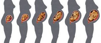
There is hardly a more beautiful moment in a woman's life than pregnancy. However, the health of the unborn baby is a natural concern for every mother. An ultrasound examination can help you get rid of worries and start getting to know your unborn child.
Nowadays it is not at all necessary to limit yourself to performing a standard two-dimensional ultrasound. Modern medicine, in addition to the usual ultrasound procedure, offers parents innovative 3D and 4D techniques. However, often mothers and fathers do not understand the difference between these diagnostic tools, which is discussed in this material.
3D and 4D ultrasound – what is it?
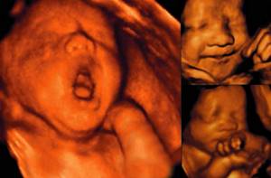
- 3D ultrasound. A three-dimensional image makes it possible to obtain a color and three-dimensional picture, in which you can see the appearance of the future baby in as much detail as possible. The three-dimensional image is static, which is the main difference between this examination and 4D ultrasound.
- 4D ultrasound. With the help of a four-dimensional picture, future mothers and fathers get an exclusive opportunity to look at their child literally in real life. Choosing this technique allows you to purchase a recording of the baby’s movements, which can be saved in the form of photographs, on disks, or flash drives for memory.
3D ultrasound - how it works
The technique of three-dimensional ultrasound examination has no fundamental differences from standard ultrasound, which is based on the penetrating ability of ultrasound, its distribution in the tissues of the body depending on the density and composition of the medium. Only the image provided by a classic ultrasound does not allow future parents to detect the baby’s spine and large bones without medical assistance. While a 3D ultrasound image can be compared with an ordinary photograph in which the child’s face is visible, it is possible to count his fingers.
You need to prepare for the fact that the duration of the procedure will increase compared to a standard ultrasound. It will take at least 40 minutes.
3D and 4D ultrasound – what to choose
Three-dimensional examination has some limitations that relate to the location of the fetus and the timing of pregnancy. For example, if the baby turns his back to the sensor, mom and dad will not be able to see his face. However, volumetric images allow better examination of some structures that cannot be studied in the usual mode. This makes it easier to understand the image and assess the child’s health status, which is very important for the timely detection of external malformations. Therefore, this examination must be prescribed if indicated. Conducting a 3D ultrasound reassures the expectant mother, as she can independently verify the absence of external pathologies and count the baby’s fingers and toes.
If a four-dimensional ultrasound examination is performed, time is added to the depth and height of the image, so you can admire the baby in real time. Ultrasound in this mode does not pose the slightest danger to the health of the child and his mother, which has long been proven by numerous clinical trials. However, it is still worth carrying out this examination as prescribed by a doctor, strictly within certain periods during which it is most valuable.
Safety
Many believe that such “modern” research is more dangerous for the child. This is not so, because the physical basis of the method remains the same, similar to conventional sonography. The child receives only half of the ultrasound waves. The pulsed nature of the effect further reduces the number of ultrasonic waves reaching the fetus.
Conversations about whether 3D ultrasound is harmful during pregnancy were born out of the myth that long-term and strong exposure is necessary to obtain a more accurate image. This is not true, since the design of modern devices does not “link” image quality and duration of ultrasound irradiation.
Importance of the study
Screening helps to identify various pathologies at an early level of their appearance. This diagnostic method makes it possible to promptly identify anomalies and developmental defects in the unborn child:
- Down syndrome.
- Cleft lip.
- Wolf's mouth.
- Heart disease and so on.
The pros and cons of 4D ultrasound are exactly the same as the 3D format.
Is 3D ultrasound safe for mother and baby?
3D-4D ultrasound are recognized as the safest diagnostic methods, which, even with frequent use, do not harm the fetus. Ultrasound examination is safe for both the pregnant woman and her unborn child.
In gynecological practice, pregnancy situations occur with complications. This is often aggravated by the expectant mother’s worries about the safety of certain diagnostic procedures for the health of her child. Women experience additional psychological stress, thinking about how problems in their body can affect the development of the fetus.
In such cases, ultrasound helps to dispel the doubts and worries of future parents. It does not harm the mother at all and is safe for the fetus. The results of 3D ultrasound provide valuable information for the doctor monitoring the pregnant woman.
If you need a 3D ultrasound or treatment, contact the specialists of our clinic in Moscow. You can make an appointment by phone. It is recommended to clarify the exact prices and availability of the necessary examination methods by phone.
Currently, in obstetrics there is no method for diagnosing pregnancy that could compete with ultrasound in terms of information content.

The first ultrasound examination is carried out to determine the presence of a fertilized egg in the uterine cavity and determine whether the size of the fertilized egg corresponds to the expected period of pregnancy. According to the recommendations of the Ministry of Health of the Russian Federation and WHO, three mandatory screening studies are performed - at 11–14, 18–22 and 30–32 weeks.
Diagnostic capabilities of 3D ultrasound
The study allows you to obtain maximum important information about the condition of the fetus. Three-dimensional ultrasound is recommended to be performed in the first trimester of pregnancy to confirm the absence of chromosomal abnormalities and other abnormalities in the development of the embryo.
3D ultrasound is indicated to exclude the following pathologies:
- Chromosomal abnormalities of the fetus.
- Anomalies in the development of the central nervous system of the embryo.
- Congenital anomalies of the facial structure.
- Congenital heart defect.
Diagnostics allows you to most accurately and quickly assess the general condition of the embryo at an early stage of development. Helps ensure that the placement of body parts and proportions are correct.
The device accurately visualizes the amniotic membrane and the body of the embryo itself. Allows you to measure the cervical fold - a parameter on the basis of which conclusions are made about the normal development of the fetus. The condition of the nasal bone is also studied using 3D ultrasound. It tells about the presence/absence of chromosomal abnormalities.
Ultrasound in three-dimensional scanning mode makes it possible to reconstruct the structure of the fetal brain. This helps to eliminate disturbances in its development.
3D ultrasound, unlike standard two-dimensional ultrasound, provides the opportunity to study the condition of the fetal face. During an ultrasound examination, the doctor may detect abnormalities such as cleft lip and cleft palate.
Such anomalies have become increasingly common in recent years and are considered one of the common causes of sudden death in newborns. 3D ultrasound allows you to examine the fetal heart with maximum accuracy and detect abnormalities in its structure. The standard ultrasound diagnostic procedure is practically powerless in this matter.
Women with complicated pregnancies are usually referred to 3D ultrasound when it is necessary to clarify the condition of the fetus. The study is carried out if there are suspicions of abnormal development of the embryo. Women who have used assisted reproductive technologies to conceive undergo three-dimensional ultrasound.
If a pregnant woman is expecting twins or triplets, doctors may recommend 3D ultrasound. In addition, a woman can undergo such a study at her own request to ensure the normal development of the fetus.
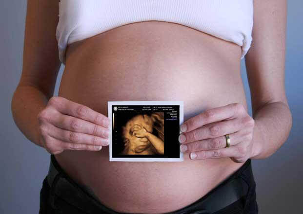
Every pregnant woman looks forward to the moment she sees her baby. Thanks to ultrasound, this moment comes earlier than 9 months. Currently, ultrasound has become a mandatory procedure in the examination of pregnant women. The first recommended ultrasound is performed at 10-11 weeks. At this stage, developmental pathologies are ruled out, the exact date is determined, and of course, the baby’s heart is listened to.
At 20-24 weeks you will already be shown your little one in motion. With the new premium device VOLUSON E8 you can see every arm and leg in detail, and your baby will definitely smile or yawn. And finally, you will find out what color dowry you should buy - the doctor will determine the sex of the child if you wish. The third ultrasound is done before birth, 32-34 weeks. The doctor will examine the baby’s organs and other important features for childbirth.
With the help of an ultrasound expert 3D, 4D you can:
- determine pregnancy in the early stages;
- determine the number of children to be carried (after the 6th week) - so that twins are not a surprise to you, and you can prepare for their appearance morally and financially, and doctors can monitor the development of the pregnancy and make the right decisions;
- determine the exact age of the fetus;
- diagnose ectopic pregnancy (in the early stages) to avoid surgical intervention and correct the situation without surgery;
- determine the pathology of the pregnancy (placental abruption, threat of miscarriage, uterine hypertonicity, etc.) - in order to maintain the pregnancy;
- characterize the physical condition of the fetus;
- identify fetal malformations in order to make an objective decision about the need to terminate the pregnancy (if the malformations are incompatible with life) or carry out the necessary treatment (if appropriate);
- prepare for some features of childbirth - find out the weight of the fetus, determine its presentation, diagnose the presence or absence of umbilical cord entanglement, determine the placenta insertion, clarify the due date;
- find out the gender of the child.
Advantages of three-dimensional ultrasound 3D, 4D expert class:
Unlike conventional ultrasound, 3D and 4D three-dimensional examination data provide additional information that makes it possible to diagnose many abnormalities in the fetus; thanks to this, important information is obtained about some malformations of the face, spine, limbs, and internal organs. The 3D, 4D format is a unique diagnostic method that makes it possible to see the child as realistically as possible on-line. Ultrasound used in medicine (obstetrics) is absolutely safe for both the expectant mother and the baby. This is officially confirmed by clinical studies and many years of experience in use during pregnancy.
It is necessary to clarify that ultrasound affects the fetus for only 1% of the total time of ultrasound; thus, the rest of the time, the sensor only receives reflected signals and processes the information received.
The resulting three-dimensional image of the baby, as well as his movements and the main moments of formation and development, can be recorded on an electronic medium.
Is 3D ultrasound safe for mother and baby?
3D-4D ultrasound are recognized as the safest diagnostic methods, which, even with frequent use, do not harm the fetus. Ultrasound examination is safe for both the pregnant woman and her unborn child.
In gynecological practice, pregnancy situations occur with complications. This is often aggravated by the expectant mother’s worries about the safety of certain diagnostic procedures for the health of her child. Women experience additional psychological stress, thinking about how problems in their body can affect the development of the fetus.
In such cases, ultrasound helps to dispel the doubts and worries of future parents. It does not harm the mother at all and is safe for the fetus. The results of 3D ultrasound provide valuable information for the doctor monitoring the pregnant woman.
If you need a 3D ultrasound or treatment, contact the specialists of our clinic in Moscow. You can make an appointment by phone. It is recommended to clarify the exact prices and availability of the necessary examination methods by phone.
How 3D and 4D ultrasound is performed during pregnancy
3D and 4D ultrasounds during pregnancy are performed no earlier than 12 weeks. The most optimal period is the second trimester of pregnancy. It is at this time that you can get the clearest image.
Women are interested in how 3D and 4D sonography is done. The procedure itself is no different from the usual one. The pregnant woman lies down on the couch, and the doctor conducts an examination through the anterior abdominal wall using a special sensor. A shorter time allows a woman to quickly turn into a position that is comfortable for her. Thus, a regular ultrasound lasts approximately 15 minutes, and 3D sonography lasts about 3 minutes.
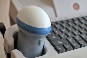
Special volumetric (three-dimensional) sensor with mechanical drive
No special preparation is required for this study. There is no need to follow a diet; filling the bladder does not affect image quality.
The clarity of the resulting “picture” in 3D can be slightly distorted by some factors: excess weight of the mother, a large amount of amniotic fluid, features of the presentation (location) and activity of the baby, scars on the anterior abdominal wall, late pregnancy.
Diagnostic capabilities of 3D ultrasound
The study allows you to obtain maximum important information about the condition of the fetus. Three-dimensional ultrasound is recommended to be performed in the first trimester of pregnancy to confirm the absence of chromosomal abnormalities and other abnormalities in the development of the embryo.
3D ultrasound is indicated to exclude the following pathologies:
- Chromosomal abnormalities of the fetus.
- Anomalies in the development of the central nervous system of the embryo.
- Congenital anomalies of the facial structure.
- Congenital heart defect.
Diagnostics allows you to most accurately and quickly assess the general condition of the embryo at an early stage of development. Helps ensure that the placement of body parts and proportions are correct.
The device accurately visualizes the amniotic membrane and the body of the embryo itself. Allows you to measure the cervical fold - a parameter on the basis of which conclusions are made about the normal development of the fetus. The condition of the nasal bone is also studied using 3D ultrasound. It tells about the presence/absence of chromosomal abnormalities.
Ultrasound in three-dimensional scanning mode makes it possible to reconstruct the structure of the fetal brain. This helps to eliminate disturbances in its development.
3D ultrasound, unlike standard two-dimensional ultrasound, provides the opportunity to study the condition of the fetal face. During an ultrasound examination, the doctor may detect abnormalities such as cleft lip and cleft palate.
Such anomalies have become increasingly common in recent years and are considered one of the common causes of sudden death in newborns. 3D ultrasound allows you to examine the fetal heart with maximum accuracy and detect abnormalities in its structure. The standard ultrasound diagnostic procedure is practically powerless in this matter.
Women with complicated pregnancies are usually referred to 3D ultrasound when it is necessary to clarify the condition of the fetus. The study is carried out if there are suspicions of abnormal development of the embryo. Women who have used assisted reproductive technologies to conceive undergo three-dimensional ultrasound.
If a pregnant woman is expecting twins or triplets, doctors may recommend 3D ultrasound. In addition, a woman can undergo such a study at her own request to ensure the normal development of the fetus.
Recommended timing
Mandatory screening ultrasound examinations during pregnancy are carried out three times: at 11 - 14, 18 - 24 and 32 - 36 weeks. Additional research is prescribed if there are indications and if there are suspicions of possible anomalies in the development of the fetus and placenta, pathology of the amniotic fluid and the course of pregnancy. Three- and four-dimensional ultrasound is recommended at 17-18 weeks.
You can see prices for services
What are 4D and 5D?
The terms 4D and 5D mean the same 3D cinema, the reproduction of which is accompanied by various effects adapted to certain scenes of the film. This could be stereo sound in close proximity to a person, drops of water simulating rain, vibration, rotation or movement of spectator seats. Often 4D and 5D cinema are considered as a type of attraction.
Thus, 4D and 5D are rather marketing terms. They do not indicate some technology for playing films on a screen, which is a level above 3D cinema. Of course, in modern cinemas that position themselves as entertainment centers showing 4D and 5D films, as a rule, the most modern and high-tech stereo cinematic systems are installed. Therefore, to a viewer who has had experience watching 3D cinema on outdated equipment and subsequently visited a 4D or 5D cinema, it may seem that 4D and 5D technologies are significantly superior to 3D.

But this will be an erroneous perception of their essence. The next time a person goes to a cinema with an even more technologically advanced stereo cinematography, but without 4D and 5D effects (that is, 3D) and, quite likely, he will immediately understand that the quality of the picture is not fundamentally determined by the format in which the film is played - 3D , 4D or 5D. What is important is how technologically advanced a particular stereo cinema system is that displays the picture on the screen.
In 4D and 5D cinemas, when all effects are activated, sometimes only short films are played - about 10-15 minutes long, since many viewers get tired of the effects that accompany the show. 4D and 5D technologies are used not so much in classic cinemas, but in those entertainment centers that position themselves as attractions. In this case, stereo cinematography systems are installed not in halls, but in separate rooms designed to be visited by a small number of people. They can be adapted to reproduce a film with very powerful effects. For example, capable of causing sensations similar to those experienced by a person riding a carousel.