What is sputum?
Sputum is a substance that accumulates on the walls of the organs of the respiratory system in case of illness. The secretion in the lungs and bronchi is always produced and is released in small quantities without irritating the cough receptors.
But as the pathological process develops, it becomes too much. Sputum varies in composition and can be:
- in case of inflammation or bronchial asthma – mucous membrane;
- for bacterial diseases - purulent;
- with pulmonary edema - serous;
- in case of tuberculosis or cancer of the respiratory system - bloody.
In any case, it must be removed from the respiratory tract. Its accumulation is fraught with bronchial obstruction. If their lumens are blocked, respiratory failure will occur, which is life-threatening.
Can fluorography be done if you have a cold or during pregnancy?
Sometimes you have to go for an examination during ARVI and when you have a cough, then the question arises whether fluorography can be performed for such diseases. If there is no severe cough, sputum, or fever, then it is acceptable to come to the clinic with snot to undergo fluorography.
It is better not to ignore such cold pathologies, since it is possible that the examination will show, for example, an increase in the pulmonary pattern, which may require further more detailed analysis.
For a cold, this procedure can help identify tuberculosis, which sometimes occurs under the guise of bronchitis with an endless dry or wet cough.
Therefore, undergoing fluorography for a cold is not only possible, but also desirable to exclude pathologies, especially if an antibiotic does not relieve pain.
Types of cough
Based on the amount of discharge, a productive and non-productive cough is distinguished. The first is accompanied by the discharge of sputum and, in fact, performs its main function. The second one is dry. At the initial stage of the disease, it occurs due to the increased viscosity of the sputum. But it can also occur under the influence of other irritants of cough receptors (inflammatory process or flowing nasal secretions).
Typically, the cough is initially nonproductive and becomes wet as the sputum thins. This is inconvenient, but necessary for recovery. However, unpleasant sensations and ignorance of the operating principle of the cough mechanism force people to make serious mistakes during treatment.
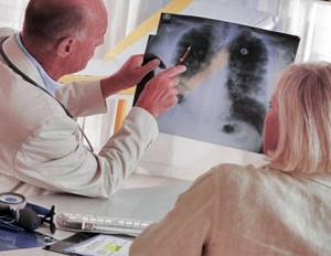
Diagnostic fluorography
However, fluorography can be prescribed for a cold accompanied by a cough and for diagnostic purposes - for example, if pneumonia is suspected.
This method is highly informative and practically no different from conventional radiography. Radiation exposure during FG is small and cannot harm the patient.
In case of pneumonia, the image will reveal foci of darkening - places of infiltration of the lung tissue with inflammatory fluid. This is decisive when making a diagnosis.
With bronchitis there will be no characteristic changes. Severe inflammation can lead to increased pulmonary pattern, but this radiological finding is not pathognomonic specifically for bronchitis. It also occurs in other pathologies:
- Bronchial asthma.
- Chronic obstructive pulmonary disease.
- Occupational pathology (workers of mines, plants, factories).
- In chronic smokers.
Sometimes fluorography for a common cold helps to identify tuberculosis, which often occurs under the guise of bronchitis with a constant dry or wet cough. In recent years, the incidence of lung cancer has increased in older people.
It is possible to perform fluorography for colds. However, you should consider the purpose for which this examination is prescribed.
Common misconceptions when treating coughs
We have collected the most common mistakes that are made when fighting a cough. By avoiding them, you can recover faster and prevent complications.
Misconception 1: Suppressing the cough reflex when producing sputum
This error is caused by a misunderstanding of the nature of cough. People believe that this is where the problem lies, and therefore they strive to eliminate the unpleasant symptom. However, coughing is not a disease, but a mechanism for clearing the airways. It helps fight the consequences of the disease in the form of sputum accumulation.
But sometimes understanding the need to clear your throat doesn’t help. The patient takes medications to suppress this reflex and relieve discomfort. With a wet cough, there is a feeling as if the person is about to choke. I want to prevent this.
But taking antitussive drugs in this case is unacceptable, otherwise sputum will accumulate in the lungs. It is necessary to clear the airways. Mucolytic and expectorant drugs will help with this.
Misconception 2: Treating a cough with antibiotics
In the minds of many, antibiotics are a miracle remedy that saves from advanced diseases. If the cough is very severe or does not go away for a long time, then it can only be cured with antibiotics. But this is not a mistake - it is a choice that can lead to serious side effects:
- suppression of intestinal microflora;
- avitaminosis;
- liver dysfunction;
- violation of renal structures.
Antibiotics are prescribed to treat complications of the disease causing the cough. And only if they are of a bacterial nature (for example, pneumonia). But the symptom itself is never caused by bacteria.
Therefore, you can take antibiotics only when indicated. It is better not to resort to them without the supervision of a doctor. Without medical knowledge, it will not be possible to properly build supportive therapy.
Misconception 3. Taking mucolytics for non-viral dry cough
Many people believe that a dry cough is always a symptom of the onset of a viral respiratory disease. But it can also be caused by other reasons. It happens that there is no excess phlegm in the body, and there is simply nothing to liquefy with mucolytic drugs.
To correctly select the necessary medications, you need to establish the cause of the cough:
- if it is nasal congestion, treat a runny nose;
- if it is an allergic reaction, take antihistamines;
- if this is a reaction to dry air, humidify it;
- if it is cough neurosis, undergo a course of psychotherapy.
In such cases, taking traditional antitussive drugs has no effect. People begin to suspect complications and take strong medications, which are harmful to the body. To prevent this, it is better to immediately consult a doctor who will find the cause of the cough.v
Misconception 4. Using ineffective folk remedies
Here we will look at four common mistakes at once, find out their nature and determine the correct procedure.
| Error | Why is this wrong | What do we have to do |
| Staying in a dry room during acute respiratory infections. | It is a well-established myth that indoor humidity interferes with healing. In fact, dry air reduces the activity of interferons necessary to fight the virus. | Intentionally increase the humidity in the room where the patient is. Household appliances or regular wet cleaning will help with this. |
| Refusal of medications in favor of herbal analogues. | People are afraid of medicines, considering them chemicals. But plants can cause no less harm (for example, an overdose of the active substance or an allergic reaction). | Take medications selected by your doctor taking into account the diagnosis and concomitant diseases. |
| Treatment with mustard plasters and cupping. | Their benefits have not been clinically proven. But the harm in the form of skin burns and excessive stress on blood vessels is obvious. | Choose treatment methods that have been proven to be effective. |
| Ignoring cough. | Many people believe that a cough, like a runny nose, goes away on its own sooner or later. But it is not always caused by a mild cold, so there is a risk of missing a serious illness. | Treat a cough after first establishing its cause. It is important to prevent it from becoming chronic, as this makes the respiratory tract more vulnerable. |
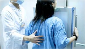
Screening fluorography
If a medical examination is necessary in the autumn-winter period, a mild cold or acute respiratory viral infection, the patient’s cough is quite likely. Could this affect the quality of the examination?
The main task of fluorography (FG, or FLG) is to identify tuberculous or tumor lesions of the lungs. That is why it is classified as a screening research method.
An X-ray of the lungs should be performed annually for all persons except pregnant women and children under 14 years of age. In conditions of low incidence of tuberculosis, fluorography is valid for two years.
If the risk of infectious or tumor damage to the respiratory system is high, fluorography is recommended every 6 months. Can fluorography be done if you have a cold?
In case of acute respiratory viral infection or a slight cough, there will be no changes in the image, and it can be safely performed during this period. Unlike the blood system, the respiratory organs do not react so sharply to the inflammatory process that it can be seen in the picture.
If you take a picture of a more serious pathology - for example, severe bronchitis, chronic obstructive pulmonary disease in the acute stage, fluorography will most likely show an increase in the pulmonary pattern. This will be a reason for further examination.
If the disease occurs with obstruction (bronchial spasm), this can lead to accumulation of sputum in certain areas. Sometimes this process is mistaken for pneumonia.
In these situations, it is better to postpone a routine preventive medical examination until complete recovery.
Proper cough treatment
The main task in treating a respiratory disease is to free the lungs from phlegm. It can build up in the first few days, causing a dry cough. But in the future, the secretion should liquefy and be removed from the body.
If this does not happen, it is necessary to take mucolytic (thinning sputum) and secretomotor (stimulating its discharge) medications. It is better to select them under the supervision of a doctor.
According to the observations of pulmonologists, sputum is best removed in the morning, lying on your side. You should not take expectorants at night, otherwise you will not be able to sleep.
If a dry cough is caused not by respiratory diseases, but by an inflammation of the throat or an allergy, the treatment strategy will be different. Here it is permissible to suppress the cough reflex. However, this does not negate the need to combat the disease that caused it.
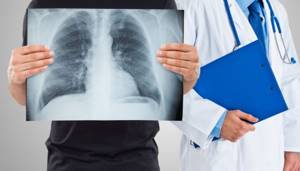
What is FG
Until this method was introduced, fluoroscopy (where organs are viewed on a screen while exposing the patient to X-rays) was used throughout the world to detect early chest disease.
Fluorography is one of the methods for examining the chest organs, which uses x-ray irradiation. The point of the examination is to take a photo of the visible image on a fluorescent screen, which is formed as a result of the passage of X-rays through the patient's body and due to the varying degrees of absorption by their organs and tissues. The method can be used not only for the chest organs, but this is its most common application. Other bodies include:
- bony skeleton of the chest;
- ribs;
- thoracic spine;
- lungs;
- mediastinal organs.
The procedure can reveal some pathologies in the early stages of development. Diseases that can be diagnosed during fluorography:
- different forms of tuberculosis;
- lungs' cancer;
- sarcoidosis;
- pneumonia, pneumonia;
- chronic lung diseases;
- mediastinal diseases.
Also, based on the results of this procedure, the doctor can suggest heart pathologies, and then refer you for a consultation with a cardiologist.
The radiation dose to patients during fluorography using new equipment has decreased several times compared to the radiation dose received using traditional equipment. It is about 0.3 mSv, while the permitted radiation dose for preventive studies is 1 mSv.
The possibilities have also increased: the doctor can enlarge some areas if he suspects any abnormalities; The image quality has improved.
How often is the procedure allowed?
Some groups of citizens must undergo mandatory 1-2 fluorography examinations per year: military personnel, citizens with chronic diseases, persons without permanent residence.
Indications
Fluorography is recommended for preventive purposes once every 2 years for the following categories of patients:
- over 16 years old;
- patients in a clinic or other medical institutions, if there has been no prior examination;
- people living with a pregnant woman or small children;
- military personnel and HIV-infected people.
In cases where it is necessary to resort to an examination of the chest organs, the first mandatory procedure on the list is fluorography.

Contraindications
There are no contraindications that would completely prohibit this procedure.
However, fluorography is not recommended in the following situations:
- the patient's condition does not allow him to be in an upright position;
- claustrophobia in the patient;
- pregnancy in the early stages (fluorography is safe only after the 2nd trimester, when the fetus has already formed vital organs);
- the period of breastfeeding (if research is still necessary, then it is worth expressing milk, and feeding the baby only with the milk that is produced afterwards);
- childhood (this procedure can be harmful to a weak body, and the resulting radiation can weaken the immune system, which will increase the likelihood of viral and infectious diseases).
Possible complications
A large volume of fluid in the pleural cavity can provoke a serious complication - acute respiratory failure. It occurs due to compression of the lung tissue by accumulating exudate. The situation can lead to the following adverse consequences:
- Poor circulation and the development of severe cardiac pathology.
- There is a high risk of displacement of organ structures in the middle parts of the chest cavity.
- Attachment of an infectious factor. Empyema of the pleura develops - a purulent process in the pleural cavity.
If an infection occurs, hydrothorax can be complicated by pleural empyema - a diffuse purulent lesion of the pleural layers.
Diagnostic methods in Medscan
The doctor detects the presence of excess fluid in the pleural cavity at the initial stages of the examination. The specialist examines the chest, performs percussion (tapping) and auscultation (listening with a stethoscope).
Hydrothorax during oncological process is confirmed by the following studies:
- Chest X-ray. The examination allows you to see not only the upper level of fluid, but also the lymph nodes affected by metastases.
- Computed tomography and magnetic resonance imaging. The methods will provide detailed information about the pathological condition. The images can show even a small amount of fluid in the pleural cavity and reveal neoplasms and metastatic foci.
- Thoracentesis. The procedure is carried out for both diagnostic and therapeutic purposes. It is necessary if a significant amount of exudate has accumulated. A special needle is used to make a puncture in the intercostal space, remove part of the fluid, and then examine it in the laboratory.
- Videothoracoscopy. The method helps to make a final diagnosis for formations of unknown origin. A thin endoscopic instrument, a thoracoscope, is inserted into the chest cavity. A video camera is attached to it, allowing you to study tissue structures in detail. If necessary, the doctor performs a biopsy to confirm the malignant process.
The Diagnostic Center of the Medscan Clinic carries out the necessary complex of laboratory and instrumental studies. Specialists will identify oncology and its consequences - hydrothorax, metastatic foci.
What causes fluid to accumulate in the pleural cavity during cancer?
Pleural effusion syndrome is a concept that combines the conditions described above with the accumulation of fluid in the pleural cavity (hydrothorax, pleurisy, empyema).
Various types of oncopathology can lead to the syndrome:
- malignant neoplasm of the lungs;
- cancer of the female reproductive organs;
- blood oncology;
- cancer of the immune system;
- melanoma (skin cancer);
- oncological process of the stomach;
- sarcoma (aggressive forms of connective tissue tumor).
Pleural effusion forms continuously. The substrate is the liquid part of the blood, which permeates the walls of small blood vessels (capillaries). After this, the resulting fluid is absorbed into the lymphatic system. Both processes in a healthy person are in balance, in dynamic equilibrium.
Damage to pleural tissue by atypical cells increases capillary permeability. When metastatic foci occur in the lymph nodes, the outflow of lymph becomes difficult. The situation is especially aggravated in the case of damage to the lymph nodes located in the chest.
An increase in effusion production and a decrease in its outflow leads to the development of hydrothorax.
The situation worsens with diseases of the heart, liver, and kidneys.
Possible risks
Most hospitals are equipped with fluorographs, special machines that give an effective equivalent dose in the range of 0.5 to 0.8 mSv, which can further affect health if the procedure is carried out too often. Digital fluorographic examination gives a radiation dose of 0.4 mSv, but such equipment is not used in every clinic.
Sources:
https://elaxsir.ru/zabolevaniya/gripp-i-prostuda/mozhno-li-delat-flyuorografiyu-pri-prostude.html https://apteke.net/article/kogda-mozhno-i-kogda-nelzya-delat -flyuorografiyu.html https://idiagnost.ru/issledovaniya/fluorografiya/chto-takoe-flyuorografiya-mozhno-li-delat-ee-pri-prostude