Red blood cells in urine in children
means that red blood cells, which are responsible for oxygen transfer and metabolism, have entered it, in other words, they are engaged in delivering nutrition to cells. When a person experiences blood loss, red cells also disappear, and the person becomes anemic. In the case of an ordinary wound, it is enough to bandage it or seal it with a band-aid.
Everything is much more difficult when the red cells leave during urination. Moreover, this happens less often with an adult body than with a child or teenager. The appearance of red blood cells indicates possible diseases and pathologies, which a urologist
.
What do elevated red blood cells mean in a urine test?
An excess of red blood cells is diagnosed both visually and through laboratory tests
- Stage 1: visual change in urine color. When the number of red blood cells in the urine is several times higher, it acquires a reddish-brown tint. This is the first sign of macrohematuria - this is the official name of this pathology.
- Stage 2: microscopic examination. The diagnosis is confirmed by urine analysis and Nechiporenko test. The norm of red blood cells in urine in women is 0-3, in men - 0-1 (in the analysis they often write “single”) and up to 6 in newborns. If there are more than 3 red blood cells in the field of view, this indicates an excess of the norm.
When testing urine according to Nechiporenko, no more than 1000 red blood cells should be detected per 1 ml of urine.
Next, it is important to determine the type of blood cells present in the material being studied.
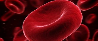
1. Unchanged red blood cells in the urine - carry hemoglobin, are red in color and have the shape of a biconcave disc.
2. Changed - colorless, ring-shaped cells that do not contain hemoglobin, they are called alkaline..
Blood in the urine is a serious reason to see a doctor immediately. The causes of this pathology can be very dangerous. Urine is formed in the renal glomeruli, where it immediately begins to be filtered. And if the analysis showed that the content of red blood cells, white blood cells or proteins in the urine is increased, then they have penetrated the glomerular membrane, which indicates a disruption of the kidneys.
What may affect the results
Eating foods that can change the color of urine (for example, beets, carrots, blueberries), have a diuretic effect (for example, watermelon, beer), foods that are too salty or too sweet, or alcohol. Some medications affect changes in the color and volume of urine, so you should carefully read the instructions before taking any drug. If the drug has an undesirable effect, then, if possible, its use should be discontinued 2-3 days before the test or the attending physician should be notified about their use.
Causes of increased red blood cells in urine
If a urine test shows a higher level of red blood cells than normal, then the source of the bleeding should be determined. There are 4 groups of reasons that can cause hematuria:
- Somatic (prerenal) - causes not related to the pathology of the urinary system;
- Renal – bleeding is caused by kidney disease;
- Postrenal – the cause of hematuria is due to pathology of the urinary tract;
- Physiological - external causes not related to the state of the internal systems of the body.
Keep in mind that an increased content of red blood cells in the urine in men and the same symptom in women can occur for various reasons; individual characteristics of anatomy and physiology should be taken into account.
Somatic
There is no renal pathology in this case, and the increased content of red blood cells in the urine is a reaction of the kidneys to a concomitant disease of another organ. Somatic causes of hematuria include:
- Thrombocytopenia. The condition is characterized by a reduced number of platelets in the blood, which leads to a blood clotting disorder. This explains the presence of blood in the urine.
- Hemophilia. A hereditary disorder of blood clotting, which passes through the glomeruli into the urine.
- Intoxication. Some toxins, when entering the body, increase the permeability of the glomerular membrane, which causes red blood cells to enter the urine. Such conditions occur due to viral and bacterial infections.
Rental
This means that the increase in red blood cells is caused by kidney disease:
- Acute and chronic glomerulonephritis. The filtering function of the organ is disrupted, causing blood cells to enter the urine.
- Kidney cancer. When tumor formation occurs, the walls of blood vessels are affected, which leads to bleeding. A urine test reveals unchanged red blood cells.
- Urolithiasis. Stones damage the mucous membrane, which is the cause of bleeding.
- Pyelonephritis. The inflammatory process makes the walls of blood vessels more permeable, due to which blood enters the urine.
- Hydronephrosis. With this disease, the pressure in the tissues of the urinary system is increased due to the obstructed outflow of urine. The kidneys are stretched, and the vessels receive microdamage.
Also, the cause of blood in the urine can be injuries and injuries to the kidneys (bruises, knife blows, falls, etc.).
Post-Rental
Post-ventral causes of hematuria are diseases of the bladder and urinary tract.
1. Cystitis. The picture is similar to pyelonephritis - red blood cells in the urine appear due to the walls of blood vessels weakened by inflammation, only in this case - the vessels of the bladder, and not the kidneys.
2. Presence of sand/stone in the bladder. The mucous membranes are injured and bleeding begins.
3. Injuries and wounds of the bladder and urethra. Such conditions often cause gross hematuria.
4. Bladder cancer. Often, blood vessels rupture due to tumors. The intensity of bleeding and the amount of blood in the urine directly depend on the diameter of the rupture.
Physiological
In addition to the above, there are a number of reasons that can lead to an increase in the rate of red blood cells in the urine. However, they are external and not related to diseases of the internal organs.
- Increased air temperature. Hematuria can be caused by intense work in a hot room or excessive exposure to a sauna.
- Severe stress negatively affects vascular permeability.
- Alcohol. Increases pressure in the vessels of the kidneys, narrowing them, and at the same time increasing the permeability of their walls.
- Excessive physical activity.
- Eating a lot of spices.
General urine analysis (Urine analysis with sediment microscopy)
You can take a general urine test at the nearest INVITRO medical office.
A list of offices where biomaterial is accepted for laboratory research is presented in the “Addresses” section. Interpretation of study results contains information for the attending physician and is not a diagnosis.
The information in this section should not be used for self-diagnosis or self-treatment. The doctor makes an accurate diagnosis using both the results of this examination and the necessary information from other sources: medical history, results of other examinations, etc. A general urine test in the INVITRO laboratory includes the determination of the following parameters: physical - color, transparency, specific gravity;
chemical - pH
, protein content, glucose, ketones, urobilinogen, bilirubin, as well as examination of sediment - epithelium, leukocytes, erythrocytes, salts, mucus, etc.
When interpreting the results of a general urine test, it should be taken into account that all indicators should not be analyzed in isolation, but in combination with each other and taking into account the patient’s complaints.
Exceeding the norm of red blood cells in urine in adults
As already written above, in people of different sexes, hematuria can have completely different causes. Therefore, the doctor takes into account gender when making this diagnosis.
Causes of blood in urine in men
In men, an increased number of red blood cells in the urine can be caused by diseases of the reproductive system:
1. Prostatitis. This is an inflammatory process in the prostate gland and it affects the blood vessels just as negatively as any other inflammation.
2. Prostate cancer. The tumor destroys the walls of blood vessels, causing blood to appear in the urine.
Hematuria in women: causes
Female genital diseases can also cause excess red blood cells in the urine:
1. Cervical erosion. This is bleeding damage to the mucous membrane, caused either mechanically (trauma), or due to hormonal imbalance, or as a consequence of an infectious disease of the genital organs.
2. Uterine bleeding. Blood from the vagina can easily leak into the urine, this is called “false hematuria.”
Normal indicators
| Index | Meaning |
| Color | light yellow, straw yellow, yellow |
| Transparency | Transparent |
| Specific gravity | 1003-1035 |
| pH | 5,0-8,0 |
| Protein | Less than 0.140 g/l |
| Glucose | Less than 2.8 mmol/l |
| Ketone bodies | Less than 1 mmol/l |
| Urobilinogen | Less than 34 mmol/l |
| Bilirubin | Not detected |
| Squamous epithelial cells | Up to 5 in sight |
| Transitional and renal epithelial cells | Not detected |
| Leukocytes | Up to 5 in sight |
| Red blood cells | Up to 2 in sight |
| Hyaline casts | Not detected |
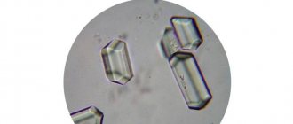
| Uric acid crystals | Not detected |
| Urats | In small quantities |
| Tripelphosphates | Not detected |
| Slime | In small quantities |
| Bacteria | Not detected |
| Mushrooms | Not detected |
Hematuria as a manifestation of isolated urinary syndrome in children
Hematuria refers to the presence of red blood cells in the urine. Does this always indicate pathology? Can erythrocyturia be observed normally? If yes, in what quantity and how often? There is no clear answer to these questions. Many people consider the presence of single red blood cells in the morning urine collected after the appropriate toilet as a variant of the norm [2, 15, 19]. At the same time, children who even occasionally show single red blood cells in a general urine test require observation and a specific examination algorithm for often several months.
When considering hematuria as a manifestation of isolated urinary syndrome (IUS), it is necessary to take into account both the degree of its severity and the possibility of combining it with other changes in urine analysis and, above all, with proteinuria.
According to the degree of severity, macro- and microhematuria are distinguished. With gross hematuria, urine becomes reddish-brown in color (the color of “meat slop”). With microhematuria, the color of urine is not changed, but when examined under a microscope, the degree of hematuria varies. It is advisable to distinguish between severe hematuria (more than 50 red blood cells per field of view), moderate (30–50 per field of view) and minor (up to 10–15 per field of view).
Hematuria should also be distinguished by duration. It can be short-term (for example, during the passage of a stone), have an intermittent course, as is the case with Berger's disease - one of the variants of IgA nephropathy, and also be characterized by a persistent, persistent course, maintaining varying degrees of severity over many months and even years ( various types of glomerulonephritis, hereditary nephritis, some types of kidney dysplasia). It can be asymptomatic (with a number of congenital and hereditary kidney diseases) or accompanied by dysuria or pain syndrome (with renal colic).
Depending on the site of origin, hematuria can be renal or extrarenal. The presence of so-called “altered” erythrocytes in urine sediment does not always indicate their renal origin, because their morphology often depends on the osmolality of the urine and the duration of stay in it until microscopy of the sediment [26]. At the same time, “unchanged” red blood cells in the urine can be of renal origin (for example, with macrohematuria due to rupture of the basement membrane in some forms of glomerulonephritis or with hemorrhagic fever with kidney damage and the occurrence of thrombohemorrhagic syndrome; as well as with kidney tuberculosis, with Wilms tumor ). In turn, renal hematuria is divided into glomerular and tubular. For glomerular hematuria, the appearance of erythrocyte casts in the urine sediment is typical, but this is observed only in 30% of glomerular hematuria [25]. The renal nature of hematuria can be more reliably established using phase-contrast microscopy of urinary sediment [13].
Glomerular hematuria (), as a manifestation of IMS, occurs mainly with non-inflammatory damage to the glomerular capillaries; more precisely, in the absence of any pronounced proliferative reaction on the part of the mesangium. The only exception is IgA nephropathy, which can occur for a long time without any extrarenal manifestations. Diseases in which hematuria is accompanied by extrarenal symptoms morphologically have a pronounced proliferative nature of the inflammatory reaction either predominantly from the mesangium or from the endothelial lining of the glomerular capillaries.
Tubular hematuria is observed in a variety of diseases of both congenital and acquired origin ().
The mechanism of occurrence of renal hematuria. To this day, there is no common understanding of the pathogenesis of renal hematuria. It goes without saying that red blood cells can enter the urinary space of the kidney only from the capillary bed, and hematuria in renal pathology is traditionally associated with damage to the glomerular capillaries. In microhematuria, red blood cells pass through anatomical pores in the basement membrane due to its increased permeability [7, 23]. Gross hematuria is caused rather by necrosis of glomerular loops [9, 14, 20, 22, 23]. The cause of hematuria may be thinning of the basement membrane with disruption of the structure of type IV collagen and a decrease in the laminin content in its dense layer, which is characteristic of hereditary nephritis [5, 7].
It is considered more likely that the main site of penetration of red blood cells through the capillary wall is the glomerulus. This is facilitated by the increased intracapillary hydrostatic pressure present in the glomerulus, under the influence of which the red blood cells, changing their configuration, pass through the existing pores [18, 22]. Permeability to erythrocytes increases when the integrity of the basement membrane is disrupted, which occurs with immunoinflammatory damage to the capillary wall. Some authors do not exclude a violation of the morphofunctional properties of erythrocytes, in particular, a decrease in their charge, in the occurrence of hematuria [6]. However, there is no correlation between the severity of changes in the glomeruli and the degree of hematuria [11]. This fact, as well as the often absence of severe hematuria in nephrotic syndrome, when the structure of the basement membrane is sharply disrupted, has given rise to a number of authors to express a different point of view on the mechanism of hematuria, namely, the main place of release of erythrocytes is the peritubular capillaries [10, 11]. These capillaries, unlike glomerular capillaries, do not have an epithelial layer and are in very close contact with the tubular epithelium; in this case, significant changes of a dystrophic nature are often found both in the endothelial cells of the capillaries and in the epithelium of the tubules [11].
Despite the existing uncertainty about the nature of renal hematuria in nephropathies, it is nevertheless important to know the place of its origin - the glomerulus or tubule. Dysmorphism of erythrocytes, detected by phase-contrast microscopy, makes it possible to distinguish renal hematuria from extrarenal hematuria [4, 8, 9, 25, 26], but does not allow to differentiate glomerular erythrocyturia from peritubular [11, 16, 21, 27]. Tubular or peritubular hematuria may be indicated by the appearance in the urine of plasma low molecular weight proteins, which are usually completely reabsorbed in the proximal tubule. These proteins include beta2-microglobulin (beta2-MG). If, during hematuria, beta2-MG is detected in the urine in an amount exceeding 100 mg in the absence or less amount of albumin in it, then such hematuria should be regarded as tubular [24]. Other markers of tubular hematuria may include retinol binding protein [12] and alpha1-microglobulin [17, 28]. Determination of the latter is preferable, since beta2-MG is easily destroyed in very acidic urine.
Diagnosis of hematuria in children. Diagnosis of asymptomatic hematuria presents the greatest difficulties for the doctor. However, the absence of one or another symptomatology at the moment does not exclude the presence of it in the anamnesis, such as, for example, past pain, or dysuria, or fever without catarrhal phenomena. The diagnostic process, as always, should begin with a detailed history. The main points to which the doctor’s attention should be drawn when collecting an anamnesis are presented. Identification of certain features of the medical history will allow the most rational examination of the patient, and analysis of the circumstances under which hematuria was detected will help to simplify it.
It is extremely important to determine the age when the debut of hematuria took place, because establishing the fact of the appearance of hematuria in early childhood allows us to consider it as a manifestation, most often, of some congenital or hereditary pathology. A carefully studied family and obstetric history will allow you to confirm this. It is important to establish whether hematuria is constant or occurs occasionally against the background of any intercurrent illness, cooling or exercise. Its severity is also of certain importance, i.e. whether it manifests itself as macro- or microhematuria. But greater significance should be attached to the accompanying proteinuria, especially when it is permanent. This always indicates a renal origin of hematuria.
When starting to examine a child with detected hematuria in a clinic, first of all, it is necessary to determine the place of its origin, that is, whether the hematuria is renal or extrarenal. Undoubtedly, if hematuria is accompanied by proteinuria, then its non-renal origin is excluded. In the absence of proteinuria, the first step in the examination should be a two-glass test (see diagram 1 on page 56). The detection of red blood cells only in the first portion indicates their external origin. In this case, examination of the external genitalia, taking smears for microscopy and latent infection, scraping for enterobiasis will help identify the inflammatory process and its cause. If signs of inflammation are detected, it is necessary to exclude its allergic nature. To do this, in addition to obtaining relevant anamnestic data, a vulvo- or urocytogram should be prescribed, which, in the presence of a predominance of lymphocytes and the detection of eosinophils, will exclude the bacterial nature of the inflammatory process. The detection of red blood cells in two portions indicates involvement of the kidneys and/or bladder in the pathological process. Bladder pathology can be suspected, in addition to relevant anamnestic data, during ultrasound examination, but only cystoscopy makes it possible to definitively verify the presence or absence of cystitis. Ultrasound examination (ultrasound) can reveal changes in the position of the shape and size of the kidneys, suggesting the possibility of cystitis, as well as a neurogenic bladder. In addition, ultrasound can detect the presence of stones. Subsequent IV urography and/or renoscintigraphy will help clarify the nature of the detected changes.
Hematuria, combined with proteinuria, as already mentioned, is of renal origin. If this pathology is established in urine tests in early childhood, after taking an appropriate history (), it is necessary to determine whether the disease is congenital or hereditary. The proposed algorithm of actions (see diagram 2 on page 57) allows at the first stage not only to outline the differential diagnosis between congenital and hereditary kidney pathologies, but also to approach the identification of diseases such as interstitial nephritis and metabolic nephropathy, for which hematuria is one of the manifestations of this pathology.
When hematuria, combined with proteinuria, appears in preschool and school age, the hereditary or congenital nature of the disease cannot be ruled out. However, the role of acquired pathology in the form of various forms of primary or secondary glomerulonephritis, interstitial nephritis, diabetic nephropathy, and pyelonephritis is significantly increasing. After a detailed history collection, examination of this group of children should begin with the collection of 24-hour urine for protein and an orthostatic test. It is preferable to collect daily urine for protein separately during the day and at night. This makes it possible to assess the importance of physical activity on the severity of both proteinuria and hematuria. Since in children of this age group, when hematuria is combined with proteinuria, the incidence of various variants of glomerulonephritis increases, it is necessary to identify a possible connection between this pathology and hemolytic streptococcus. To do this, it is not enough to detect its presence by taking swabs from the throat; it is necessary to establish the appearance and increase in the titer of antistreptococcal antibodies (ASL-O), as well as the activation of the complementary system.
An obligatory step in the examination of this group of patients is an ultrasound scan of the kidneys. Despite the normal ultrasound characteristics of the kidneys in the presence of isolated urinary syndrome in the form of hematuria with proteinuria, regardless of their severity, a positive orthostatic test requires intravenous urography. The latter will eliminate kidney dystopia, the presence of their immobility, and also finally resolve the issue of the absence of pathological kidney mobility. For a functional examination, it is often enough to confine ourselves to performing the Zimnitsky test, and to clarify the state of the tubulointerstitium, a test with Lasix [1]. If certain abnormalities are detected by ultrasound of the kidneys, in addition to the above, it may be necessary to perform a Rehberg test, as well as renoscintigraphy.
Thus, before deciding on the need to use invasive examination methods in children with IMS, manifested in the form of hematuria, it is necessary to conduct the above basic examination on an outpatient basis. This will, on the one hand, prevent unnecessary hospitalization, and on the other, reduce the stay of children in a specialized bed if a more in-depth examination is required.
Literature
- Arkhipov V.V., Rivkin A.M. Diagnosis of the functional state of the kidneys using furosemide. Guidelines. St. Petersburg, 1996.
- Burtsev V.I., Turchina L.P. Hematuria // Clinical Medicine, 1997, No. 6, p. 66–69.
- Duman V. L. Phase-contrast microscopy of urinary sediment in the differential diagnosis of hematuria. Collection of proceedings of the IV annual nephrology seminar. St. Petersburg, 1996, p. 150–152.
- Zaidenvarg G. E., Savenkova N. D. Study of erythrocyte dysmorphism with phase-contrast microscopy, pH, urine osmolality in children with hematuria. Materials of the 1st Congress “Modern methods of diagnosis and treatment of nephrourological diseases in children.” M., 1998, p. 94.
- Ignatova M. S., Fokeeva V. V. Hereditary nephritis: diagnosis, pathogenesis, genetics, prognosis // Klin. honey. 1994, no. 2, p. 51–55.
- Nikolaev A. Yu., Shcherbin A. A. et al. The mechanism of hematuria in hematuric nephritis // Ter. archive, 1988, No. 6, p. 34–37.
- Papayan A.V., Savenkova N.D. Clinical nephrology. St. Petersburg 1997, p. 197–201.
- Prikhodina L. S., Malashina O. A. Modern ideas about hematuria in children // Nephrology and Dialysis, 2000, No. 3, p. 139–145.
- Savenkova N. D., Zaidenvarg G. E., Lisovaya N. A. Hematuria in children. Lecture. St. Petersburg, 1999.
- Shulutko B.I. Pathology of the kidneys. L., 1983, p. 80.
- Shulutko B.I. Nephrology. St. Petersburg 2002, p. 106–110.
- Bernard AM, Moreau D., Lanweys R. Comparison of retinal binding protein and beta2-microglobulin in urine in the early detection of ambular proteinuria // Clin. Chim. Acta. 1982; 126:1–7.
- Birch DF, Fairley KF Haematuria - glomerular or nonglomerular? // Lancet. 1979; vol. 2, p. 845–846.
- Bohle A. at al. Morphological contribution on gross hematuria in mild mesangioproliferative glomerulonephritis without crescents // Klin. Wochenschr. 1985; 63:371–378.
- Dodge WF, West EF et al. Proteinuria and hematuria in schoolchildren: epidemiology and early natural history // J. Pediatrics. 1976; 88:327–347.
- Gibbs DD, Linn KJ Red cell volume distribution curves in the diagnosis of glomerular and nonglomerular hematuria // Clin. Nephrol. 1990. 33: 143–147.
- Hofmann W., Rossmuller B., Guder WG, Edel HH A new strategy for characterizing proteinuria and haematuria from a single pattern of defined proteins in urine // Eur. J. Clin. Chem. Clin. Biochem. 1992; 30: 707–712.
- Jai-Trung L., Hiroyoshi W., Hiroshi M. et al. Mechanism of hematuria in glomerular disease // Nephron. 1983 Vol. 35. P. 68–72.
- Froom P. et al. Significance of microhaematuria in young adults // British Med. J., 1984; vol. 288, p. 20–21.
- Kincaid-Smidt P. et al. Acute renal failure and tubular necrosis associated with haematuria due to glomerulonephritis // Clin. Nephrol. 1983; 19: 206–210.
- Kitamota Y., Tomita M., Akamine M. et al. Differentiation of hematuria using a uniquely shaped red cell // Nephron. 1993, 64: 32–36.
- Lin JT et al. Mechanism of hematuria in glomerular disease. An electron microscopic study in a case of diffuse membranous glomerulonephritis // Nephron. 1983, 35: 68–72.
- Mouradian JA, Sherman RL Passage of erythrocyte throuhg a glomerular basement membrane gap // N. Engl. J. Med. 1975, 293(18):940–941.
- Peterson PA et al. Differentiation of glomerular, tubular and normal proteinuria: determinations of urinary excretion of beta2-microglobulin, albumin and total protein // J. Clin. Invest. 1969, 48: 1189–1198.
- Rizzoni G. et al. Evaluation of gomerular and nonglomerular hematuria by phase contrast microscopy // J. Pediatrics. 1983, 103: 370–371.
- Sandoz P. Verbesserte Urinsedimentdiagnostik durch Differenzierung der Eritrozytenmorphologie // Ther. Umschau. 1988, 12:851–856.
- Stapleton FB Morphology of urinary red blood cells: a simple guide in localizing the site of hematuria // Pediatr. Clin. North Am. 1987, 34(2):561–569.
- Yu H. et al. Alpha-1 microglobulin: an indicator protein for renal tubular function // J. Clin. Pathol. 1983, 36: 253–259.
A. M. Rivkin , Candidate of Medical Sciences, Associate Professor N. A. Lisovaya , Candidate of Medical Sciences
SPbGPMA, St. Petersburg
Contact information for authors for correspondence
Additional examination if indicators deviate from the norm
Patients with abnormalities in the general urine test are usually consulted by general practitioners, urologists, gynecologists, and surgeons.
The list of studies required to make a final diagnosis depends on the patient’s underlying disease and which indicators are not normal. Sources:
- Zakharova I.N., Osmanov I.M. et al. Clinical urine analysis: historical significance for the development of medicine. Medical advice, magazine. No. 2. 2021. pp. 131-140.
- Druzhinina T.V., Alimova I.L. Possibilities of interpreting a general urine test during medical examinations of minors. Smolensk Medical Almanac, magazine. No. 3. 2016. pp. 78-82.