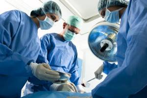
Laparoscopy is one of the methods of surgical intervention in the abdominal cavity. The procedure can be carried out both for diagnostic purposes and to remove organs partially or completely, and to extract the stones they contain. For example, removal of stones from the gallbladder, or removal of the organ itself, most often occurs during laparoscopic intervention. Laparoscopy of an organ is considered a safer surgical intervention for the patient than full abdominal surgery, and the accuracy and success of its implementation directly depend on the skill level of the surgeon.
How the gallbladder works, what are the dangers of diseases and pathologies of the organ
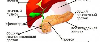
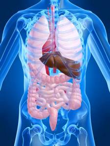
The gallbladder is a hollow organ in the form of a pear-shaped pouch. It is located in the abdominal cavity, in a special recess on the surface of the liver, with which it forms a close functional relationship. The organ is directly involved in the process of digesting food: it is responsible for the accumulation of bile. In turn, bile is produced by the liver parenchyma. While the liver structures produce bile, it is collected in the bladder.
Content:
- How the gallbladder works, what are the dangers of diseases and pathologies of the organ
- The essence of the laparoscopy method: how and why it is performed
- Advantages of laparoscopic intervention over laparotomy
- Indications for the procedure: in what cases is laparoscopy prescribed?
- When is gallbladder surgery prohibited?
- Requirements for preparing for the procedure
- How to remove stones from the bladder cavity
- Laparoscopy of the gallbladder: how the organ is removed
- What happens after surgery
- Life of a patient after gallbladder removal
- Consequences and possible complications after surgery
When food enters the body, the bile duct releases the collected fluid into the duodenum, where it, together with intestinal and pancreatic enzymes, begins the process of digesting the bolus of food coming from the stomach. In addition, the walls of the bladder produce mucus and produce anticholecystokinin, a hormone responsible for relaxing the muscles of the bladder walls. The capacity of the organ is about 60-80 milliliters.
Structurally, the gallbladder consists of a bottom, walls (middle part) and a neck. The latter passes into the narrow cystic duct. There is a functional bend in the area of the organ’s neck, so the neck is located at a certain angle relative to the body.
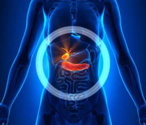
An abnormal diet, congenital structural abnormalities and general abnormalities in the functioning of the digestive tract often become a catalyst for the development of a number of gallbladder diseases. Their consequences are dangerous not only due to disruptions in the processes of food digestion. Since all elements of the digestive system are closely interconnected, disturbances in the functioning of the bladder, first of all, negatively affect the liver. In some cases, stagnant processes in the organ cause inflammation of the liver tissue, up to biliary cirrhosis.
The gallbladder and bile ducts are susceptible to the development of the following diseases:
- cholelithiasis: cholelithiasis, when stones form in the cavity of the bladder or ducts;
- dyskinesia: disorders of the contractile function of the muscular muscles of the organ and ducts;
- cholecystitis: inflammatory and necrotic processes in the walls and cavity of the bladder;
- cholangitis: acute or chronic inflammation of the bile ducts;
- cancerous tumors, benign neoplasms.
Contraindications to cholecystectomy
There are often situations when cholecystectomy cannot be performed:
- near-death state;
- decompensated nosologies - bronchial asthma, hypertension, myocarditis and others;
- malignant neoplasia;
- endocrine or metabolic disorders with severe course - in particular, diabetes mellitus;
- blood pathology.
The final decision whether to perform cholecystectomy rests with the attending physician. He is obliged to warn the patient what he can eat after removal of the gallbladder when using a specific method of operation, when to get out of bed after it, etc.
The essence of the laparoscopy method: how and why it is performed
Doctors, describing the operation of laparoscopy, focus on the fact that it is low-traumatic, safe, effective and quite fast when compared with abdominal surgery.
Laparoscopy, in this case, can be understood as an operation to remove a bladder, or as a process of removing stones located in its cavity and ducts, which is carried out during laparoscopic access to the abdominal cavity. It is the particular access that the physician uses during the process that distinguishes this operation from other types of surgical interventions. Such access is made possible through the use of a specific instrument - a laparoscope.
Conventional abdominal operations are performed through an incision in the anterior abdominal wall made with a scalpel or other special instrument, that is, laparotomy. At the same time, the size of the dissection can be quite significant, up to ten or more centimeters. After such an operation, surgical sutures are placed on the incision, leaving a noticeable scar.
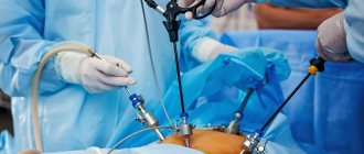
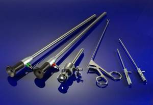
A laparoscope is a miniature video camera equipped with a light that is inserted into the patient's abdominal cavity. To do this, the surgeon does not need to make large incisions in the abdominal wall - a few small punctures are enough. The image captured by the camera is displayed on the device’s monitor. The doctor is able to perform an operation based only on this image. The puncture length is usually no more than 20 millimeters. In addition to the laparoscope, special tubes - trocars or manipulators - are inserted into the abdominal cavity. Through the cavities of these tubes, the surgeon inserts medical instruments into the abdominal cavity and can control them.

Trocars have special devices that allow the doctor to cut adhesions, cauterize blood vessels, apply clamps, and perform other necessary surgical procedures.
In general, to perform laparoscopy, the surgeon will need 3 small punctures. The operations themselves, their technique and essence, both during laparoscopy and laparotomy, are no different.
Why can laparoscopy of stones and gallbladder be prescribed? The attending physician refers the patient to surgery if there is a need:
- remove the gallbladder;
- remove the concretions located in it;
- control the quality of the previously performed operation.
Recently, the term “gallbladder laparoscopy” is more often used to mean the removal of an organ. According to doctors, if there are too many stones in the cavity and they are small in size, it makes sense to remove the entire organ, since it has already undergone pathological changes and will no longer be able to function normally. Moreover, if only the stones are removed, there is a high probability that the bladder will periodically become inflamed in the future, provoking other diseases. If there are few stones and they are small in size, it makes sense to use other methods of getting rid of them, for example, crushing with ultrasound or resorption with drugs containing ursodeoxycholic acid.
Possible complications
Thanks to proven techniques, cholecystectomy proceeds safely, but in some patients complications are possible for various reasons. Medical tactics depend on them (for example, nutrition a month after removal of the gallbladder in the presence of adhesive disease should still be light). The most common complications:
- injury during bile duct surgery;
- leakage of bile;
- suppuration of a postoperative wound;
- intra-abdominal bleeding;
- thromboembolic conditions;
- formation of adhesions.
Complications require immediate correction.
On our website you will find answers to all questions, for example, what drugs are used for treatment after removal of the gallbladder or how the postoperative period goes in the elderly.
Related services: Surgical operations Laparoscopy
Advantages of laparoscopic intervention over laparotomy
General abdominal surgery is prescribed mainly in cases where it is not possible to solve the patient’s problem using laparoscopy.
Laparoscopy is the more preferable method of invasive treatment because:
- during its implementation, a slight violation of the integrity of the tissues of the anterior abdominal wall is required, usually three or four punctures up to 20 millimeters long;
- pain after laparoscopy subside within 24 hours, provided that the operation was performed professionally;
- the patient can walk and move 4-6 hours after the end of the procedure (we are not talking about physical activity or complex movements);
- hospital stay after laparoscopy lasts from 1 to 10 days;
- the rehabilitation period lasts significantly less than after laparotomy;
- scars remaining from punctures are hardly noticeable;
- the risk of a postoperative hernia is minimal.
Gallbladder removal surgery
With a traditional cholecystectomy, a fairly large incision of about 15-20 cm is made on the anterior abdominal wall, through which the gallbladder is removed.
Laparoscopy of the gallbladder has a number of advantages compared to traditional cholecystectomy:
- The gallbladder is removed through small, neat punctures instead of a large incision: after the operation you are left with almost imperceptible spots instead of an extensive scar
- You recover quickly: up to 2-3 days in hospital and up to 7 days outpatient
- Laparoscopy of the gallbladder occurs with virtually no blood loss
- The risk of complications after surgery (suppuration, formation of adhesions, etc.) is practically zero
- After surgery, you will need much less or no pain medication
Indications for the procedure: in what cases is laparoscopy prescribed?
Surgical intervention, even at such a gentle level, must be justified by objective reasons. The attending physician may prescribe surgery if the patient has the following indications for it:
- in the presence of stones in the bladder cavity, without accompanying symptoms;
- in case of exacerbation of cholecystitis, in the first two days of the attack;
- if the patient has stones in the bile ducts and obstructive jaundice is diagnosed;
- established chronic calculous cholecystitis;
- identified polyps and neoplasms in the bladder.
Indications for cholecystectomy
The gallbladder is a kind of “storage” of bile, which is required for trouble-free digestion. But often problems develop from the bladder itself, which is why abdominal surgery to remove the gallbladder is practiced, which is referred to as “cholecystectomy.” Indications:
- cholelithiasis;
- noncalculous cholecystitis;
- cholesterosis – “impregnations” of cholesterol in the gall bladder walls;
- polyposis;
- metaplasia;
- gallbladder disorders accompanied by pain that cannot be eliminated by conservative methods.
The most common reason for cholecystectomy is the presence of one or more stones in the gall bladder. Removing stones without removing the gallbladder is becoming less and less common.
The formation of stones in the gallbladder is an indication for cholecystectomy also because they can provoke a series of complications - the most common are:
- perforation of the gallbladder;
- biliary peritonitis - an inflammatory process in both layers of the peritoneum due to irritation by bile that gets on them;
- empyema of the gallbladder - its suppuration.
When is gallbladder surgery prohibited?
Contraindications for laparoscopy are specific, due to the atypical technique of the operation itself. For example, laparoscopy is not prescribed for patients who have already undergone abdominal surgery, since there is a high risk of the instruments touching and damaging the adhesions on the internal organs, which can lead to damage to the organs themselves.
Other contraindications include:
- respiratory dysfunction, respiratory failure: during the injection of air into the abdominal cavity, the diaphragm may shift, which creates additional difficulties with breathing;
- severe cardiac and pulmonary pathologies;
- peritonitis;
- pregnancy in the third trimester;
- obesity of the second and third stages;
- dysfunction of blood clotting, if it cannot be corrected;
- acute pancreatitis;
- the presence of fistulas between the intestines and bile ducts;
- installed pacemaker.
Requirements for preparing for the procedure

A surgical intervention of this nature requires special preparation of the patient.
2 weeks before the date of surgery, you must pass a whole list of tests:
- general blood analysis;
- coagulogram;
- blood biochemistry;
- analysis to determine the Rh factor and blood group;
- for women - a smear on the vaginal flora;
- PCR for HIV, syphilis, hepatitis A, B, C.
In addition, the doctor who will perform the operation will definitely require electrocardiography results.
All tests should be within normal limits. If any indicators are abnormal, the operation cannot be performed until the patient undergoes a course of treatment. If repeated tests show normal values, then the patient can be allowed to proceed to the next stages of preparation.
A person should inform the doctor about all chronic diseases he has, especially diseases of the respiratory organs, endocrine and digestive systems. Taking any medications 2-3 weeks before the scheduled surgery must be agreed with the surgeon. 10 days before the appointed date, stop using vitamin E, anticoagulants, and Aspirin.
On the day before surgery, after 18:00 it is forbidden to eat, and after 22:00 - to drink liquid. For 3-4 days before the procedure, you should avoid fatty, fried, hot and spicy foods, smoked meats and pickles. Before going to bed, you need to do a cleansing enema, and repeat the procedure in the morning.
The operation is performed strictly on an empty stomach, so eating breakfast or drinking liquids in the morning is prohibited.
Before the procedure begins, the surgeon tells the patient in general terms how laparoscopy will proceed and how long the operation will last. On average, removing stones takes from 40 minutes to an hour, removing a bladder takes 1.5-2 hours.
Removal of the gallbladder in the Stolitsa network of clinics means:
High tech
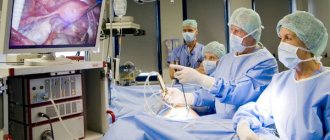
To effectively carry out low-traumatic operations to remove the gallbladder, we use modern premium PENTAX video equipment. Thanks to a powerful optical system, it allows the doctor to examine the condition of the gallbladder in detail and safely remove it.
Professionally
The experience and professionalism of the doctor is the key to a successful operation. After all, without a highly qualified specialist, even the most premium equipment is just devices. Only an experienced doctor can perform laparoscopic surgery professionally and safely for your health. Over the years of successful practice, our specialists have performed a huge number of laparoscopic operations to remove the gallbladder and have restored health to hundreds of people. By contacting us, you can be sure that our specialists will thoroughly understand your situation: they will conduct a detailed preliminary diagnosis and determine exactly how necessary it is to remove the gallbladder. Before the operation, we enter into a formal contract with you. You receive all the necessary documents. The contract price includes payment for the operation itself, the cost of anesthesia and postoperative observation in the clinic. After the operation, you will receive a sick leave certificate from the first day of hospitalization, which you can extend if necessary either in our clinic or at your local clinic. After discharge, you receive a discharge summary with a protocol of the operation and detailed recommendations from the doctor for treatment during the rehabilitation period.
Comfortable
In our cozy rooms you will feel comfortable, because they have everything you need: a button to call a nurse at any time of the day, a separate bathroom, air conditioning, refrigerator, TV, microwave and Wi-Fi. After the operation, your condition will be continuously monitored by our specialists. Good nutrition and rehabilitation procedures will help you quickly regain strength after surgery and return to a full life. Get your health back. Make an appointment now.
How to remove stones from the bladder cavity
The procedure is carried out under general anesthesia - thanks to this, it is possible not only to ensure the absence of pain in the patient, but also to achieve maximum relaxation of the abdominal muscles.
Having put the patient into a state of medicated sleep, the anesthesiologist places a special probe in his stomach, which allows him to remove the contents of the organ - liquid or gases. This step is mandatory, as it eliminates the possibility of accidental vomiting, in which the contents of the stomach can enter the respiratory tract and cause suffocation.
After the probe is installed, a mask is applied to the patient’s mouth and nose, which is connected to the ventilator system - during the entire operation he will breathe with its help. Carrying out laparoscopy without the use of artificial ventilation is almost impossible, since the gas pumped into the abdominal cavity puts pressure on the diaphragm, and it, in turn, compresses the lungs, and the patient’s independent breathing becomes difficult.
Having successfully completed all the described preparatory measures, the surgeon and assistants begin laparoscopic stone removal. A puncture is made in the fold of the navel, through which sterile gas is first injected into the abdominal cavity to straighten the folds of the organs and expand the volume of the cavity, and then a laparoscope is inserted.
Another 2-3 punctures are made along the line of the right hypochondrium, where trocars with instruments are inserted. Then the doctor examines the location and appearance of the bladder, and, if necessary, cuts the adhesions between it and surrounding organs. Next, the wall of the bladder is cut, the tip of the suction is inserted into it, and the contents of the organ are removed. The wall is sutured, the abdominal cavity is washed with an antiseptic. The doctor removes the trocars and laparoscope and places sutures at the incision sites on the skin.
In what cases is the gallbladder removed?
Your doctor may refer you for gallbladder removal surgery if you:
- Choledocholithiasis is a form of gallstone disease in which stones form in the lumen of the common bile duct
- Bile duct blockage
- Acute cholecystitis - if the disease occurs against the background of an existing gallstone disease, the doctor may decide on urgent surgery
- Gallstone disease (GSD) with attacks of biliary colic or with the manifestation of “minor” symptoms of the disease: bitterness in the mouth, aching painful sensations in the hypochondrium on the right and a feeling of heaviness after eating in the same area
- Asymptomatic gallstone disease
- Gallbladder calcification
- Gallbladder polyps with cholelithiasis or at risk of developing a tumor
- Perforation of the gallbladder - this disease can be caused by malignant tumors, severe abdominal trauma and a number of systemic diseases
- Cholesterosis – deposition of cholesterol on the walls of the bladder due to cholelithiasis
- Development of secondary pancreatitis
These are the main problems for which the gallbladder is removed. The extent to which removal is necessary in your case will be determined by your attending physician based on the results of a thorough preliminary diagnosis and your general condition. It is important to know: if you do not perform the operation on time, sooner or later serious complications will arise that can cost you your life. Therefore, do not delay your visit to the doctor. Make an appointment and undergo preliminary diagnostics.
Laparoscopy of the gallbladder: how the organ is removed
The operation to remove the bladder requires compliance with similar preparatory measures before the start - the patient is also immersed in a medicated sleep, a probe is inserted into his stomach, and he is connected to an artificial respiration apparatus.
A laparoscope is inserted into the abdominal cavity through an incision in the area, and 2 or 3 trocars are inserted through punctures along the line of the right hypochondrium. Having determined the location of the bubble, the doctor, if there are adhesions, cuts them. Next, the surgeon assesses the degree of tightness and fullness of the organ. If it is very tense, the surgeon first cuts its wall and, using a special suction, removes some of the fluid from the cavity of the bladder.
A clamp is applied to the bladder, after which the bile duct, the common bile duct, is released from the tissue and cut. The next tissue to be isolated is the cystic artery. Clamps are applied to it, between which the vessel is cut. The surgeon sews up the lumen of the artery.
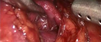
Having freed the organ from the duct and artery, the doctor begins to separate it from the hepatic cavity. All bleeding vessels are cauterized as the organ is cut off from them. After complete separation from surrounding tissue, the physician removes the gallbladder through a puncture in the navel.
The surgeon carefully examines the abdominal cavity so as not to miss any remaining bleeding vessels, bile or other pathological structures. When all the vessels are coagulated and the altered tissues are removed, an antiseptic liquid is injected into the abdominal cavity for flushing. Then it is sucked out, the laparoscope, trocars, and instruments are removed from the punctures. The incisions made are sutured, except for one - a special drainage tube remains in it for the next 1-2 days so that the remaining antiseptic can freely leave the abdominal cavity.
If at some point the surgeon realizes that it is impossible to successfully remove the bladder using laparoscopy, the operation may proceed to laparotomy.
Prevention of consequences
It should be understood that any surgical intervention is traumatic for the body, and it needs time to adapt and recover. Therefore, you should follow the recommendations of doctors.
- You should not adhere to any long-term diet after removal of the gallbladder in the absence of problems with stool and abdominal pain: a varied healthy diet is necessary.
- If you have diarrhea or bloating, you should limit your intake of fatty, spicy foods, drinks containing caffeine, and the consumption of dairy products, as well as treating bile outflow dysfunction.
- It is necessary to control weight and hormonal levels in the presence of relevant diseases.
An expert on the site Pokhmelye.rf, gastroenterologist Daniela Purgina, refutes the typical misconception regarding cholecystectomy.
It is a very common belief that after removal of the gallbladder, you need to constantly take some kind of medication to drain bile. In fact, there is no need for this. Taking choleretic drugs is completely unjustified, since the bladder has been removed and there is nowhere to drive the bile from; it will go from the liver through the ducts to the intestine. Ursodeoxycholic acid drugs are prescribed only if there is a risk or stones have been identified in the bile ducts.
What is postcholecystectomy syndrome
The operation to remove the gallbladder is scientifically called cholecystectomy. Such a surgical intervention can go without problems, but it can cause some complications.
If, after cholecystectomy, problems with the outflow of bile occur, accompanied by characteristic symptoms, doctors call this postcholecystectomy syndrome (PCES). Symptoms may occur immediately after surgery or some time later.
This pathological condition is most often associated with dysfunction of the sphincter of Oddi, which plays a key role in the normal outflow of bile and pancreatic juices. The fact is that the contractility of this sphincter of the duodenum is impaired. According to studies, the disease is diagnosed in approximately 10–15% of patients after gallbladder removal. Manifestations may vary from person to person, but in any case you will need to see a doctor and begin treatment to avoid more serious complications.
What causes PCES to develop?
The development of postcholecystectomy syndrome is associated with various problems that arise in the gastrointestinal tract after removal of the gallbladder. One of the most common causes is disruption of the normal functioning of the sphincter of Oddi, which is located on the wall of the duodenum. This structural element of the digestive system is a circular muscle responsible for the supply of bile and pancreatic enzymes to the intestinal cavity. Upon a signal, the sphincter of Oddi opens, which is accompanied by the outflow of bile and pancreatic juices into the intestines, and also closes upon a signal.
Pathological changes in the sphincter of Oddi develop in most patients precisely after surgery to remove the gallbladder. As doctors explain, the mechanism of the disease works like this:
- In the absence of a bladder, signals about its filling do not arrive to the sphincter, which leads to the formation of constant tension in the orbicularis muscle and the development of congestion in the pancreatic gland and hepatic ducts.
- An uneven outflow of bile leads to a decrease in its bactericidal effect in the small intestine, and bacteria begin to multiply excessively in it. These microbes are opportunistic, that is, they should even be present in small quantities in the intestines, but when there are a lot of them, problems arise (gas formation, pain in the navel).
Among the most common causes of symptoms of PCES are also:
- the presence of residual calculi (stones) in the bile ducts after surgery;
- progressive bile duct cyst;
- biliary dyskinesia;
- stone formation as a result of obesity or diabetes.
Sometimes the cause of this pathological symptom complex may be severe postoperative pain, which is not relieved by non-narcotic analgesics, and the accumulation of effusion (fluid) in the area of the surgical intervention.
Rarely (about 5% of clinical cases) the true reasons for the development of changes in the functionality of the sphincter of Oddi remain unknown to doctors.
How does PCES manifest itself?
As already mentioned, the symptoms of postcholecystectomy syndrome can vary. Just in case, after surgery to remove the gallbladder, listen to your body - and if alarming phenomena occur, consult a doctor. Bad signs can appear either a few days or several weeks after surgery.
The most common manifestations of PCES include:
- dyspeptic symptoms, which are manifested by a feeling of bitterness in the mouth, nausea, flatulence;
- diarrhea or constipation;
- aching, cramping pain of varying intensity in the right hypochondrium, which radiates to the right shoulder and collarbone;
- bloating, excess gas formation;
- hypovitaminosis conditions associated with insufficient absorption in the intestine;
- jaundice and chills with an increase in body temperature of 38°C and above (if stones remain in the bile ducts after surgery).
The disease can take various clinical forms. Sometimes after removal of the gallbladder the following consequences occur:
- relapses of stone formation in the biliary tract;
- strictures (narrowing of the lumen) of the common bile duct;
- papillitis (inflammation of the papilla of the excretory duct in the duodenum;
- the formation of adhesions that disrupt the outflow of bile fluid in the subhepatic space;
- inflammation of the pancreas;
- ulceration of the gastroduodenal digestive tract.
Pain in this area can occur not only after surgery, but also with the entire organ. Read the article about how to identify the disease by the nature of the pain and how to treat pain in the gallbladder.
A case from the practice of gastroenterologist Daniela Purgina, an expert on the website Pokhmelye.rf.
I rather remember a whole group of cases where a patient with asymptomatic cholelithiasis had his gallbladder removed, and after the operation, postcholecystectomy syndrome developed or, even worse, hologenic diarrhea (that is, diarrhea with bile). So the tactics should be in the maximum effort to preserve the organ.
Modern diagnostics
To confirm postcholecystectomy syndrome, the patient needs a series of examinations. As a result, the doctor will be able to determine the main causes of the development of the pathological condition and the degree of functional disorders of the digestive system. Unfortunately, most patients do not immediately turn to specialists.
Diagnostics usually includes the following steps:
- Conversation with the patient, detailed discussion of complaints, study of the medical history and life of the patient, identification of the leading pathological signs from all complaints.
- Clinical examination of a person, allowing to determine the localization of the source of the disease and assess the extent of the disorders.
- A set of laboratory tests, which includes general and biochemical blood tests with assessment of the functioning of the liver and pancreas, analysis of thyroid hormones, tests for viral hepatitis.
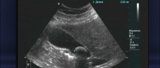
- Ultrasound examination of the organs of the hepatobiliary zone (standardly includes the liver, gall bladder and bile ducts. Since the gall bladder has already been removed, only the ducts and liver are examined).
- Esophagogastroduodenoscopy.
- If necessary, endoscopic examination of the bile ducts (endosonography, endoscopic retrograde cholangiopancreatography), manometry of the sphincter of Oddi, MRI and abdominal organs is also performed.
A complete and detailed diagnosis of suspected PCES allows the doctor to draw conclusions about why the disorders began, how far they have gone, whether there are any serious complications, and what therapy is suitable for treatment. Read on to learn how to treat post-operative problems.
How to be treated
The main thing in the treatment of PCES is to eliminate the factors that triggered the development of symptoms. The main principles of such therapy are:
- eliminating the influence of factors contributing to the occurrence of pathological manifestations;
- symptomatic treatment of dysfunctions of the organs of the hepatobiliary region;
- prevention of possible complications.
As a rule, in addition to drug intervention, doctors recommend in the first stages an increase in the frequency of food intake to 5–7 times a day to normalize the outflow of bile. Any strict diet is usually not required; this is discussed individually.
In order to eliminate the symptoms of the disease, the patient may be prescribed:
- analgesics and antispasmodics, since inflammation is accompanied by pain and spasm of the sphincter of Oddi;
- proton pump inhibitors to reduce acidity in the upper gastrointestinal tract if there is inflammation there;
- drugs to normalize the flow of bile;
- antacids to combat high acidity;
- non-absorbable antibiotics to eliminate bacterial overgrowth in the small intestine.
Sometimes medications cannot correct the problem, in which case surgery is indicated. Surgical treatment methods are aimed at restoring the patency of the bile ducts and normalizing the functioning of the sphincter of Oddi.
How to avoid complications after gallbladder removal
Doctors are well aware that postcholecystectomy syndrome is a common occurrence. However, today there are no special measures to prevent it. When you have your gallbladder removed, you must remember that PHES may not catch up with you months later.
In order to reduce the risk of its occurrence, you should follow the principles of a healthy lifestyle: a balanced diet, giving up bad habits, adequate sleep and moderate physical activity. People with high cholesterol levels and endocrine system disorders should be regularly examined by doctors.
The prognosis for cure for PCES directly depends on how effectively the problem that caused its development was dealt with.
What happens after surgery
Upon completion of all surgical procedures, the anesthesiologist removes the patient from the state of anesthesia. For 4-6 hours the patient cannot move at all, and only after 6 hours will he be allowed to roll over, sit down, stand up and walk. From this moment on, you are allowed to drink liquids - for now only non-carbonated clean water. On the first day after surgery, food is prohibited. The next day, the patient’s diet includes fruits, low-fat cottage cheese, light meat broths, and chopped meat. At the same time, you need to eat often, 5-7 times a day, but in small portions. After the first two days, on the third day you can gradually return to a more familiar menu, but the patient should not consume foods that increase the secretion of bile and gas formation.
Already from the third or fourth day, the patient switches to diet 5. The diet after surgery excludes onions, garlic, many spices, and fatty foods.
The postoperative period usually lasts until the tenth day. In the first two days, the patient may feel pain in the puncture sites, above the collarbone and in the right hypochondrium. By the fourth day they usually go away, otherwise you need to tell your doctor about the unpleasant sensations.
Physical activity of any kind is prohibited in the first 10 days after surgery. Around the eleventh day, in a clinic setting, the stitches are removed from the punctures.
A person must receive a sick leave certificate - it includes a period of hospital stay, and about 10-12 more days, which must be spent at home, in bed, to recover after the operation. The total sick leave is up to 20 days.
After cholecystectomy
You should prepare in advance for postoperative recovery. Most people go home the day after a cholecystectomy, but complications may increase the length of your hospital stay. In some cases, the surgeon may have to make an incision in the abdominal wall to remove the gallbladder. In this case, the patient will have to stay in the hospital for a longer period. It is not always possible to say exactly how the operation will proceed. If you have to stay in the hospital longer, you should immediately take personal items with you, for example, a toothbrush, comfortable clothes, books or magazines, so that you don’t get bored.
Arrange for someone to take you home after discharge and look after you. Ask a friend or family member to drive you home and stay with you the first night after surgery.
Thanks to cholecystectomy, the pain and discomfort caused by gallstones will go away. Conservative treatment, such as changing your diet, does not prevent the formation of gallstones. Cholecystectomy is the only way to prevent the formation of gallstones.
After cholecystectomy, some patients experience mild diarrhea, which then goes away. Most patients do not experience digestive problems after cholecystectomy. The gallbladder is not an organ necessary for healthy digestion.
How quickly a patient can return to normal activities after cholecystectomy depends on the surgical procedure and overall health. Patients who have undergone laparoscopic cholecystectomy can return to work within a few days. Patients who have had an open cholecystectomy need several weeks to recover and return to work.
Life of a patient after gallbladder removal

Rehabilitation after surgery is generally quick. A person fully recovers after about half a year, taking into account the mental aspects of the rehabilitation period.
General normalization of well-being occurs within 2-3 weeks after the procedure, but the patient must strictly adhere to all rehabilitation requirements. The first month after bladder laparoscopy, sports training should be avoided. For the first six months, lifting weights is prohibited. Strict diet No. 5 is indicated for 3-4 months.
If necessary, to speed up the process of tissue restoration and wound healing, the doctor may recommend physiotherapy.
Diet after gallbladder removal has some restrictions. For the first 3-4 months, the patient strictly follows table No. 5, after which raw vegetables, as well as unchopped fish and meat, can be introduced into his diet.
This stage lasts 2 years, after which the patient is gradually allowed to eat previously prohibited foods, but in moderation.
Preparatory drug therapy
If necessary, to prevent complications, as well as improve the functional state of the structures of the hepatobiliary system, the doctor may prescribe preparatory drug conservative therapy, which usually lasts several days. Such treatment may include antibacterial agents of various groups, antispasmodics, choleretics or cholekinetics, which improve the process of formation and outflow of bile from the structures of the hepatobiliary system. The doctor determines the duration of such preparatory treatment, the type of medications, and their dosage individually, which depends on the severity of the inflammatory process, as well as the individual characteristics of the patient.
Proper preparation before removal of the gallbladder is the key to successful completion of the operation, as well as reducing the duration of the postoperative period.