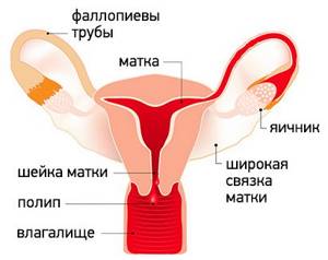
Uterine polyps are local benign neoplasms growing from the endometrial mucosa. They look like tubercles located on a wide base, or round or oval formations on a thin stalk. There may be one or several of them in the uterine cavity; in the latter case they speak of polyposis. The size of polyps varies from a few millimeters to 8 or more centimeters.
- Signs of a polyp in the uterine cavity and their diagnosis
- When is polyp removal necessary?
- Methods for removing endometrial polyps
- What is hysteroscopy of the uterus for polyps
- Types of hysteroscopy
- Indications for hysteroscopy
- Features of the event
- Preparation
- Basic recovery recommendations
- Complications after polyp removal
- Contraindications
Signs of a polyp in the uterine cavity and their diagnosis
Often uterine polyps do not manifest themselves, but some patients have the following symptoms:
- Menorrhagia - heavy menstruation.
- Bleeding outside of menstruation.
- Spotting after sexual intercourse.
- Bleeding during menopause.
- Infertility and miscarriages.
- With large polyps, women may notice copious whitish discharge, discomfort and pain during intercourse, as well as periodic cramping pain in the lower abdomen.

To make a diagnosis, it is necessary to undergo a comprehensive examination, which includes the following procedures:
- Gynecological examination - particularly large polyps can be palpated during manual examination; cervical polyps can be detected during examination in the speculum.
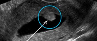
- Ultrasound of the pelvis - in the uterus, growth of the endometrium with a local increase in its mucous layer is detected. The structure of this neoplasm is homogeneous.
- Separate diagnostic curettage followed by histological examination of the obtained material.
- Hysteroscopy - allows you to examine the endometrium from the inside, visualize single or multiple polyps and determine their location. The procedure is also therapeutic in nature, since it allows for the simultaneous removal of all identified tumors and their subsequent histological examination. This is the most effective method for diagnosing a polyp in the uterus.
What is this?
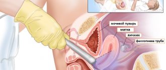
Hysteroscopy is a minimally invasive diagnostic and therapeutic procedure that allows an endoscope to study the structure of the uterine cavity (endometrium) to diagnose the disease or further carry out therapeutic manipulations. The examination is carried out using an ultra-thin optical instrument - a hysteroscope, which is inserted through the vagina. This examination is the “gold standard” for diagnosing endometrial lesions.
Cervical endoscopy is indicated for:
- Presence of signs of endometrial damage on ultrasound or GHA (polyposis, hyperplasia, neoplasm).
- Infertility, unsuccessful IVF attempts.
- Intrauterine synergies.
- Endometriosis.
- The presence of residual elements of the ovum after abortion.
- Anomalies in the development of internal genital organs.
- Submucosal myomatous node (endoscopy of fibroids allows you to determine its size, quantity and direction of growth).
Contraindications to the procedure include progressive pregnancy, active inflammation, uterine bleeding, cervical stenosis, malignant tumor, exacerbation of infectious pathologies and the patient’s serious condition.
When is polyp removal necessary?
The choice of patient management tactics and determination of the need to remove the polyp is determined individually. If a woman is of reproductive age and has no symptoms of a polyp, conservative management tactics can be chosen. In this case, the patient should undergo regular monitoring.
In all other cases, removal of polyps is indicated:
- Presence of symptoms of the disease.
- Pre- and postmenopausal periods, even if there are no symptoms.
How much does it cost to treat polyps in the uterus?
The cost of treatment differs in different clinics. It primarily depends on the type of formation and the choice of treatment method. Thus, with conservative therapy, it may include control examinations to monitor the effectiveness of therapy. If the patient’s condition does not improve during therapy, surgical removal of the polyp may be required. Different surgical treatment methods also have an impact on cost. The use of expensive equipment increases the price. In fact, it is possible to find out how much it will cost to remove a polyp only after a complete examination of the patient. In addition, prices are influenced by the clinic’s pricing policy and the level of qualifications of doctors.
Methods for removing endometrial polyps
Surgical removal of endometrial polyps is called polypectomy. It involves curettage of the endometrium followed by histological examination of the resulting material. Curettage can be performed blindly, under visual control using ultrasound or hysteroscopy. The latter method is the most preferable because it allows you to control the completeness of polyp removal and avoid the development of surgical complications (uterine perforation, excessive curettage with damage to the basal layer of the endometrium, etc.).
If treatment does not help, and the patient is in menopause, she may be offered surgery to remove the uterus in the following cases:
- Persistent relapses of multiple polyposis.
- High risk of malignancy of polyps.
- The presence of severe symptoms that significantly worsen a woman’s quality of life.
Complications
The most important unpleasant consequence of the operation is the recurrence of the disease - the appearance of a new polyp. Even the most modern techniques associated with the destruction of the polyp bed and curettage do not lead to 100% remission. In 10-12% of cases, the tumor appears again (as of 2005).
Other possible complications include the following:
- Formation of scars and adhesions. As a result of frequent removal of polyps or their multiplicity, epithelial tissue is replaced by connective tissue. As a result, the canal itself becomes narrower, difficulties with conception arise, and infertility may develop.
- Infection. During surgery, the immune status decreases and the body becomes more susceptible to pathogenic viruses and bacteria. The risk of infection at the site of the removed polyp is especially high.
- Malignant degeneration of tissue. A cancerous tumor may develop if the polyp is not completely removed. The remaining cells begin to grow and can give rise to a malignant neoplasm.
- Bleeding when the wall of the cervical canal is injured. Treatment depends on the extent of the damage, and further surgery may be required.
- Allergic reaction, swelling. Corrected by taking antihistamines. As a rule, they pass without consequences.
- Hemametra – internal bleeding. The difficulty of diagnosis lies in the fact that the woman does not observe any discharge. This occurs due to spasm of the cervix - blood cannot leave the organ. Possible nagging pain, pallor of the integument. Treatment is carried out by taking antispasmodics or suctioning the blood using a probe.
Important! Minor discharge in the first days after surgery and minor discomfort in the lower abdomen are not causes for concern.
Types of hysteroscopy
There are diagnostic or surgical hysteroscopy. Diagnostic involves only examining the endometrium and endocervix for the presence of pathologies. The study is performed on an outpatient basis and does not require serious anesthesia (local anesthesia and sedation are sufficient) and a long recovery period. Therefore, it was called office hysteroscopy. Based on its results, the doctor draws up a plan for further management of the patient.
Surgical hysteroscopy allows for diagnosis and immediate treatment of identified pathologies. It is performed in an operating room and requires full anesthesia. The procedure is prescribed either when the diagnosis has already been established, or when the likelihood of detecting a pathology is extremely high.
We specialize in surgical hysteroscopy because we believe that invasive diagnostic techniques can be successfully replaced by safer tests. This will help make the correct diagnosis without the risk of surgical complications.
Features of the event
Therapeutic hysteroscopy is carried out on a hospital basis, including a one-day hospital in an operating room. An hour before the start of the intervention, premedication is performed - a sedative injection is given. After being delivered to the operating room, the patient is placed in a gynecological chair and given intravenous anesthesia. Next, proceed to the operation:
- Dilation of the cervical canal.
- A certain amount of sterile liquid is injected into the uterine cavity to “straighten” its walls.
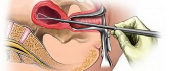
- A hysteroscope is inserted through the cervical canal. As it is introduced, a step-by-step examination of all parts of the uterus is performed. First, the cervical canal is examined, then the uterine cavity and its angles.
- If necessary, surgical procedures are performed - taking a biopsy, removing polyps, dissecting synechiae, etc.
- At the end of the operation, fluid is removed from the uterine cavity.
Office hysteroscopy – what is it in gynecology?
The hysteroscope for office procedures is equipped with a very thin optical tube with a diameter of no more than 3 mm
Office hysteroscopy is a purely diagnostic procedure that never turns into a therapeutic one. A hysteroscope is a doctor’s eye, and nothing more. Office hysteroscopy is performed in a gynecological office - hence the name; the procedure does not require anesthesia , since there is no dilation of the cervical canal.
How is office hysteroscopy performed?
Office hysteroscopy is performed in a doctor’s office on a gynecological chair. The doctor expands the walls of the vaginal vault and inserts a very thin hysteroscope tube into the cervical canal (cervix). The doctor examines the lining of the uterine cavity, establishes (or confirms the diagnosis) and issues a conclusion.
What is the difference between the “office hysteroscopy” procedure and “therapeutic and diagnostic hysteroscopy”
Office hysteroscopy never turns into medical hysteroscopy. And its meaning is only to “look” to confirm the diagnosis. In Russia, this diagnostic method is not used often. Usually, having established a diagnosis by ultrasound, a diagnostic and treatment hysteroscopy is performed, when the doctor simultaneously “looks and removes.” Therapeutic and diagnostic hysteroscopy is always associated with an operation, is performed in an operating room under intravenous anesthesia and requires, in addition to a gynecological surgeon, the presence of an anesthesiologist. The operation is called separate diagnostic curettage (SDC), when pathological biomaterial is taken separately from the uterine cavity and cervix and sent for histology to exclude the presence of atypical cells. If atypia is excluded, the treatment process is carried out by a gynecologist.
Depending on the purpose of the study, there are: • office (diagnostic) hysteroscopy - only diagnostics are performed. In practice it is not used often. • therapeutic and diagnostic (surgical) hysteroscopy – during hysteroscopy, surgical manipulations are immediately performed, as a rule, this is what happens in practice. • hysteroresectoscopy is a type of diagnostic and treatment (surgical) performed with a hysteroscope with a special attachment for the removal instrument.
Office hysteroscopy and polyp removal
“Office hysteroscopy and removal of polyps” - the use of this wording only speaks of the patient’s ignorance (which is not his fault). The word “removal” excludes an outpatient appointment with a gynecologist; this is always an operation either using a hysteroresectoscope, or a full separate diagnostic curettage. The completeness of the surgical intervention is determined by the doctor. There is no point in entering the uterine cavity twice: first to look (do office hysteroscopy ), and then for diagnostic and treatment purposes. Polyps are easily diagnosed by ultrasound.
Office hysteroscopy of the uterus
Office hysteroscopy of the uterus can be performed to confirm the diagnosis made by ultrasound.
Indications for the diagnostic procedure:
• suspicion of internal endometriosis of the uterine body ; • submucosal fibroid node – can be identified on ultrasound; • synechia (fusion) in the uterine cavity ; • remnants of the fertilized egg – can be identified on ultrasound; • endometrial pathology – identified by ultrasound (conditionally) ; • suspicion of malformations and anomalies of the uterus.
Office hysteroscopy with endometrial biopsy
Endometrial biopsy is a procedure for collecting biomaterial and its histological examination for atypical cells. According to the degree of reliability, they are distinguished:
- pipel biopsy - using aspirate from the uterine cavity, the presence of atypical cells without localization is assessed;
- biopsy during office hysteroscopy – the area of collection of biomaterial from the endometrial mucosa is specified. , office hysteroscopy, unlike pipel biopsy, allows for targeted sampling from the problem area. But since the optics are thin, the viewing area of the uterine cavity is quite narrow.
Both biopsy methods are diagnostic tests only . If it is not possible to obtain expanded information during RDV surgery, this research method may be justified. But undoubtedly it does not have the completeness that can be obtained with diagnostic and treatment hysteroscopy (in this case, both diagnosis and treatment occur simultaneously), as a result of which the cavity and cervix are examined. Endometrial biopsy during office hysteroscopy does not provide such information.
Prices for office hysteroscopy at the Medservice clinic
| SERVICE | COST, RUB. | WHAT IS INCLUDED |
| Consultation on hysteroscopy | For free | |
| Initial appointment with an obstetrician-gynecologist | 2 000 | Ultrasound, examination, consultation |
| Office hysteroscopy in a doctor's office on an outpatient basis | ||
| Office hysteroscopy | 9 950 | Visualization of the cavity and cervix with issuance of a conclusion |
| Histology | 2 000 | Study of biomaterial for atypical cells |
| TOTAL office hysteroscopy | 13 950 | |
| Hysteroscopy in the operating room | ||
| Tests on the day of surgery | 4 150 | Hospital complex, coagulogram, smear |
| Anesthesiology benefits | 3 900 | Intravenous anesthesia "Propofol". Control of vital functions |
| Diagnostic hysteroscopy without separate diagnostic curettage | 9 950 | Visualization of the cavity and cervix with issuance of a conclusion |
| Treatment and diagnostic hysteroscopy with separate diagnostic curettage without histology | 15 950 | Visualization of the cavity and cervix with issuance of a conclusion and curettage of the cavity and cervix |
| Treatment and diagnostic hysteroscopy with separate diagnostic curettage and histology from 2 places | 19 950 | Visualization of the cavity and cervix with the issuance of a conclusion and curettage of the cavity and cervix. Histology from 2 zones |
| TOTAL diagnostic hysteroscopy without separate diagnostic curettage | 20 000 | |
| TOTAL therapeutic and diagnostic hysteroscopy with separate diagnostic curettage without histology | 26 000 | |
| TOTAL therapeutic and diagnostic hysteroscopy with separate diagnostic curettage and histology from 2 places | 30 000 | |
BEFORE CLOSING THIS SECTION, MAKE SURE YOU HAVE FOUND THE ANSWER TO YOUR QUESTION.
IF NOT, WE WILL GUIDE YOU TO THE RIGHT PAGE:
YOUR GOAL IS A DIAGNOSTIC BIOPSY OF THE UTERINE CAVITY [using the paper-scoring method]
[targeted during office hysteroscopy]
OR TREATMENT AND DIAGNOSTIC BIOPSY OF THE UTERINE CAVITY
OR TREATMENT AND DIAGNOSTIC BIOPSY OF THE CERVIX
click on the desired answer
Call online Make an appointment
Preparation
Preparation for hysteroscopy is similar to other surgical interventions in gynecology. First, an examination and, if necessary, certain treatment are carried out. The following tests are performed:
- General blood and urine analysis.
- Coagulogram.
- Blood type and Rh factor.
- Vaginal smear for flora.
- ECG.
- Test for STIs.
- Tests for HIV and parenteral hepatitis.
Hysteroscopy is planned at the beginning of the menstrual cycle (before day 10), since endometrial tumors are clearly visible during this period.

Immediately before surgery, it is recommended to follow these rules:
- Two days before hysteroscopy, avoid sexual contact.
- On the day of the operation, take a shower and then put on underwear made from natural fabrics.
- Remove jewelry and contact lenses.
- Immediately before entering the operating room, empty your bladder.
- Do not eat for at least 6 hours before anesthesia.
Basic recovery recommendations
- If an antibiotic is prescribed, take it for as long as your doctor recommends.
- To relieve pain after hysteroscopy, you can take antispasmodics or analgesics.
- Avoid sexual intercourse and heavy physical activity until the bleeding stops, but not less than 10 days.
- Perform hygiene measures in the shower. Taking a bath is not recommended.
- Toilet your genitals as needed, but at least 2 times a day.
- During the recovery period, it is not recommended to use tampons and menstrual cups; discharge should come out of the vagina freely.
Most women generally feel satisfactory after therapeutic hysteroscopy. The main complaints are nagging pain in the lower abdomen and weakness. The full recovery period takes about two weeks. However, if complications develop - fever, heavy bleeding, foul-smelling discharge, you should immediately consult a doctor.
Complications after polyp removal
As after any gynecological operation, the following complications may develop after hysteroscopy:
- Damage to the wall or cervix - treatment may require additional surgery to close the perforation.
- Damage to the cervix - may require stitches.
- Bleeding.
- Infectious complications.
It should be noted that complications after hysteroscopy are much less common than after other gynecological interventions performed “blindly”. But the possibility of their development cannot be completely excluded.
Contraindications
- The presence of decompensated chronic diseases that may interfere with anesthesia, for example, cardiovascular pathology.
- Presence of STIs and vulvovaginitis.
- The presence of diseases accompanied by a tendency to bleeding.
Euroonco specialists closely monitor the condition of patients not only in the postoperative period, but also at the initial stage. In this regard, we perform the procedure only after we are convinced that the patient has no contraindications. You can make an appointment for a consultation about uterine polyps by calling:
Book a consultation 24 hours a day
+7+7+78
What is a polyp in the uterus
A uterine polyp is a benign space-occupying formation that forms in the uterine cavity against the background of local hyperplasia of cells in the growth layer of the endometrium. The pathology can affect patients of different age groups. Detectability is approximately 6-20%.
The sizes of endometrial polyps range from 1-2 millimeters to several centimeters.
The formation is attached to the uterine wall through a thin stalk or a wide base. Nutrition is provided by a vessel in the center of the polyp. Pathological structures can be either single or multiple. When several formations are detected, they speak of polyposis. Polyps can cause intermenstrual bleeding, abdominal pain, and problems with conception. Usually pathology is diagnosed during ultrasound. In gynecology, polyps are considered potentially malignant formations that need to be removed surgically.