Indications for use
Although cardiography is a routine research method, it also has indications.
To determine the cause of pain or discomfort in the chest area, the patient consults a therapist or cardiologist. The doctor initially collects anamnesis, examines, measures blood pressure and pulse, auscultates the heart, and then sends it for examination to find out what the cardiogram shows. Indications for ECG:
- chest pain (suspicion of angina or myocardial infarction);
- dyspnea;
- discomfort in the heart after viral or bacterial infections;
- pathological heartbeat, interruptions in the functioning of the heart muscle.
An ECG is mandatory in the following cases:
- when hospitalized in an inpatient department of any profile;
- before surgical interventions;
- during preventive examinations of adults;
- for schoolchildren when choosing a group of physical education classes.
An electrocardiogram of the heart is used both for the primary diagnosis of pathological conditions and for monitoring the dynamics of the disease. When prescribing drugs, the doctor relies on both the patient’s subjective sensations and ECG data, which reflect actual changes in the cardiovascular system.
Preparation rules
- Before having an electrocardiogram, you need to get a good night's sleep. Also, you need to go to a medical facility in a calm state. Please know that you will not be given any injections before your appointment. During the examination you will not experience any discomfort at all. Often before the procedure the patient is allowed to lie quietly for 5-10 minutes;
- The ECG room should be at normal room temperature so that the patient feels comfortable. Otherwise, trembling in the body from the cold can distort the data of the organ’s functioning;
- Patients who experience severe shortness of breath are advised to monitor while sitting in a chair. In such patients, the heart rate is recorded better in this position;
- Women should not use greasy creams on the breast area before monitoring. This may reduce skin conductivity, resulting in distorted measurement data;
- Try to wear clothing that can be easily removed, as the electrodes must be attached to bare skin during the procedure. It is better for women not to wear tights, since ECG electrodes are also attached to the ankles;
- On the day of the electrocardiogram, you are allowed to eat two hours before the procedure. You can drink as much as you want.
If you complain about suspicious symptoms in the chest area, you can contact a doctor at the ProfMedPomosh clinic, who will write you a referral for an ECG. The ECG procedure itself is performed by a functional diagnostics doctor or nurse.
Execution technique
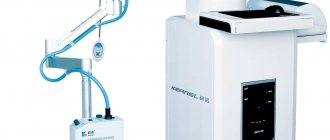
Carrying out cardiography does not require particularly complex skills, so middle and junior medical personnel know how to make a heart cardiogram. A device for such manipulation is a cardiograph. It can be stationary and permanently located in a specially equipped room, which each clinic has, or mobile - for convenient ECG recording at the patient’s bedside.
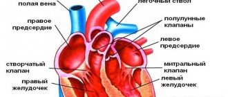
When performing an ECG, the patient lies on his back. The points where the electrodes are applied are freed from clothing and moistened with an isotonic sodium chloride solution to improve conductivity. Electrodes in the form of plates are attached to the limbs: red - on the right hand, yellow - on the left, green - on the left leg and black on the right. Six electrodes in the form of suction cups are installed on the chest. They are called chest leads (V1-V6), and electrodes from the limbs are considered basic (I, II, III) and amplified (aVL, aVR, aVF). Each of the leads is responsible for a specific area in the heart. If pathological processes along the posterior wall of the heart muscle are suspected, additional chest leads (V7-V9) are used.
It is important that before a scheduled electrocardiogram the patient does not drink alcohol or coffee. When removing it, it is undesirable to move or talk, as this leads to distortion of the examination results.
The cardiogram is recorded as a graph on special paper or in electronic form. It is important to record at least four cardiac cycles to obtain objective data on the condition of the heart. The film is signed indicating the full name, gender (male, female), date of the study, and age of the patient, since an adult and a child have different values of normal parameters. After this, the recording is transferred to the doctor, who interprets the ECG in detail.
Features of the event
- The patient exposes his body and lies down on the sofa. In this case, it is necessary to behave completely calmly;
- The health worker degreases the places where the electrodes will be attached with alcohol;
- A special gel is applied to the electrode plates, which conducts current well, as can be seen in the photo;
- The algorithm for applying electrodes is simple: electrodes are attached in the chest area, on the hands and ankles. In this case, the plates are fixed either with adhesive tape or with special suction cups;
- The algorithm for taking an ECG is as follows: the electrodes receive electrical impulses from the heart and transmit them through wires to the device itself, which processes them;
- Then the specialist turns on the device and begins recording ECG data. The device produces a long tape with a graph in the form of teeth of different heights. The monitoring data is deciphered by a doctor who will determine the diagnosis and prescribe treatment.
At the ProfMedPomoshch clinic you can get an ECG with interpretation and learn everything about the work of your heart.
Various techniques and indications for them
A classic ECG helps to see how the myocardium and conduction system of the heart behave at the current moment. In many cases (preventive examinations, normal pregnancy), a regular cardiogram is sufficient. But situations arise when the patient complains of pain or shortness of breath only during stress or physical activity, or at a certain time of day, and the film does not show characteristic changes in rhythm or pathological waves. In such episodes, additional types of cardiography are used.
With angina pectoris, it is not always possible to record changes on the ECG, so you have to use a stress ECG or treadmill test. This method involves performing physical exercises (treadmill or bicycle ergometer) while recording a cardiogram.
Indications for performing a stress test:
- diagnosis of angina pectoris and determination of its functional class;
- monitoring the effectiveness of treatment of coronary artery disease and angina pectoris.
In addition, there are a number of contraindications to this procedure:
- acute period of myocardial infarction;
- unstable angina;
- arrhythmia, severe blockades;
- heart failure in the stage of decompensation.
Another specialized type of ECG is Holter (24-hour heart monitor). To perform this procedure, electrodes and the recorder itself are attached to the patient’s body, which is small in size and measures electrical potentials around the clock. Read more about this type of cardiography in the article “Method of Holter ECG monitoring”.
How does it work
Electrodes placed on the patient's body receive weak electrical currents that originate in the heart. As they become stronger, they are recorded using a galvanometer. Any changes in ripple are transmitted to a special device, which produces data in the form of a broken line on a paper tape.
The next important stage of the examination is the decoding of the data that the doctor deals with. The fact is that the teeth on the tape are sized according to special standards. If there are any deviations from the norm, then there is a health problem. And only a specialist can correctly decipher the cardiogram and establish an accurate diagnosis.
In order for the results of an ECG to be reliable, there is no special preparation, but some rules must be followed.
Decoding the results
Decoding the electrocardiogram of the heart is an important and crucial stage in making a diagnosis and prescribing treatment. For correct interpretation, it is necessary to understand the essence of the teeth and lines on the graph.
The ECG printout contains three important elements:
- tooth – concavity or convexity of a line. Encrypted with Latin letters P, Q, R, S, T;
- interval includes segments and teeth;
- segment – the distance between two teeth.
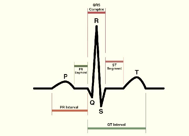
When describing a cardiogram, the duration of the intervals, the height of the teeth, the position and shape of the segments are taken into account. Important factors are the film recording speed with which the electrocardiograph works (usually 25 or 50 mmsec) and artifacts (patient movement during the procedure, isoline drift):
- Wave P – displays processes in the atrium, normally positive, up to 2.5 mm high and lasting 0.1 s.
- Q wave – shows impulses in the interventricular septum, duration – 0.03 s.
- The R wave is the highest, displaying the impulses of the ventricles themselves.
- The S wave is negative and shallow, indicating the completion of the impulse in the ventricles.
- The T wave reflects the repolarization of the ventricles.
The next important indicator of a normal ECG is sinus heart rate. Criteria: the P wave is in front of all QRS, equal to PQ (0.12-0.2 s) in all leads and the heartbeat is 60-80 beats/min.
Next, the electrical axis of the heart (EOS) is determined, which characterizes the conductive and fiber organization of the organ. It can be vertical (+70 +90 degrees), horizontal (0 +30) and normal (+30 +60).
Who is doing
A doctor of any specialty has at least a minimal understanding of how to read a cardiogram of the heart and be able to recognize the signs of serious conditions. Most often, cardiograms are deciphered by therapists or cardiologists, because they prescribe this study. Paramedics and EMTs read the films to quickly make decisions about medical support or admission to a cardiac hospital. Many clinics employ doctors who only interpret cardiograms (functional diagnostics doctor) and write a conclusion for the study performed.
Modern cardiographs at the end of the recording provide a preliminary result of the study indicating the size of the intervals and waves, heart rate, position of the electrical axis of the heart and signs of such pathologies: blockade, arrhythmia, hypertrophy of the myocardial walls. This makes the doctor’s job easier in counting and measuring segments, but sometimes the program misinterprets the results. The doctor rechecks the pathological signs on the ECG and makes the correct conclusion.
In some cases, the conclusion of an electrocardiogram of the heart does not completely resolve the diagnostic issue. The doctor may ask to see previous films and findings from other examinations. When making a diagnosis, data from the medical history, course of the disease, and medication intake are taken into account.
Is it possible to interpret the results yourself?
Many patients want to know how to independently decipher a cardiac cardiogram, because they often want to quickly find out the result of the study in order to reassure themselves. But it is better to entrust this task to a doctor, having received competent advice, although some ECG data are easy to interpret even for beginners. The procedure is easier to do if the recording is of high quality and there are no artifacts on the film.
To understand how to read a heart cardiogram, you need to know about the parameters of rhythm and heart rate. To determine the number of contractions, count the number of large squares on the film between the two nearest R waves. At a speed of 50 mms, divide 600 by the number of squares, and at 25 mms, divide 300 by the number of squares.
Afterwards the EOS value is indicated. As mentioned earlier, the position of the axis can be normal, horizontal or vertical. Normal: vertical in thin people, horizontal in hypersthenics (stocky, with a wide chest). Deviation of EOS is interpreted as hypertrophy of the myocardial walls, blockade of conduction pathways or other pathologies.