Diagnosis of hair and scalp diseases in doubtful cases includes a number of studies, the most informative of which is a biopsy. A biopsy removes cells or tissue from the body for diagnostic purposes. Biopsy includes histological and immunohistochemical examination of the biopsy specimen.
Based on the results of histology and IHC, treatment is most accurately selected based on what changes in the tissues are identified.
A biopsy of the scalp can definitively clarify the diagnosis, including androgenetic alopecia, alopecia areata, and scarring alopecia at the tissue level.
Histological examination of the skin
- Will help determine the number of damaged, “miniaturized follicles”, thinning hair, the preservation of hair follicles with a decrease in hair density
- Allows you to determine the presence, localization and severity of inflammation.
- Determine the integrity of hair follicles or the presence of scar tissue/fibrosis, which determines the possibility of hair restoration.
- Allows you to determine the condition of blood vessels and nerves on the scalp (the preservation of blood supply and innervation is determined).
Complications of skin biopsy
Skin biopsy is usually simple and complications are rare. Typically, the more skin sample removed, the higher the chance of complications. The following complications may occur.
- Bleeding
Intraoperative or postoperative bleeding can occur in anyone, but it can be especially problematic in people who have a bleeding tendency or are taking blood thinning medications such as warfarin or aspirin.
- Infection
Bacterial wound infection affects approximately 1–5% of excisional biopsies. This is, however, extremely rare with minor punches, shaving or post-operative biopsies. Ulcerated or crusted skin lesions, biopsy site, and patient characteristics such as diabetes, advanced age, or use of immunosuppressants may contribute to an increased risk of infection.
- Nerve injury
The blade may cut the superficial sensory nerve, causing pain or numbness. This is most likely when the skin is thin, such as on the face or back of the hand. The risk of motor nerve disruption is extremely rare, but may occur during skin cancer surgery in facial danger areas. These include the temporal, marginal mandibular, zygomatic branches of the facial nerve and the auxiliary spinal nerve (at Erb's point).
- Scarring
There is usually a significant permanent scar at the biopsy site. Certain areas of the body, such as the center of the chest, are prone to developing excessive or hypertrophic scars. It is also more common in Afro-Caribbean skin types.
- Persistence or recurrence of skin lesions
Many biopsies are intentionally partial and are intended for diagnostic purposes only. With an excisional biopsy, there is a risk of not removing the complete lesion, which may recur later.
- Problems with anesthesia
Allergy to local anesthetics is possible, but also extremely rare. Vasovagal reactions are more common and can result in patient fainting and potential injury. Palpitations are another side effect associated with epinephrine, commonly found in local anesthesia.
- Wound disorder
This is a rare complication of wound suturing. This most likely occurs in areas of the body where there is a lot of stress on the scar (eg, chest, back), immediately after a stitch is removed, or as a result of infection. Avoiding exercise and using straps and dissolvable sutures can help prevent this.
Immunohistochemical study
- Allows you to determine the sensitivity of tissues to the main sex hormones - estrogens, androgens, progesterone. This analysis is important for determining the possibility of developing androgenetic alopecia, for clarifying the diagnosis, as well as determining the need for treatment with antiadrogen drugs or estrogen drugs/analogs
As you know, androgens are the cause of the most common hair loss problem - androgenetic alopecia. The immunohistochemistry method allows you to evaluate the sensitivity of hair follicles to androgens, and thus give a long-term prognosis to what extent this problem will bother the patient.
Filling out the request form
The clinician should ensure that the pathology request form includes basic patient information (including age and identifying information), biopsy site and type, and time and date. Left and right are best written out completely to avoid mistakes
In addition to this, it is essential that the pathologist is provided with clinical information and a range of possible diagnoses. For better clinicopathological correlation, clinical information should include description of duration, symptoms and dermatological description.
The sampler must be labeled with patient identification, biopsy location, time and date, and verified against the request form. When multiple biopsies are taken, Roman numerals are best used to match request forms to their corresponding specimen pots.
How does the procedure work?
The biopsy procedure involves taking a skin sample, which is examined under a microscope to clarify the suspected diagnosis. To avoid painful sensations during the procedure, anesthesia is first performed, an anesthetic is injected into the selected area of the skin. The process of taking material for research is carried out using the punch method, which is carried out with a special instrument, positioning it perpendicular to the surface of the skin. This method captures the deeper layers of the skin and subcutaneous fat. After collecting the material, the wound is sealed with medical biological glue.
Choosing the type and location for biopsy
It is essential that the biopsy site is chosen carefully, otherwise the pathological diagnosis may be incorrect or misleading. Here are some guidelines to help you find the best location, some general tips and mistakes to avoid depending on the type of skin lesion.
For suspected skin cancer:
- Punch biopsy - usually provides the pathologist with the best sample of skin to determine the pattern of growth and depth of invasion. A 3mm fence will be sufficient in most cases.
- Avoid taking a biopsy from the center of the lesion if it is ulcerated. It will be more difficult to close the wound if it is bleeding, plus the tissue may be largely necrotic, making it difficult to obtain a proper tissue sample.
- If there is a lot of deposits, first carefully remove it and take a biopsy from the immediate skin.
- It is generally not recommended to try to completely remove skin cancer using a needle biopsy.
For most inflammatory skin conditions:
- A core biopsy usually provides a good view of the entire skin for the pathologist, and a 4-mm punch is usually sufficient.
- As lesions develop over time, they will show more (or less) beneficial properties on histological examination, so when choosing a biopsy site, the age of the lesion is an important aspect to consider.
- If vasculitis is suspected, the best lesion to biopsy is fresh (24 to 48 hours old).
- In general, it is best to take a biopsy from the center of a larger, well-developed area. This should be taken from the raised edge of the ring board.
- Avoid areas that are scratched/crossed out as they will show non-specific changes.
- If possible, avoid areas that have been treated with topical steroids or other anti-inflammatory agents.
For ulcers, erosions and blisters:
- The skin adjacent to erosions and ulcers usually provides the most useful diagnostic information.
- When blisters are present, the biopsy is best taken from the edge of the blister, including two-thirds of the normal adjacent skin.
- A small, intact blister can show more useful information than the corner of a large one.
- Punch biopsy for immunofluorescence is best performed on periliated skin.
Preparation
To prepare for a scalp biopsy, it is necessary to briefly stop medications that increase bleeding, such as anti-inflammatory drugs, aspirin, anticoagulants (check with your doctor).
At the moment, the procedure is as safe as possible, painless and quick, and the results of a skin biopsy can be ready in 2 weeks.
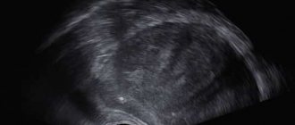
These images show a variant of the norm - hair follicles are normal in size, terminal hairs in the anagen stage predominate. Anagen/telogen 13/1, terminal/vellus 12/2, hairs are grouped into follicular units. There is no inflammatory infiltrate. There is no fibrosis. The condition of the stroma (nerves, blood vessels) is normal.
You can find the price list for these studies here
Indications for the procedure
A skin biopsy is ordered to make a specific diagnosis. Without confirmation, it is impossible to prescribe correct treatment that will have minimal risks. The indications are as follows:
· Skin diseases. These are eczema, psoriasis and others.
· Systemic diseases with skin manifestations. For example, cutaneous lupus erythematosus.
· Fungal and infectious diseases of the skin. For example, folliculitis, erythrasma, mycoses, pyoderma and more.
· Skin cancer. Melanoma, squamous cell or basal cell carcinoma.
Lymph node biopsy
If there is a suspicion that melanoma has spread from the primary site through the lymphatic system, a biopsy of the sentinel and regional lymph nodes is performed.
When choosing a fine-needle aspiration biopsy, CT or ultrasound is used. This control allows you to very accurately take a tissue sample from large lymph nodes with a fine-needle syringe.
This technique is gentle for the patient, but does not always allow obtaining a sufficient amount of tissue for examination.
Therefore, a surgical biopsy is also applicable, allowing the removal of an enlarged lymph node through an incision.
To detect metastases in regional lymph nodes, sentinel lymph node biopsy is used. The sentinel node is detected using the radioisotope method by injecting a radioactive drug into the area where melanoma is located.
Using a radioactivity detector, the specialist examines the lymph nodes. Having discovered a node with the maximum level of radioisotope, it is excised and examined. If there is an enlarged lymph node close to the melanoma, a biopsy is performed. In these circumstances, there is no need to look for a sentinel node.
The tumor is considered to have not spread when no melanoma cells are found in the sentinel lymph node.
If they are detected, lymph node dissection is performed, which involves removing the remaining lymph nodes with subsequent assessment of their condition. This is important for deciding on further treatment tactics for the patient.
What should you expect during and after the procedure?
During a puncture biopsy, insertion of the needle is accompanied by short-term acute pain. During a surgical biopsy, local anesthesia and general anesthesia help relieve pain.
The introduction of a local anesthetic to numb the skin is accompanied by a light injection, after which the sensitivity of the skin disappears for a while. There is a feeling of slight pressure as the biopsy needle is inserted.
Before the procedure, the doctor may prescribe the patient a mild sedative in tablets. In addition, sedatives are administered intravenously, if necessary, throughout the procedure.
There may be some soreness where the needle was inserted for a few days after the biopsy. For severe pain, analgesics can be used.
How to care for your skin after a biopsy may vary, but you can usually remove the bandage one day after the procedure and then shower or bathe.
Up
When is the procedure prescribed?
There are many dermatological diseases, among them:
- Infectious lesions;
- Chronic allergic diseases;
- Connective tissue pathologies;
- Autoimmune diseases;
- Neoplasms.
It happens that diagnosing dermatoses is not easy. And here a biopsy comes to the rescue, which can confirm diagnoses and conduct differential diagnostics. The procedure is prescribed to patients with suspected following pathologies:
- Skin vasculitis, scleroderma, dermatomyositis;
- Tuberculosis and sarcoidosis of the skin;
- Deep mycosis;
- Lymphoma of the skin;
- Plaque parapsoriasis;
- Dermatoses;
- Suspicion of cancer.
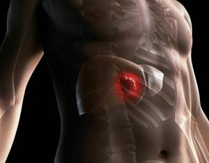
Tests for liver cancer
What does the diagnostic equipment look like?
There are quite a large number of biopsy techniques. Moreover, the equipment used is very different.
In a needle biopsy, a tissue sample is removed using a needle.
The biopsy needle is usually several centimeters long, and the syringe barrel is the diameter of a wide paper clip. The needle is hollow inside, allowing the tissue sample to be grasped and removed.
Several types of needles are used when performing a biopsy:
- Thin needle attached to a syringe: Thinner in diameter than needles used to draw blood from a vein.
- Cutting automatic needle with spring mechanism: consists of an internal "thick" needle, which is inserted into a locking cell, covered with a protective sheath and attached to a spring mechanism.
- Vacuum aspiration biopsy device: allows you to obtain a fairly large tissue sample.
A puncture biopsy is often performed under the guidance of CT, fluoroscopy, ultrasound or MRI.
Mammograph
It is a rectangular apparatus equipped with an X-ray tube. The device is used exclusively for x-ray examination of the mammary glands and has special additional devices that ensure that radiation reaches only the gland. A device is attached to the X-ray machine to position and compress (squeeze) the breast, which helps obtain images from different angles.
Ultrasound scanner
consists of a console that includes a computer and electronic equipment, as well as a video display and an ultrasound probe used for scanning. The sensor is a small, portable device that resembles a microphone and is attached to the scanner using an electrical cord. The ultrasound sensor sends high-frequency sound signals inaudible to the human ear and picks up echoes reflected from the internal structures of the body. The principle of operation of the device is similar to sonars that are used on submarines. In this case, an image instantly appears on a monitor resembling a television or computer screen. Its appearance depends on the amplitude (strength), frequency and time it takes for the sound signal to return from the patient's body to the transducer, as well as the type and condition of the tissue through which the sound travels.
Computer tomograph
usually a massive rectangular apparatus with a hole, or short tunnel, in the middle. During the procedure, the patient is placed on a narrow table that slides inside the tunnel. The X-ray tube and electronic X-ray detectors are located opposite each other inside a ring-shaped structure called a gantry. In a separate office there is a computer workstation where the resulting image is processed. There is also a doctor or technologist here who monitors the operation of the tomograph and the progress of the examination.
Vacuum device for aspiration biopsy
is an instrument in which a tissue sample is taken into a needle under pressure.
When performing a surgical biopsy
the pathological area is often marked with special thin wires or contrast material, which is introduced into the patient’s body under the control of ultrasound, CT or X-ray.
Other sterile instruments are also used during the procedure: syringes, forceps, scalpels, sponges and microscopic glasses or tissue containers.
Up
Clinicopathological correlation
Skin diseases and conditions can sometimes be very difficult to accurately diagnose. In these cases, clinical and histopathological data together form a more complete picture for making the correct diagnosis. This is called clinicopathological correlation. Many organizations hold regular multidisciplinary meetings (MDMs) in which a panel of experts reviews clinical information, clinical photographs, and pathology slides to determine the best diagnosis and treatment for the patient.
Source:
https://www.dermnetnz.org/topics/skin-biopsy/