This is a question many expectant mothers ask themselves. The American College of Radiologists lists x-rays as generally safe during pregnancy. The impact of X-rays on pregnancy is minimal if the necessary precautions and safety precautions are followed. Therefore, we can say with confidence that a single study will not harm the unborn child, because the dose of radiation received will be minimal.
If you suspect you are pregnant, you and your doctors need to consider whether an X-ray is really necessary. After all, sometimes the harm from not doing an x-ray can exceed the possible harm to the child. You need to weigh both of these factors.
Therefore, there is a generally accepted rule: X-rays, mammography and other radiation examinations during pregnancy should be carried out only according to strict indications, if the risk of harm from failure to perform the examination exceeds the harmful effects on the fetus! If you follow this rule, you will not wonder why x-rays are dangerous during pregnancy.
What are the dangers of lung x-rays at the beginning of pregnancy?
Its influence in the early stages of pregnancy is negligible, because the absorbed dose of radiation during pregnancy is only 0.1-0.2 mSv. It will not bring any tangible harm to the unborn child, but there will be no benefit from it either. The impact of mammography on pregnancy should also not be overestimated. However, if you are not sure that there is no pregnancy, it is better to refrain from any studies related to ionizing radiation (x-rays, mammography, fluorography, etc.).
In the first months of pregnancy, the fetus is most sensitive to external influences, however, the radiation dose from an X-ray of the lungs is minimal and will not harm the child. However, the effect of X-rays on early pregnancy has been little studied, and if you are unsure, it is better to refuse this study.
Indications for X-ray
Dentists treat teeth without x-rays only in cases of obvious caries in the absence of hidden causes. An x-ray will help the doctor determine the cause of toothache, the location and form of the inflammatory process. This diagnosis is necessary when:
- extensive focal infection (deep caries of a group of teeth);
- hidden pathologies of the crown or root part of the tooth;
- inflammation of periodontal tissue;
- filling root canals for pulpitis;
- problematic growth of wisdom teeth;
- formation of a cyst, granuloma or other neoplasm;
- suspected periodontitis;
- fracture or tooth root injury.
Timely and high-quality dental treatment will help reduce the risks of the negative impact of infection and bacteria on the development of the child. In many cases, such treatment can only be carried out after an image has been obtained. Even an experienced dentist will not be able to accurately determine the position of the roots, dental canals or the condition of the pulp chamber without x-rays.
Protective measures
So, can pregnant women have a dental x-ray? Yes, but only if you take all precautions and take the photo using modern equipment.
Be sure to notify your doctor before taking an x-ray and tell him the exact duration of your pregnancy. The safety of the procedure depends on this.
Avoid taking x-rays using a film x-ray machine. It is better to choose clinics where a computer visiograph is used. The radiation beam of such equipment acts locally, thanks to which it gives 10 times less radiation exposure compared to conventional equipment.
Before the procedure, cover your chest, stomach and legs to the knees with a protective lead apron. Such metal does not transmit gamma radiation. Do not remove the apron for 5 seconds after the x-ray, during which time the rays decay.
Where to get a dental x-ray in Moscow
Many public dental clinics use outdated X-ray machines that produce dangerous loads. Therefore, during pregnancy it is better to give preference to private clinics with modern equipment.
For example, you can undergo an examination at the TopSmile aesthetic dentistry clinic. We take pictures using safe and high-quality equipment. The emitted waves are so weak that they cannot affect the development of the child. The patient does not have to go from office to office; all procedures, including x-rays, are performed at the dental chair.
You can sign up for a consultation in any way convenient for you. Fill out the feedback form on the website or call +7. If you have any questions, you can get an express consultation with the chief physician, just write to WhatsApp.
There are contraindications. Specialist consultation is required.
Standard question about x-rays during pregnancy
QUESTION. Doctor, I did an x-ray of myself (for example, lungs, head, bones, etc.), and then I found out that at that time I was pregnant in the early stages. Are you very concerned about what harm has been done to the baby? Should we be afraid of deformities? Should I have an abortion?
ANSWER. When a fetus comes into contact with ionizing radiation for medical purposes, one should not talk about a direct teratogenic effect on the fetus (that is, an effect that necessarily causes developmental disorders), since the dose received is too small. We can only talk about an increased probability of diseases occurring in the unborn child (this is called the stochastic effect), but this probability increases only by a few fractions of a percent. So there is no need to worry seriously. A careful examination of the problem shows that similar minimal harm is caused by other types of radiation that we constantly encounter in life. So, we are exposed to radiation when we sunbathe in the sun, fly on an airplane, are high in the mountains, or walk along a pavement made of radioactive (yes!) granite. Finally, there is a normal natural radiation background (it would be better without it). In this sense, life is not a very useful thing at all. To summarize, the answer is this: don’t worry, there is no direct harm to the unborn child, but you need to remember about the mandatory screenings and listen to your gynecologist.
Why is CT harmful?
The principle of CT scanning is the effect of X-ray radiation on the body. This radiation is practically harmless to an adult, if you do not exceed the permissible amount of research. However, exposure of a developing fetus to X-rays can result in embryonic death or congenital deformities.
Therefore, if it is possible to conduct an MRI instead of a CT scan, the doctor should choose the first option. CT scan is indicated only in cases where the life of the expectant mother is at stake.
MRI in our center
Power 1.5 Tesla
High image quality
Study for patients up to 250 kg
Burn to disc for free
Second medical opinion on the image
Remember that, if necessary, the obtained X-ray images can be re-analyzed by the right specialist! You can order a repeat interpretation of an X-ray, CT or MRI using the National Teleradiology Network. A team of highly specialized radiologists will help clarify your diagnosis and provide a detailed description of the submitted x-rays and other studies within 24 hours.
Pavel Popov
Candidate of Medical Sciences, Member of the European Society of Radiologists
General contraindications for CT scanning
Computed tomography is one of the most accurate and informational methods for diagnosing almost all diseases of the human body. Moreover, the tomograph is capable of diagnosing ailments of the human body at the earliest stages. Therefore, doctors do not recommend neglecting this method. Still, not everyone can use the innovative method. There are still contraindications. Let's list them:
- women at all stages of pregnancy;
- young children (up to 14 years old);
- people who are overweight (more than 150 kg);
- nursing mothers;
- people with kidney and heart failure;
- patients with diabetes (preparation required).
The last three categories cannot undergo CT scanning using iodinated contrast agent.
But what should pregnant women do if it is impossible to make an accurate diagnosis without a CT scan? Let's figure it out.
When is an X-ray of the cervical spine prescribed?
An X-ray of the cervical spine is prescribed by a specialist. The study is carried out at:
- various neck injuries;
- discomfort in the cervical region;
- the occurrence of pain in the upper extremities;
- dizziness and headaches;
- coordination disorders;
- the appearance of blurred vision.
X-rays are also prescribed if there are suspicions of various diseases. It is used to diagnose degenerative and infectious diseases of the spine.
Kinds
The disease is divided into types according to which sinus is affected by the inflammatory process. The name of the type of disease comes from the name of the paranasal sinus. Since sinusitis is not uncommon in pregnant women, doctors can easily identify this pathology in a short time and select the optimal treatment regimen.
1. Frontal sinusitis - inflammation affects the frontal sinuses, causing severe pain mainly in the frontal region.
2. Ethmoiditis – damage to the ethmoid labyrinth. Not a common form of the disease.
3. Sphenoiditis - inflammation of the mucous membrane of the sphenoid sinuses.
4. Sinusitis - inflammation of the maxillary (maxillary) sinuses. This form of the disease occurs most often and is dangerous due to the rapid development of complications.
When is a hand x-ray prescribed?
X-rays are most often prescribed for hand injuries. The main symptoms are pain and obvious signs of tissue damage (swelling, bruising, etc.). X-rays are also recommended if the following symptoms appear:
- discomfort in the hand both during activity and at rest;
- visible changes in the joints of the hand;
- inflammatory processes;
- suspicion of osteomyelitis;
- visible growths on the hand;
- abnormalities in the development of joints and bone tissue of the hand.
X-rays are performed in an outpatient setting. The image can be taken in several projections (depending on the symptoms and location of the pathology). Using X-rays, you can identify various pathologies:
X-ray of joints is one of the most popular methods for identifying pathologies of the musculoskeletal system. The images allow you to detect various signs indicating pathologies (for example, calcium deposits, proliferation of cartilage tissue, and so on). X-ray of joints helps in making diagnoses, makes it possible to choose the right treatment tactics and evaluate its effectiveness. It is actively used in modern medicine, not inferior to modern diagnostic techniques.
Causes
The risk of sinusitis, especially in the third trimester, due to swelling of the nasal mucosa increases in every woman. In addition, there are certain unfavorable factors that greatly increase the likelihood that sinusitis will develop in pregnant women. Such provocateurs of the disease include:
- disturbances in the structure of the nose, and especially the curvature of its septum;
- presence of untreated carious teeth;
- frequent allergic reactions, which are accompanied by severe runny nose;
- proliferation of adenoids;
- neoplasms of benign and malignant nature in the nasal cavity;
- chronic inflammatory diseases of the pharynx;
- traumatic injuries to the face in the area of the nose and paranasal sinuses.
Sinusitis almost always appears after colds and viral infections, if they have not been treated by a doctor. This is due to the fact that reduced immunity is not able to resist pathogens, which is why they are able to actively reproduce.
What can a doctor see on an x-ray?
He can see:
- total number of teeth;
- anatomical features of the structure of the maxillofacial region;
- level, bone height;
- possibility of installing implants.
X-rays are also done for the purpose of establishing a diagnosis, when the doctor cannot make one based solely on the patient’s complaints.
X-rays are also done to monitor the doctor’s work. It shows how:
- canals are sealed;
- tooth removed;
- an implant was installed.
When is a joint x-ray prescribed?
X-rays are prescribed by a doctor. The following symptoms may be a reason to contact a specialist:
- joint dysplasia;
- mechanical injuries;
- osteoporosis;
- change in skin color;
- swelling of the joint;
- sprain;
- acute pain syndrome localized in the joint;
- various congenital pathologies.
X-rays are contraindicated for patients in serious condition, pregnant women and people with metal implants in the joints.
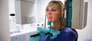
How they do it
No special preparation is required for CT scanning. Before the manipulation, you must remove all metal objects (jewelry, belts, removable dentures, etc.). Otherwise, they may distort the results or damage the equipment.
Diagnosis is carried out in a standing position. The patient is put on a lead vest to protect internal organs from radiation. She approaches the machine and places her chin on a special stand. The head must be fixed in the correct position for the image to be as clear as possible. You cannot move while performing the procedure.
The doctor turns on the machine and the jaw is irradiated with cone-shaped X-ray beams. A special sensor moves around the subject’s head and reads information. The doctor can adjust the strength of the radiation. The scanning duration is only a few seconds. The manipulation is painless and does not cause the patient any discomfort or pain.
Computed tomography is considered an indispensable method for examining hard-to-reach areas of the jaw and deep tissues. But during pregnancy it is rarely performed, only with the permission of a doctor. If possible, testing should be delayed until after birth.