Tumor markers or tumor markers are specific substances that are metabolic products of tumor cells, as well as cells associated with the development of cancer. Blood or urine samples are used to detect them. Most often, tumor markers are simple or modified molecules belonging to the group of glyco- or lipoproteins. Some of them are characterized by high tumor specificity, that is, they are present in a certain type of cancer, others are found in various types of tumors.
- Why do you need an analysis for the tumor marker CA 50?
- Indications for analysis for tumor marker CA 50
- Norms and interpretation of results
- What do the results mean?
The tumor marker CA 50 is a glycolipid. Its structure is similar to the tumor antigen CA 19-9. For neoplasms of the gastrointestinal tract, pancreatic cancer and colorectal cancer, the percentage increase in the concentration of CA 50 and CA 19-9 is similar. However, CA 50 has a high diagnostic value, since it is elevated in Lewis antigen-negative Le (ab-) patients in whom the concentration of CA 19-9 does not increase in the presence of carcinoma.
Elevated levels of the tumor marker CA 50 are also observed in 50-70% of patients with primary cancer of the liver, lungs, cervix and endometrium, prostate, ovaries, kidneys and mammary glands, as well as in some patients with melanoma (usually formed on the skin) and lymphoma.
Why do you need an analysis for the tumor marker CA 50?
A high concentration of tumor antigens may indicate the development of a malignant process. When combined with other diagnostic tests, tumor marker tests help doctors identify certain types of cancer and plan treatment. The main goals of the analysis are:
- Diagnosis of cancer. A high concentration of CA 50 may indicate the development of pancreatic cancer, colorectal cancer and, to a lesser extent, other types of malignant tumors.
- Choice of treatment method. Determining the level of tumor antigen sometimes helps to choose the most effective treatment tactics.
- Monitoring the effectiveness of treatment. Changes in the concentration of CA 50 in the blood help in assessing the effectiveness of antitumor therapy.
- Early diagnosis of cancer recurrence. Relapse is the resumption of a malignant process after treatment. Tumor marker tests can often detect cancer recurrence earlier than other diagnostic tests.
In some cases, determining the concentration of tumor markers in the blood or urine is recommended for people with an increased risk of developing certain cancers.
Types of tumor markers
The list of tumor markers contains about 200 compounds. But of these, no more than 20 species are of diagnostic value.
Most often, blood is tested for:
- PSA is a prostate specific antigen.
It is found in small quantities (0–4 ng/ml) in healthy prostate tissue. With an elevated PSA level (more than 10 ng/ml), there is a high (but not 100%) probability of a malignant tumor. The indicator may also increase with benign formations.
- CEA - cancer embryonic antigen.
It is synthesized by the cells of the fetal digestive system. In adults, the concentration of CEA increases (more than 5 ng/ml) with malignant tumors in the lungs, stomach, intestines, female genital organs, pancreas, thyroid and prostate glands.
A slight increase in the indicator can be observed in chronic pathologies of the kidneys and liver, in autoimmune diseases, and in smokers.
- AFP - alpha-fetoprotein.
With a high probability, hepatocellular carcinoma is diagnosed, and during pregnancy it reports the presence of pathologies in the development of the fetus. The concentration of AFP may increase with benign liver tumors and gonadal cancer.
- CA 125
Specific marker of ovarian cancer. Almost always, a concentration of more than 40 IU/ml is evidence of a malignant process.
- SA 15-3
Allows detection of breast cancer. If the level exceeds 38 IU/ml, they are referred for additional examinations.
- SA 19-9
Tumor marker for pathologies of the digestive system. If its concentration is above 37 IU/ml, it means that a malignant or benign tumor is developing in the gastrointestinal tract.
- CA 242
Marker for colon and pancreatic cancer. Exceeding 39 IU/ml indicates the presence of a malignant or benign formation in the organs.
- UBC
Used to detect bladder tumors. Normally, its concentration does not exceed 15 ng/ml.
- B2-MG
Exceeding the norm (0.9-2.0 ng/ml) indicates a risk of lymphoma, leukemia, multiple myeloma.
- hCG
An hCG test is usually performed to confirm pregnancy and detect abnormalities in fetal development. It also allows you to suspect an ectopic pregnancy. And in the absence of signs of pregnancy, it may indicate trophoblastic disease (hydatidiform mole, chorionic carcinoma), germ cell tumors of the ovaries and testicles.
Only an oncologist can correctly interpret test results. Therefore, after receiving the test results, you need to make an appointment for a consultation.
It is worth considering that some factors can distort the results. The concentrations of many markers increase in chronic inflammatory diseases. Smoking and alcohol abuse doubles the CEA content. In women, the level of CA-125 increases significantly during the menstrual period. In men, the PSA concentration exceeds the norm after medical manipulations on the prostate.
Norms and interpretation of results
The upper limit of normal concentration of the tumor marker CA 50 in the blood is 23 U/ml. This tumor marker has no obvious tumor or organ specificity. It can be isolated from various carcinomas of the gastrointestinal tract. High concentrations - over 100 U/ml - are most often found in pancreatic and cholangiocellular carcinoma.
An elevated CA 50 level does not always indicate the development of cancer. Sometimes (in 16-18% of cases) the concentration of a tumor marker above 100 U/ml can be observed in benign diseases of the pancreas or cirrhosis of the liver.
Types of tumor diseases
Cancer is a very old disease, which was first described back in 1600 BC. e. The name cancer itself was introduced by Hippocrates. These malignant tumors arise due to the transformation of normal cells, which begin to multiply uncontrollably. If the immune system does not begin to recognize the presence of a tumor in time, then metastases develop in all tissues and organs. Benign tumors, in turn, are distinguished by the fact that they do not metastasize, and therefore they do not pose a danger to life, however, at the same time, the possibility of degeneration of a benign tumor into a malignant one remains. The final diagnosis can be made after histological examination. Without the necessary treatment, malignant tumors can lead to the death of the affected person.
Symptoms of a tumor disease can vary and depend on the location of the tumor. In the early stages, the tumor does not cause any sensation, and pain appears only in the later stages. The most common symptoms are:
- loss of appetite;
- weight loss;
- exhaustion;
- anemia;
- immunopathological conditions;
- the occurrence of unusual compactions and swellings;
- inflammation;
- jaundice;
- bleeding.
With metastases, depending on where they develop, bone pain occurs and bone fractures often occur; cough (sometimes with blood); the liver and lymph nodes enlarge, etc.
Malignant tumors are divided into the following types (depending on the cells from which they are formed): carcinoma, sarcoma, melanoma, lymphoma, leukemia, glioma, teratoma, choriocarcinoma, etc.
According to international studies, the most common types of cancer are identified:
- Lung cancer ranks first in both the number of cases and the number of deaths from it. The first symptoms appear in the form of hemoptysis, chest pain, weakness, decreased performance, and cough. The main cause of the disease is smoking tobacco. There are four stages in the development of the disease:
- on the first, the tumor is up to 3 cm and is located in only one segment of the lung;
- in the second, the tumor is up to 6 centimeters, also located in one part of the organ. Metastases develop;
- in the third, the tumor reaches a size of more than six centimeters and is already beginning to move to the adjacent lobe of the lung. Metastases have more obvious manifestations;
- at the fourth stage, the cancer extends beyond the organ and spreads to neighboring organs.
Treatment for lung cancer can be carried out using various methods and is prescribed depending on the state of health, the extent of the disease and many other factors.
- Mammary cancer . This disease ranks second in terms of the number of patients and fifth in terms of mortality from it. Mostly the female half of the population suffers from breast cancer. The risk group includes women who have not given birth, as well as those who had their first birth after 30, and smokers. Also an important role is played by the presence of a family history, diabetes mellitus, hypertension and obesity. Symptoms of breast cancer:
- seals;
- various discharge from the nipple;
- change in the structure or color of the breast.
- The third place in the number of cases is colon cancer.
- Although stomach cancer ranks fourth in terms of incidence of diseases, it is in second place in terms of mortality.
What do the results mean?
A single test for the CA 50 tumor marker is not enough to diagnose cancer. The results of the study are considered taking into account the following factors:
- complete medical history;
- physical examination;
- results of other laboratory tests;
- results of instrumental diagnostics.
If the analysis was carried out as part of monitoring the effectiveness of ongoing anticancer treatment, its results are compared with the results of studies performed before the start of cancer therapy. A decrease or return to normal in the CA 50 level may indicate the effectiveness of treatment. An increase in the concentration of a tumor marker may mean that the tumor is not responding to treatment, is growing, or is reoccurring. A slight increase, as a rule, has no diagnostic value. Typically, the doctor pays attention to the tendency for indicators to increase over time.
Chemotherapy treatment may cause a temporary increase in tumor marker levels. This occurs due to the massive death of tumor cells, which secrete large amounts of specific substances.
Book a consultation 24 hours a day
+7+7+78
Our clinics in St. Petersburg
Structural subdivision of Polikarpov Alley Polikarpov 6k2 Primorsky district
- Pionerskaya
- Specific
- Commandant's
Structural subdivision of Zhukov Marshal Zhukov Ave. 28k2 Kirovsky district
- Avtovo
- Avenue of Veterans
- Leninsky Prospekt
Structural subdivision Devyatkino Okhtinskaya alley 18 Vsevolozhsk district
- Devyatkino
- Civil Prospect
- Academic
For detailed information and to make an appointment, you can call +7 (812) 640-55-25
Make an appointment
A benign or malignant neoplasm secretes a special cancer antigen. In this regard, when different tumors arise in the body, their own tumor markers appear. Tumor marker studies are also carried out in case of diagnosis of the disease and its treatment. It will allow doctors to choose subsequent tactics and, if necessary, prescribe additional drugs.
In what cases is research necessary?
The examination is usually prescribed not for the initial detection of the disease, but to monitor treatment. The level of the marker determines changes in the tumor during therapy. But sometimes such diagnostics are also indicated for primary pathologies of the mammary glands. These include:
- presence of lumps in the chest;
- change in skin and or nipple color;
- constant pain;
- deformation of the nipple area;
- presence of discharge.
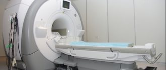
Before testing blood for markers, an instrumental examination (CT, MRI or biopsy) is prescribed. A referral for analysis is issued after cancer is detected.
What are the features of the research procedure?
To donate blood correctly, you must follow several rules that will reduce the likelihood of errors. Primary requirements:
- a short rest before the procedure;
- giving up alcohol, smoking, and taking medications the day before the examination;
- The test should be taken on an empty stomach; only drinking clean boiled water is allowed.
The blood collection procedure is no different from the usual one. The sample is taken from a vein according to standard rules.

What recommendations should be followed if the marker value is elevated?
If during the study a borderline value of CA 15 3 is detected and no pathology is detected, certain rules should be followed that will exclude further development of the disease:
- visit a mammologist twice a year - the doctor will conduct a preventive examination and give recommendations on lifestyle;
- choosing the right bra – it should be loose, products made from synthetic materials are contraindicated;
- special diet - vegetables and fruits should predominate in the diet, fatty, fried and smoked foods should be excluded;
- Excessive physical overload is not allowed - increased activity negatively affects the parenchyma of the mammary gland, sport should be moderate;
- avoiding stress – nervous tension often provokes cancer of the reproductive system. Try not to worry about trifles. If necessary, visit a psychologist.

Bad habits must be avoided - smoking and alcohol are prohibited. An important component of the prevention program is regular sex life with a permanent healthy partner.
What examination should be performed if markers are detected?
Sometimes women, when borderline tumor marker values are detected, stop being examined, hoping for a benign course of the pathology or its absence, but this is a mistake. Even if you have cancer during this period, there is a high chance of recovery, the main thing is to start treatment on time.
If there are deviations from the norm, it is recommended:
- Visit a mammologist - the doctor will evaluate the test results, advise on lifestyle and prescribe further examination.
- CT or MRI of the breast - these methods show the presence or absence of lumps. If they are detected, the following research method is required.
- A biopsy is the removal of a microscopic piece to determine the type of cells in the lesion. It is carried out at the final stage, after which a final diagnosis is made.
If there are no changes in the breast, an examination of the internal organs is prescribed. First, a survey CT, MRI or ultrasound of the abdominal cavity is done. When a pathological focus is detected, targeted diagnosis is carried out.