Before scanning an x-ray, you need to become familiar with some of the process features and digitization methods. At first glance, the simple scanning process in the case of an x-ray is problematic.
Scanning X-rays may be needed to send them by email, for example, to obtain advice from a qualified specialist or for posting on forums, or for the purpose of storing them electronically in your own archive.
If you know in advance that you will seek a remote consultation with a specialist, you can always ask for this image in electronic form.
If you have film on hand and it is not possible to get a digital copy, then the easiest and most reliable way is to contact special scanning centers that have special equipment for such purposes. The cost of such a service is not small, but it is quite affordable, however, if there are no organizations nearby that provide such services, it is easier to make a digital copy yourself.
What to choose?
Copy shop
In modern conditions, almost all clinics and medical centers, along with film images after the examination, give the patient a digital version. This means that for further consultations there is no need to carry a large film with you; digital media is sufficient.
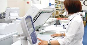
If a digital version was not received after the x-ray, and you are thinking about how best to scan the x-ray, if possible, use the services of a copy center. In this case, you are guaranteed to get a high-quality result that will not provoke unnecessary questions from the doctor, which means it will help to correctly diagnose the disease or monitor the success of the treatment.
New standard for radiological equipment
The standard for equipping heavy medical equipment, which is currently in effect, was formed in 2011–2012. Then MRI and CT scans were installed in outpatient centers for adults. Many pediatric outpatient centers are introducing CT scans for the first time.
all adult clinics will be uniformly equipped with modern diagnostic equipment . Each head building is equipped with an X-ray CT scanner, a magnetic resonance imaging scanner, an X-ray densitometer (allows you to identify bone tissue pathology), and mammographs, including those with tomosynthesis function (allows you to create a three-dimensional image of the entire breast to screen for early signs of breast cancer).
Thanks to the new equipment, in two years 90 percent of research can be carried out digitally.
“Fully digital equipment means transparency of data and its accessibility, it is the possibility of consultations, sending information and access of a patient or citizen through a personal account to all his data,” noted Sergey Morozov.
The key innovation is the installation of X-rays and mammographs in each branch of the adult clinic. Thanks to this, Muscovites will be able to undergo diagnostics close to home.
Children's clinics and city hospitals will undergo modernization and planned replacement of radiation diagnostic equipment.
In order to complete all these tasks by 2021, they plan to completely replace analogue devices with digital ones, as well as purchase another 487 units of digital equipment for retrofitting and replacement, including 30 CT scanners, 31 MRI scanners, 108 mammographs, 299 X-ray machines.
How to open a disk with magnetic resonance imaging results
Magnetic resonance scan data is usually presented in a non-standard DICOM format. It is unlikely that they can be opened by existing picture viewing applications. The extension is used exclusively for medical images. In addition to the picture, it contains other components (information about the object under study, age, scan numbering, marking of the method of operation of the tomograph).
In order to view an MRI from a disk, you will need a special program, offered for free and for money. There is a lot of different software available for download.
You need to find out what files are on the media, find the installation utility and run it. The specific procedure depends on the type of security used.
If you ask your doctor in advance how long the MRI procedure lasts and interpret the results, you can save time. Experts will record the results on disk and send them by email.
Often the software module used to view tomograms is divided into a large black area and a toolbar. There must be an option that allows you to open several images at the same time.
How to send us a 3D photo via the Internet?
In this article, we will share with you detailed instructions on how to transfer a disk with 3D tomography to the Orthosmile dental clinic via the Internet. Which will significantly save your time and money.
Some general information: Computed tomography of the maxillofacial region (3D image of teeth) is a modern method for examining the main parameters of the jaw region.
Using the latest orthopantomographs, it is possible to reproduce the structure of the jaw in 3D projection, assess the volume and height of the maxillofacial bone, the stage of tooth decay and gum damage by various diseases, such as periodontal disease, and conduct a study for the presence of an inflammatory process and cysts in the patient’s maxillary sinuses.
There are no distortions in the resulting images, which allows the implantologist to qualitatively plan the course of implantation, as well as pre-determine treatment tactics, without obliging the patient to be present. When drawing up a preliminary treatment plan, the structure of the jaw with different scanning depths and in several planes is taken into account.
Computer dental tomographs do not cause significant radiation exposure to the client’s body, so they can be used when examining both pregnant women and children.
NOTE!
To determine all possible types and types of implantation, the patient needs to undergo a 3D computed tomography scan of the maxillofacial region, and send the data to us. It is necessary that the CT scan be done in one of the following programs. These programs are certified by the Republic of Belarus and we work in particular with:
- Galileos
- iCat Vision
- Planmeca
These programs are especially common in the CIS countries. You need to find out which dental centers in your city perform 3D computed tomography, and what programs they use for viewing CT scans.
When the dental center uses a different program to view CT scans, you must call us first and inform us about it.
Transferring 3D computed tomography (CT) via the Internet
1 step
The patient has a CD in his hands with computed tomography (CT) data recorded on it. Insert the disk into the computer and open it. We see approximately the following:
Depending on what program was used when recording the CT scan, there may be much more files on the disk, which is natural.
Step 2
We select all the files that we have and press the right mouse button - in the menu that appears, select - “Add to archive”. See the picture below for an example. (Important! If you do not have such an item “Add to archive”, then you need to select the item where the word “zip” is in the text and then do everything according to the instructions).
In the “Archive name and parameters” window that appears again, click the Browse button. This is depicted in the figure below:
Then in the “Archive Search” window you need to select where to save the archive. (For example, it can be saved to the “Desktop”). At the bottom of the window that opens, you must fill in the “File name” field. There we print your full name. (Petrov A.A.) and click OK. CT files are automatically archived. See the picture below:
Step 3
We find the resulting archive on the Desktop.
Then on the Internet we go to the site for transferring files - https://www.wetransfer.com/
If this is your first time visiting this site, you will be greeted with a welcome window with a brief description of the features and offers (see the figure below). At this stage you need to click on the “Skip” button.
In the first window you need to click “I agree” - see the figure below:
In the second window, click “Add files” - see the following picture:
The following window appears in which we select the file with our archive (on the “Desktop”).
Then, by clicking on the “Friend's email” field, enter the recipient's email address there. This email address is being protected from spambots. You must have JavaScript enabled to view it. - look at the picture below:
Then you should click on the “Your email” field and type your Email (to make it clear who the CT came from and to be able to give a response). We see this in the figure below:
Now you need to fill in the “Message” field. In it you can briefly state your question and what you would like to change. After everything you need to click the “Transfer” button. See the picture below:
After clicking, we see this window:
In the window that opens, you can see what percentage of the necessary information has already been transmitted. When the file is downloaded, the program informs you about it. We will receive a letter with a computed tomography scan to our email address.
ATTENTION!
While downloading a file, do not close the browser window under any circumstances, do not disconnect from the Internet, or turn off the computer. Otherwise, the file download will be interrupted.
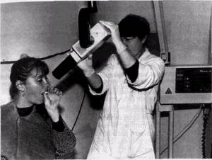
How do you take a panoramic dental photograph?
Before taking an x-ray, the patient needs to remove all metal products located in the neck and head area.
Let's consider the stages of the procedure:
- The patient is asked to stand inside the orthopantomograph.
- Push the device plate tightly towards your chest, grasping the handles to fix the position.
- You should stand without moving to avoid distortion of the picture.
- If necessary, the radiologist will ask you to change the rotation and angle of the head.
- The device will begin to move around your head. This takes no more than 20-30 seconds.
A person clamps a plastic tube with his teeth, while keeping his lips closed.
How to load tomograms onto the screen
When the MRI image is opened, it will become clear what the picture is. But often an absolutely inexplicable mixture of dark colors is shown.
There are several ways to view tomograms:
- Sagittal. Scans are the easiest to understand. The method looks like a longitudinal section of the area under study, inspection is carried out in profile;
- Transverse. The most difficult option for people who are not professionally involved in deciphering MRI results. The method forms many horizontal parallel planes. The MRI image is always black and white. To recognize organs, detected pathologies are based on shades of color (contrast).
The programs make it possible not to depend on the level of computer RAM, but if you want to expand the functionality of the software, technical characteristics are of no small importance.
The application allows you to perform the following operations:
- You can change the spatial orientation and view the image from any angle;
- Changing brightness and contrast parameters;
- Scaling an object;
- Carry out the necessary measurements of length, height, width, volumetric indicators.
The MRI viewer allows you to save MR scans in DICOM format as WMV video files or JPEG drawings. Copying to the clipboard is available for subsequent use in presentations, drawing up conclusions, for the most detailed presentation of the results of a magnetic resonance examination.
Historical reference
November 8, 1895 is considered the day of the discovery of X-rays by professor and physicist Wilhelm Roentgen. Thanks to his research, the special properties of “X” (x) radiation were recorded, which by 1919 had become quite widely used in medicine. But during the first attempts to use X-rays in dentistry, doctors encountered a problem: the images of teeth were distorted in size and configuration, which made it difficult to detect a particular pathology and, as a result, prescribe the necessary treatment. A world-class scientist, dentist, Professor A. Tsieshinsky, who made a significant contribution to the development of world dentistry, was recognized by the International Federation of Dentists (FDI) and awarded the Miller Gold Medal, approached the solution to this problem. In 1907, he published the “Radiological Atlas of Dental X-rays,” the first publication in the world in this field, and also published his works on the topic “Fundamentals of isometric dental radiography,” which allowed doctors of that time to professionally take targeted periapical (intraoral) images. The method he proposed improved over time and has not lost its significance to this day. However, the use of this method was not enough to diagnose periodontal pathologies and detect hidden carious cavities. This became clear at the beginning of the last century, and in 1920 the scientist Raper developed a technique for interproximal (bite) radiography. Despite its advantages, it also had a drawback: when taking a contact photograph, a certain projection magnification appeared, so the measurements did not correspond to real values. The scientist Harndt managed to obtain an almost identical image in 1957, who used long-focus radiography to study the marginal parts of the alveolar process, in which the divergence of the rays is minimized, and when passing through the object the direction is close to parallel. Now this method is widely known as “parallel radiography”.
Currently, targeted X-ray images continue to be widely used in dentistry both for diagnostic purposes and for monitoring dental treatment at different stages, making it possible to determine the effectiveness of each of them and the entire treatment process as a whole.
In what cases is a panoramic x-ray performed?
An orthopantomogram is necessary for all types of dental care. The procedure does not cause pain, takes little time, and has virtually no contraindications.
Panoramic radiography is done in the following cases:
during implant treatment: to assess the condition of the bones and select a design. An incorrectly determined distance to the mandibular canal threatens to impair the sensitivity of the chin and lower lip;
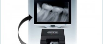
About the equipment
Modern X-ray scanners are extremely fast and digitize radiographs within seconds.
Thanks to the different sizes of the devices, they can cope with X-ray films of a wide variety of formats.
The optical resolution of the device allows you to obtain a clear and precise image, transferring the smallest details of the image into an electronic document, because any little thing can have diagnostic value.
It should be said that this equipment is expensive and before you go to the nearest copy center with your photo, you should make sure that they have it.
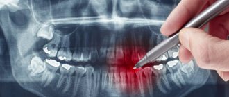
Indications
Indications for targeted radiography include pain in the tooth area, caries, pulpitis, periostitis, periodontitis, tooth root cysts, gum disease - periodontitis, gingivitis, as well as tooth trauma, control of the growth and condition of wisdom teeth, and the need for tooth extraction.