Many of us (60-95 percent) asked the question: “What is better to do: fluorography or x-ray?” And in general, is there a difference between these two methods of medical research and pathology diagnosis?
For information:
- approximately 50% of people identify these concepts;
- 30% believe that doctors use different radiation for FLG and radiography;
- 20% of people generally think that these are two radically different methods.
It's time to understand these 2 concepts!
X-ray and fluorography: difference in dose
To understand which of the above research methods to give preference to, you should know everything about their differences.
The difference between modern X-ray equipment and previous models is a much lower radiation dose. But in this technique there is no concept of “maximum possible radiation dose,” since images are always taken strictly in accordance with established medical requirements. Therefore, if a person’s life is at stake, then the procedure should be done as many times as the treatment requires.
Radiation safety during X-ray dental examinations (Part 2)
M. A. Chibisova Doctor of Medical Sciences, Professor, Head of the Department of Radiology in Dentistry, non-state educational institution "St. Petersburg Institute of Postgraduate Dentistry" (SPbINSTOM), chief physician of the group's radiology diagnostic service
Radiation safety of the population is the state of protection of the present and future generations of people from ionizing radiation harmful to their health (Federal Law “On Radiation Safety of the Population” No. 3-FZ of January 9, 1996, Art. 1). Aspects of radiation safety of personnel and patients, issues of labor protection when working with sources of ionizing radiation are regulated by a number of regulatory documents listed above. Monitoring compliance with radiation safety measures is entrusted to the service of Rospotrebnadzor of the Russian Federation.
In the process of working with sources of ionizing radiation, several principles must be observed: the principle of regulation - not exceeding the permissible limits of individual radiation doses of citizens; the principle of justification is the prohibition of the use of ionizing radiation, in which the benefit obtained for humans and society does not exceed the risk of possible harm from this exposure; optimization principle - limiting the levels of exposure of personnel and patients below dose limits by maintaining radiation doses at such low levels as technically possible to achieve, provided that the required volume and quality of diagnostics are ensured. The principle of justification when conducting x-ray examinations implies the priority use of alternative (non-radiation) methods, x-ray examinations must be carried out according to strict indications, the choice of the most gentle methods of x-ray examinations, the risk of refusing an x-ray examination must obviously exceed the risk from radiation during its conduct. The doses received by the patient from each x-ray examination must be entered into a personal sheet for recording medical radiation doses, which is a mandatory attachment to the outpatient card.
Protection against ionizing radiation is aimed at reducing the physical dose of radiation below the maximum permissible dose.
The following types of radiation protection are distinguished: 1. Protection by screens:
- stationary devices (brick, barite concrete, lead, leaded glass, etc.);
- non-stationary (aprons, gloves, collars, etc.).
2. Protection by distance. 3. Time protection:
- reduction of research time;
- reduction of staff working hours;
- reduction in the number of studies.
To assess the radiation hazard of ionizing radiation, the concept of effective dose is introduced - a value used as a measure of the risk of long-term consequences of irradiation of the entire human body and its individual organs and tissues, taking into account their radiosensitivity. The unit of effective dose is the sievert (Sv), but the derivatives millisievert (mSv) and microsievert (µSv) are commonly used in everyday practice.
In accordance with SanPiN 2.6.1.1192-03 and NRB-99/2009, maximum permissible radiation doses have been introduced for various categories of personnel and patients. For employees directly involved in X-ray diagnostic studies (group A personnel), the permissible effective dose is 20 mSv per year on average for any consecutive 5 years, but not more than 50 mSv per year. For group B personnel (employees who, due to working conditions, are exposed to ionizing radiation) - 5 mSv per year on average for any consecutive 5 years, but not more than 12.5 mSv per year. For the population, i.e. practically healthy individuals for whom X-ray examination is carried out for preventive purposes or for scientific research - 1 mSv on average for any consecutive 5 years, but not more than 5 mSv per year.
For patients, limits on annual radiation doses for diagnostic (as well as therapeutic) purposes are not established. This is due to the voluntary nature of the study, the benefits of which for the patient’s health must exceed the amount of radiation damage. When the accumulated dose of medical diagnostic radiation to a patient reaches 500 mSv, measures must be taken to further limit his exposure if radiation procedures are not dictated by vital indications. Monitoring of patient dose loads is carried out during each x-ray examination.
Dental devices with regular film without an intensifying screen and panoramic devices are allowed to be placed only in the X-ray department (room) of a medical or dental institution.
In addition, for pregnant women, studies are carried out in such a way that the dose received by the fetus does not exceed 1 mSv for two months of undetected pregnancy. If the fetus receives a dose exceeding 100 mSv, the doctor is obliged to warn the patient about the possible consequences and recommend terminating the pregnancy. Studies in pregnant women should be carried out only when clinically indicated, if possible in the second half of pregnancy.
For persons (non-staff) assisting in supporting patients (seriously ill patients, children) during X-ray procedures, a dose limit is set at 5 mSv per year. When performing X-ray dental examinations, the relevant requirements must be met.
Dental devices and pantomographs operating with a highly sensitive image receiver (without a darkroom), and dental devices with digital image processing, the workload of which does not exceed 40 (mA x min.)/week, can be located in the premises of a dental institution located in a residential building , including in premises adjacent to residential premises, subject to the requirements of radiation safety standards within the premises in which X-ray and dental examinations are carried out.
If several devices for X-ray dental examinations are installed in the room, then the anode voltage switching system should provide for the possibility of operating only one device at a time.
Composition and area of premises for X-ray dental examinations: 1. Room for X-ray diagnostics of dental diseases using radiography with a dental apparatus working with ordinary film without an intensifying screen:
- Treatment room - at least 8 m2.
- Photo laboratory - at least 6 m2.
2. Room for X-ray diagnostics of dental diseases using radiography with a dental apparatus working with a highly sensitive film and/or digital image receiver, including a radiovisiograph (without a darkroom):
- Treatment room - at least 6 m2.
3. X-ray diagnostic room using panoramic radiography or panoramic tomography (digital volumetric tomography, 3DCT):
- Treatment room - at least 8 m2.
- Control room (may be absent when using devices equipped with means of protecting personnel workplaces) - at least 6 m2.
- Photo laboratory (may be absent when using devices with digital image processing) - at least 8 m2.
When installing more than one dental X-ray device in a treatment room, the area of the room must increase depending on the type of device, but not less than 4 m2 for each additional device.
X-ray and dental equipment (domestic or imported) is permitted for delivery and operation if there is a registration certificate from the Ministry of Health and Social Development of the Russian Federation and a sanitary-epidemiological conclusion. A dental institution conducts x-ray examinations only if it has a license for the corresponding type of medical activity. An institution using X-ray dental equipment must have the following documentation: a sanitary and epidemiological certificate for the type of activity (operation, storage, testing, etc. of the X-ray machine (devices) in the X-ray room (rooms); design documentation for the X-ray room; technical passport for the X-ray room ; instructions on labor protection, including requirements for radiation safety, for the prevention and elimination of radiation accidents; sanitary rules, other regulatory and instructional documents regulating radiation safety requirements.
The administration of the dental institution determines the list of persons working on dental X-ray machines, provides the necessary training and instruction, and appoints a person responsible for radiation safety, accounting and storage of the X-ray machine, and for radiation control. The room where X-ray examinations are carried out must have a set of mobile and personal protective equipment for staff and patients:
- Large protective screen with a viewing window for devices working with regular film without an intensifying screen, panoramic devices, pantomographs (when the control panel and treatment room are located in the same room, when working with X-ray dental devices with highly sensitive image receivers, it is allowed to use X-ray protective curtains instead of a screen) - 1 PC.
- One-sided lightweight protective apron (for staff) - 1 pc., protective collar (for staff) - 1 pc.
- Protective dental apron (for the patient) or a protective cape (cape) and an apron for protecting the gonads (for the patient) - 2 pcs.
Personnel working on X-ray machines must be trained in the rules of working on this device and trained in ensuring radiation safety of personnel and patients, which must be confirmed by relevant documents. Persons over 18 years of age who do not have medical contraindications are allowed to work on an X-ray dental apparatus, after training, instruction, testing of knowledge of the safety rules for conducting work, instructions in force in the institution and classified by order of the administration of the institution as category A personnel.
The administration of the dental institution ensures continuous individual dosimetric monitoring of employees working on dental and panoramic X-ray machines. In order to protect the patient’s skin during X-ray procedures, the length of the device tube must provide a skin-focal distance of at least 10 cm for a device with a rated voltage of up to 70 kV and 20 cm for higher values of anode voltage. Ways to reduce radiation exposure to staff and patients: replacement of outdated equipment, high-quality maintenance of equipment, availability of a full set of personal protective equipment, use of highly sensitive films, transition to digital technologies, optimization of research protocols, use of specialized programs for reconstruction and image processing.
X-Ray Protection
Radiation protection for X-ray rooms when conducting intraoral examinations and for X-ray dental rooms (the radiovisiograph is located in the dental office):
- For patients - an X-ray protective dental apron FRZS-“R-K” (the attenuation factor of X-ray radiation by X-ray protective material, expressed in lead equivalent value, not less than: at U=100 kV 0.35 RV). This apron is designed to protect the patient's body, including the gonads, pelvic bones and thyroid gland from the radiation beam during dental or cranial examinations. The most reliable form of patient protection when taking dental x-rays.
- For personnel - a protective one-sided lightweight FRZOL-“R-K” apron (the attenuation factor of X-ray radiation by X-ray protective material, expressed in lead equivalent value, not less than: at U=100 kV 0.25 RV). This apron is designed to protect the front of the human body from the throat to the shins during X-ray examinations - with or without a stand. In the second case, it is recommended to wear an X-ray protective collar over the apron.
Radiation protection for X-ray rooms when conducting extraoral panoramic X-ray examinations on an orthopantomograph, cephalostat and digital volumetric tomograph (3DCT):
- For patients - a protective dental apron for panoramic examinations (for an orthopantomograph) FRZS-“R-K” (the attenuation factor of X-ray radiation by X-ray protective material, expressed in lead equivalent, not less than: at U = 100 kV 0.35 Rv). This is a specially designed apron model to protect the patient's body front and back along the spine during extraoral dental examinations.
- For pediatric patients - a protective one-sided heavy children's FRZOT-“R-K” apron (the attenuation factor of X-ray radiation by X-ray protective material, expressed in lead equivalent value, not less than: at U=100 kV 0.35 RV). This apron is designed to protect the front of the body, including the shoulder girdle, from the throat to the shins. Recommended for use when taking photographs of teeth, head and limbs, complete with a collar to protect the thyroid gland. When performing extraoral panoramic examinations, pediatric patients are recommended to additionally use an X-ray protective skirt (0.35 Rv). Unlike an apron, it covers the body area on all sides, and the double wrap adds protection to the front.
Radiation, electrical and fire safety techniques when working with radiovisiographs
Ensuring radiation, electrical and fire safety during X-ray dental examinations and the rules of work on X-ray computerized installations (radiovisiographs) are regulated by Sanitary rules and norms SanPiN 2.6.1.1192-03 “Hygienic requirements for the design and operation of X-ray rooms, devices and the conduct of X-ray examinations, clause 11 and paragraph 12." The placement and permanent protection of premises for X-ray dental examinations are determined by the type of X-ray equipment and the size of the workload of the device.
Dental devices and pantomographs operating with a highly sensitive image receiver (without a darkroom), and dental devices with digital image processing, the workload of which does not exceed 40 (mA-min.)/week. and anode voltage of 70 kV, can be located in the premises of a dental institution located in a residential building, including in those adjacent to residential premises, subject to the requirements of radiation safety standards for the population within the premises in which X-ray dental examinations are carried out.
Composition and area for X-ray dental examinations: a room for X-ray diagnostics of dental diseases using radiography with a dental apparatus working with a highly sensitive digital image receiver, including a videographer (without a darkroom) - a treatment room with an area of 6 sq. m (not less). Ventilation requirements for rooms for X-ray dental examinations must comply with the ventilation requirements for dental departments. In medical practice, domestic X-ray and dental equipment can be used, manufactured according to technical specifications agreed with the Russian Ministry of Health and passed a hygienic assessment. Imported X-ray equipment is allowed for delivery and operation if there is a registration certificate from the Ministry of Health of Russia and a sanitary-epidemiological conclusion. The room where X-ray dental examinations are carried out using a radioviseograph must have a set of personal protective equipment for staff and patients: a protective one-sided apron - light (for medical personnel - a dentist and his assistant) - 2, a protective dental apron (for the patient) - 2, a cape (drape) protective and collar (for the patient) - 1.
In order to protect the patient’s skin during X-ray procedures, the length of the device tube must provide a skin-focal distance of at least 10 cm for a device with a rated voltage of up to 70 kV and 20 cm at higher values of anode voltage
The administration of a dental institution is obliged to determine the list of persons working on dental digital X-ray machines, provide the necessary training and instruction, and appoint by order of the institution a person responsible for radiation safety, accounting and storage of the X-ray machine, and for radiation control. The administration of the institution is responsible for ensuring the radiation safety of staff and patients. Personnel working with radiovisiographs must be trained in the rules of working with this device, trained in ensuring radiation safety of personnel and patients, and have a document from an institution accredited in these matters. Persons over 18 years of age who do not have medical contraindications are allowed to work on an X-ray dental apparatus, after training, instruction, testing of knowledge of safety rules for conducting work, instructions in force in the institution and classified by order of the administration of the institution as category A personnel.
The administration of a dental institution is obliged to ensure constant individual dosimetric monitoring of employees working on dental X-ray machines. In order to protect the patient's skin during X-ray procedures, the length of the device tube must provide a skin-focal distance of at least 10 cm for a device with a rated voltage of up to 70 kV and 20 cm at higher values of the anode voltage. To ensure safe conditions for conducting x-ray examinations, measures must be taken to protect against the effects of electricity, lead and other non-radiation factors, as well as fire and anti-epidemic measures must be taken. Electrical safety of technical equipment, including personal computers of personnel workstations, is ensured by the use of electrical sockets with a grounding contact. Electrical appliances and dental devices can be connected to grounding through plug sockets with an additional grounding contact (European standard). The presence of a grounding strip is not required if the design of the device provides a grounding conductor.
The network resistance must correspond to the rated power of the X-ray power supply with a three-phase rectification circuit. Mobile dental X-ray machines must remain stable when the floor is tilted up to 15°. The moving parts of the device must have a clamping force limiter of up to 300 N. The movement of X-ray devices must be carried out in accordance with the load standards when moving heavy objects. Each X-ray room (including a mobile X-ray unit) must be equipped with carbon dioxide fire extinguishers of the OU-2 type and have free access to fire extinguishing equipment. (A filled transformer tank is not considered a fire hazard). The number and location of fire extinguishers is agreed with the fire safety authorities.
Radiation exposure and protection of patients and personnel during X-ray examinations in dentistry
When radiography of the teeth of the upper jaw on a dental apparatus, the zone of a direct diverging beam of rays includes not only the maxillofacial area, but also the neck, chest and abdominal cavities - organs and tissues located far beyond the boundaries of the area under study. The tube of the 5D-1 device does not sufficiently shield the primary radiation. The cross-sectional area of the primary beam is often 2 times that required for intraoral radiographs. In 5D-2 X-ray machines, aperture of the primary beam ensures the minimum possible divergence angle. When performing intraoral radiographs, the patient's torso is shielded with a standard protective apron made of leaded rubber. However, its large mass, bulkiness, and large cutout around the neck make it difficult to use, especially when examining children. In addition, the thyroid gland remains unprotected. Therefore, specially designed protective screens made of leaded rubber or new models of lightweight protective aprons with a collar for the thyroid gland have been proposed, providing ease of use and sufficient neck protection.
In order to reduce the cumulative effect of ionizing radiation, especially in children, repeated radiographs are taken after 3 weeks, and if several images are taken, then no earlier than after 5 weeks. Taking into account the peculiarities of the centering of the X-ray beam during panoramic radiography and panoramic tomography (zonography) of the dental system, the body is exposed to much less radiation than when performing intraoral contact radiographs of the teeth. During these studies, the thyroid gland is protected by an apron made of leaded rubber, and the patient’s torso is protected by a double-sided apron or drape.
Dosimetric values
Of the dosimetric quantities that can be used to estimate the dose load on a patient, the following are interesting: Absorbed dose of ionizing radiation (dose of ionizing radiation) - D - the energy of ionizing radiation transferred by it to a unit mass of the irradiated substance. Its unit of measurement is the gray (Gy). 1 Gy is equal to the absorbed dose of ionizing radiation, at which the energy of ionizing radiation of 1 J is transferred to a substance weighing 1 kg; 1 Gy = 1 J/kg. 1 Gy = 100 rad (from the English Radiation Absorbed Dose).
Equivalent dose of ionizing radiation (equivalent dose) - H - absorbed dose taking into account the quality factor of ionizing radiation. For X-ray radiation, the absorbed dose (D) and equivalent dose (H) are equal. The unit of equivalent dose is the sievert (Sv). 1 Sv = 100 rem (biological equivalent of an x-ray). Effective dose - Nef - is the equivalent dose of such uniform irradiation of the whole body, which can cause the same long-term (stochastic) effects as non-uniform irradiation. In other words, if there is (for example, in dental practice) uneven irradiation of the body, then it is necessary to use the concept of an effective equivalent dose, which makes it possible to establish an equally effective equivalent dose of uniform irradiation.
Effective dose - Nef - is the equivalent dose of such uniform irradiation of the whole body, which can cause the same long-term (stochastic) effects as non-uniform irradiation
For all types of radiography, radiation exposure to patients is assessed using an effective equivalent dose (EDD), which is measured in microsieverts (µSv) and is determined by measuring the exposure of vital organs that are most sensitive to the effects of ionizing radiation (lens of the eye, cerebral hemispheres, salivary glands, tongue, active bone marrow, thyroid and mammary glands). During dental radiography, leaded rubber aprons are used to protect the mammary glands and gonads. To protect the thyroid gland, it is advisable to use special leaded screens - collars. Protection of medical personnel from x-ray radiation includes strict adherence to operating and safety rules in the x-ray room. When the device is turned on, the X-ray room employees must be behind a protective screen or behind the room wall. Fixation of the film in the mouth when taking intraoral photographs is carried out by the patient himself. The use of X-ray room personnel for this purpose is strictly prohibited.
Radiation safety of patients during X-ray examinations in dentistry
In 70-80% of cases, the clinical diagnosis is established on the basis of X-ray data (M. M. Solovyov, 2000). The share of x-ray examinations in dentistry relative to other types of x-ray procedures is quite large. Often dental x-ray examinations occur at the age of 18-20 years. This implies the need to control doses during x-ray examinations in dental practice. During dental radiography, lead aprons are used to protect the lungs, heart, mammary glands and genital area; an apron to protect the thyroid gland - special screens - in the form of a collar. In this case, equivalent doses are reduced by 10-100 times, so the use of aprons-collars is mandatory. However, the total effective equivalent dose is reduced by only 15-20%, since the cerebral hemispheres, pituitary gland, salivary glands, tongue and lens of the eye are not protected. During orthopantomography, the dose loads are relatively low (load for the tongue and salivary glands) and are comparable to those during intraoral radiography of individual areas of the oral cavity (Fig. 1, 2).
When performing an X-ray examination in dentistry, the following is necessary: 1. Patient protection: In children and adults, it is desirable to perform an orthopantomogram (or digital volumetric tomography - 3DCT) and reduce X-ray examinations to a minimum;
- an apron made of leaded rubber (preferably with a collar - to protect the thyroid gland);
- all extraoral images are performed with intensifying screens, and intraoral images are taken on highly sensitive screenless film;
- radiography is carried out preferably with “hard” (not lower than 60-70 kV) radiation and the shortest possible exposure;
- mandatory use of an aluminum filter (at least 1 mm thick);
- X-ray examinations are performed only according to strict indications (the number of X-ray examinations is reduced to a minimum in children and pregnant women); Repeated X-ray examinations - if possible, no earlier than after 10-15 days.
2. Personnel protection: when working, it is necessary to move away to the side opposite to the working radiation beam, at a distance along the length of the cord to the control button;
- full compliance with the rules of design, operation and safety of work in the X-ray room;
- mandatory use of protective equipment (screens, aprons) by personnel;
- when the X-ray machine is turned on, you must be no closer than 1.5-2 m from the X-ray tube and, of course, behind a protective screen or behind the office wall;
- during intraoral dental radiography, films are fixed in the mouth by the hands of the person being examined or by the patient himself using a positioner;
- systematic dosimetric monitoring is required.
Effective doses for x-ray examinations in dentistry
Effective doses for x-ray examination (for patients according to the Testing Laboratory Center of the Federal Research Center of St. Petersburg Research Institute of Radioactive Geography of the Ministry of Health of Russia dated July 22, 2011 and December 21, 2012, studies were performed on x-ray machines: panoramic x-ray machine “Orthophos XG 3D / Ceph” with cephalostat (Moscow, 79); (Kamennoostrovsky, 42b); (Suvorovsky, 57); three-dimensional dental computed tomograph - targeted 3DX Accuitomo Morita (Nevsky, 82); panoramic 3DKT Galileos "Sirona" (Nevsky, 82); (Komendantsky, 17) and radiovisiographs, "Sirona" using X-ray dental devices such as "Trophy-Kodak", "Sirona" (Fig. 3-6).
During one office visit, the patient is allowed to take 5-6 targeted images
The effective dose for one study when producing a digital orthopantomogram is 0.055 mSv (for a patient under 15 years old - 0.024 mSv); digital teleradiogram - 0.007 mSv. The effective dose when performing one zone on a three-dimensional dental computed tomograph is 0.102 mSv (for a patient under 15 years old - 0.068 mSv).
Effective dose of one digital intraoral x-ray of a tooth on a radiovisiograph “Trophy-Kodak”, “Sirona”: lower jaw - 0.002 mSv (for a patient under 15 years old - 0.001 mSv); upper jaw - 0.005 mSv (patients under 15 years old - 0.003 mSv).
In a year, you can take up to 100 images using a radiovisiograph (for adult patients). During one office visit, the patient is allowed to take 5-6 targeted images.
Maximum effective dose per year for radiologists and x-ray laboratory technicians when working in stationary x-ray rooms; for dentists and medical assistants when working on a radiovisiograph in x-ray dental rooms is 2 rem = traffic limit 20 mSv per year.
The maximum effective dose to the population received from preventive X-ray diagnostic studies is 1 mSv per year. 0.01 rem = 0.1 mSv, 0.1 rem = 1.0 mSv, 1 Sv = 100 rem, 1 rem = 10 mS.
Rice. 1. Digital orthopantomogram. Chronic generalized periodontitis of moderate severity. Rice. 2. Teleroentgenogram of the skull in a lateral projection, hyperplasia of the lymphoid ring of the nasopharyngeal vault. Rice. 3. 3DCT. Chronic osteomyelitis in the area of the angle of the lower jaw on the right after removal of the 48th tooth. Rice. 3. 3DCT. Chronic osteomyelitis in the area of the angle of the lower jaw on the right after removal of the 48th tooth. Rice. 4. 3DCT. Chronic granulomatous periodontitis of teeth 26, 27, 28, odontogenic changes in the mucous membrane of the left maxillary sinus. Rice. 4. 3DCT. Chronic granulomatous periodontitis of teeth 26, 27, 28, odontogenic changes in the mucous membrane of the left maxillary sinus. Rice. 5. 3DCT of the dentition, cyst-like formation of the right maxillary sinus. Rice. 5. 3DCT of the dentition, cyst-like formation of the right maxillary sinus. Rice. 6. 3DCT of the dentition, cyst-like formation of the right maxillary sinus. Rice. 6. 3DCT of the dentition, cyst-like formation of the right maxillary sinus.
Conclusion
In addition to solving the problems of organizing x-ray examinations in dental clinics and observing radiation safety when performing x-ray examinations, it is necessary to widely introduce innovative methods of radiation diagnostics for practical use - three-dimensional dental computed tomography (3DCT) when conducting diagnostic examinations in outpatient dentistry, maxillofacial surgery and otorhinolaryngology. Three-dimensional dental computed tomographs are installed in private and public dental clinics; they allow moving to a new level of three-dimensional diagnostics, which significantly expands the possibilities of visualizing the dental system, maxillofacial area, maxillary sinuses and temporomandibular joints. The use of modern methods of radiation diagnostics makes it possible to optimize the diagnostic and treatment process, reduce the time required for examining dental patients, reduce the individual dose of radiation exposure, and improve the quality of dental care.
- Vasiliev A. Yu., Vorobyov Yu. I., Truten V. P., Chibisova M. A. et al. Radiation diagnostics in dentistry. - M., Medicine, 2007. - 495 p.
- Gerasimova L.P., Zaripova N.R., Verzakova I.N., Rayanova R.A. X-ray diagnostics in therapeutic dentistry. Tutorial. - M., 2011. - 196 p.
- Hygienic requirements for the design and operation of X-ray rooms, devices and the conduct of X-ray examinations. Sanitary rules and regulations. SanPiN 2.6.1.1192.-03. Ministry of Health of Russia / Moscow. - 2003. - 76 p.
- Control of effective radiation doses to patients during medical X-ray examinations / Guidelines MUK 2.6.1.1797-03 Ministry of Health of Russia. - M., 2004. - 23 p.
- Radiation diagnostics in dentistry: national guidelines / Ch. ed. volume Vasiliev A. Yu. - M.: GEOTAR-Media, 2010. - 288 p. (Series “national guide to radiation diagnostics and therapy” / chief editor of the series Ternova S.K.).
- Malakhovsky V. N., Trufanov G. E., Ryazanov V. V. Radiation safety of X-ray studies. - St. Petersburg: ELBI-SPb, 2007. - 104 p.
- Standard instructions on labor protection for personnel of X-ray departments dated January 28, 2002, No. 19. - M., 2002. - 60 p.
- Chibisova M. A. Licensing of dental clinics and offices for activities in the field of using sources of ionizing radiation. - SPb.: MEDI publishing house, 2007. - 40 p.
X-ray: how not to harm your health?
Medicine notes that you can protect yourself from X-ray equipment with a protective shield. This role may include an “apron” for the abdomen, a “collar” for the neck, a “skirt” to protect the abdominal cavity and genitals, and a “cap” for the head. All these protective screens have a solid lead layer.
At young childbearing ages, experts recommend protecting the genitals and abdominal area from radiation, since the greatest negative impact of the device affects the germ cells and blood.
Particular attention should be paid to children. Their protective screen should cover the entire body, leaving only the area being examined.
X-ray images make the examination quite informative and allow you to observe the dynamics of the body’s reaction to treatment. Doctors do not recommend doing several x-rays in one day (for example, additional fluorography or mammography). It is also important that the patient has a radiation passport, where the radiologist enters the dates of the examination and the doses received.
Fluorography: what is the danger
Fluorography is a diagnostic equipment for examining the chest, where the radiation dose is about 0.8 mSv. For comparison, you can use an X-ray of a tooth, where the beam strength is 0.1 mSv.
The high radiation dose during fluorography is due to the fact that the device screen has less sensitivity compared to X-ray equipment.
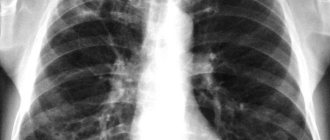
This diagnostic technique is valued for its ability to take photographs to detect tuberculosis, pneumonia and other pulmonary pathologies. In medical institutions it is used everywhere, even when preventive and initial examinations . In order not to expose a person to unnecessary radiation, it is recommended to do fluorography no more than once a year.
What is fluorography?
Fluorography – photographing an image on a fluorescent screen. It is formed due to the passage of x-rays through the patient’s body (for example, the chest). The latter become visible and focus on the film. Thanks to its ability to transform and change shape.
FLG is mainly used for preventive examinations for:
- oncology;
- pathologies of the bronchi and lungs;
- tuberculosis.
The result of a fluorographic examination is images of organs located behind the chest. The photo gives an idea of the current state of human organs: are they in normal condition or require immediate treatment/removal.
Which procedure should I choose?
When specialists prescribe one of the types of diagnostics, they always focus on the goals that such a study will help solve. It should not be forgotten that X-rays are not advisable for pregnant women and children. osteopath or chiropractor, who can establish a diagnosis by palpation can refuse X-ray diagnostics
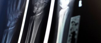
When choosing between x-rays and fluorography, you need to consider the differences:
- X-ray gives high accuracy;
- fluorography irradiates more strongly;
- Fluorography makes it possible to obtain an image of the lungs;
- X-rays take local pictures and record the dynamics of changes;
- the x-ray is taken immediately on a special film;
- The fluorography image is displayed immediately on the screen, then a photograph is taken from it;
- X-rays are more expensive than fluorography.
Based on a number of these differences, the doctor decides which type of examination to choose in a particular situation.