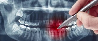
Advantages of radiography of the nasal bones
X-ray diagnostics of the nasal bones has a number of advantages over other methods of ENT examination. When compared with computed tomography or magnetic resonance imaging, the following advantages can be identified:
- X-rays take no more than 10 minutes. MRI and CT scans are time consuming.
- The examination conditions are much less dangerous with radiography, since the patient assumes a sitting position. This is a very important point to prevent blood from entering the airways during nosebleeds. For example, when performing an MRI, the patient must remain in a supine position in an isolated machine for at least 20 minutes.
- Endoscopic diagnosis of the nasal passages in the acute stage of the disease also loses its relevance in comparison with x-rays. The latter does not cause any discomfort in the patient, as it is a non-invasive examination technique.
Indications and contraindications for sinus x-rays
Indications for radiography are very wide. Most often, an otolaryngologist prescribes a referral for an x-ray in the following cases:
- pain in the forehead, which tends to intensify when tilting the head;
- chronic nasal congestion;
- purulent or other discharge from the nasal passages;
- frequent nosebleeds;
- swelling in the nose area.
In this regard, potential candidates for X-ray irradiation are patients with suspected sinusitis, neoplasms, cysts, and foreign bodies in the nose. The study is mandatory in case of injuries to the facial skeleton, nasal fractures, curvatures of the septal cartilage, and is also used to assess the effectiveness of the treatment.
An absolute limitation to performing x-rays of the sinuses is pregnancy. All other categories of citizens, including children, are allowed to undergo irradiation only after examination and with the permission of the profile specialist.
Preparing for an X-ray of the nasal bones
The first step is to stabilize the patient’s general condition, administer local anesthesia, and stop nosebleeds. Immediately before the diagnostic procedure, it is necessary to remove jewelry and metal prostheses. No other preparation is required.
It is important to follow all instructions from the x-ray technician and not move during the examination. This will allow you to do
high-quality images and establish an accurate diagnosis. If the patient is unconscious, medical personnel hold the patient in the required position.
X-ray methods for examining the nose and paranasal sinuses
An ENT disease similar to inusitis
is currently the most common ENT pathology.
Patients with inflammatory processes of the paranasal sinuses (SNS) currently make up about 40% of all patients hospitalized in ENT hospitals. The main method for diagnosing diseases of the urinary tract is radiological.
This is due to the fact that the air-containing sinuses of the nose are located deep in the bones of the facial skull, communicating with the nasal cavity only through small openings - anastomoses.
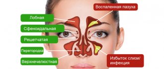
It is customary to distinguish the following paired SNPs located in the bones of the same name (Fig. 1):
- maxillary;
- frontal;
- lattice;
- front and rear;
- wedge-shaped.
In some cases, the paranasal sinuses also include air cavities, which are rare in some patients and located in the thickness of the nasal septum of the nasal concha (turbinate bullae). These cavities, lined with epithelium, almost never communicate with the nasal cavity and therefore cannot be considered true sinuses.
In a newborn child, only the maxillary and ethmoid sinuses are developed (Fig. 3), which are cavities with a volume of less than a milliliter. By 2–3 years the sphenoid sinus becomes pneumatized, and by 5–6 years the frontal sinus becomes pneumatized. The final development of the sinuses is achieved by the age of 20.
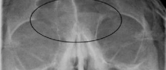
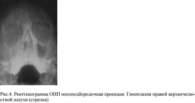
Underdevelopment of the frontal sinuses or their hypoplasia occurs in every 10th person. Underdevelopment of other sinuses is observed quite rarely. There may be unilateral hypo- and aplasia of the maxillary, sphenoid and frontal sinuses, which must be noted in the description of the radiograph. This creates difficulties in diagnosing sinusitis and puncture of such a sinus (Fig. 4). Excessive pneumatization of the SNP is possible, most often the frontal and sphenoid.
An X-ray of the skull does not allow us to assess the condition of all SNPs. To obtain a high-quality image of the SNP, it is necessary to conduct the study in a special projection.
The following types of projections are distinguished:
- nasomental;
- nasofrontal;
- mental or axial.
These projections are needed to shift the image of the pyramids of the temporal bones and the entire base of the skull below the bottom of the maxillary sinuses (nasomental position) or above them into the orbit (nasofrontal position). If this condition is not met, the pyramids of the temporal bones simulate fluid levels in the maxillary sinuses.
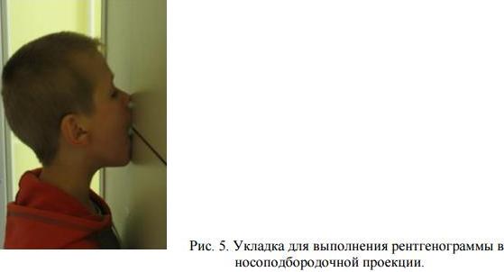
Nasomental projection
performed with the patient in an upright position (sitting, standing). The patient is asked to open his mouth and press it against the screen (Fig. 5). The central ray is directed perpendicular to the cassette and passes in the sagittal direction at the level of the outer corners of the orbits. All anterior sinuses (frontal, ethmoid, maxillary) are clearly visible. In the projection of the open mouth, you can see the sphenoid sinus (Fig. 1).
Sometimes, in this position, the pyramids of the temporal bones still overlap the lower parts of the maxillary sinuses and it is recommended to perform a chin (anterior semi-axial) projection, when the sitting patient touches the cassette with his chin.
The line connecting the external auditory canal and the chin (mental) is perpendicular to the plane of the cassette. The central ray runs through the wings of the nose parallel to the mental line. The ethmoid sinus in this case is poorly visible due to the layering of the anterior cells on the posterior cells and on the slopes of the nose. The frontal sinuses appear enlarged.
Nasofrontal projection
used to study the frontal and ethmoid sinuses. The patient presses his forehead and tip of his nose against the cassette. The central beam passes perpendicular to the cassette through the back of the head.
Currently, the nasomental projection is most often used.
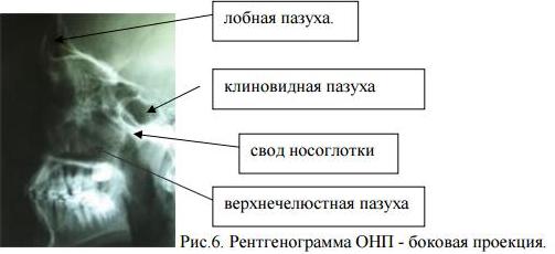
A survey radiography of the skull in a lateral projection allows one to assess the depth and condition of the walls of the frontal, sphenoid sinuses, maxillary and nasopharynx (Fig. 6).
Sometimes special installations are used for targeted examination of a specific sinus: X-ray according to G.M. Zemtsov to identify the sphenoid sinus in the projection of the open mouth (nasochin position with maximum tilting of the head), according to Ya.A. Fastovsky targeted examination of the ethmoid labyrinth and others.
There are difficulties in performing X-ray examinations on mobile and excitable patients and young children. It is necessary to hold them in the desired position or conduct the examination under anesthesia. In this case, the attending physician must determine how much X-ray examination is necessary in making a diagnosis.
Sometimes the required projection is prevented by stiffness of the joints (spine, temporomandibular) or muscle rigidity (for example, stiffness of the neck muscles with meningitis).
Normally, during X-ray examination, air-containing SNPs appear as light areas with a clearly defined dark outline corresponding to their bony border. Typically a negative X-ray image is described, so areas containing more white are called darker. Pneumatized SNPs correspond to orbital transparency. If the contents of the sinuses are darker than the contents of the orbits, then they speak of darkening, which means the presence of a pathological process
. In this case, it is necessary to characterize the darkening according to several parameters. The first of these is the size or degree of filling of the sinus with a pathological process.
They are distinguished (Fig. - Scheme 8):
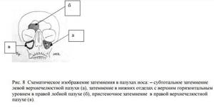
- total (complete) darkening of the sinus
, almost complete -
subtotal
(Fig. 8 a), which correspond to the complete filling of the sinus with contents, which can be either exudate or edematous soft tissue. - darkening in the lower sections with an upper horizontal level
means the presence of liquid, exudate (Fig. 8 b). - parietal darkening
, following the contour of the sinus walls, corresponds to thickening of the mucous membrane (Fig. 8 c).
If normally the thickness of the mucous membrane in the SNP is 120 - 1000 microns and is not visible on an x-ray, then with inflammation and allergic edema it can increase tens and hundreds of times, giving parietal darkening.
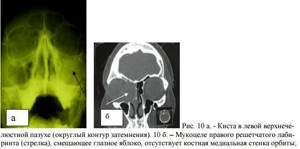
- In the sinus there may be limited darkening
emanating from one or two walls (Fig. 10 a).
In this case, it must have a designated shape
(round, oval, irregular),
size
(in centimeters or millimeters) and
contours
(smooth or uneven).
Such limited processes correspond to polyps, cysts, tumors
.
The second mandatory characteristic of darkening is intensity
, which means the degree of delay of the X-ray beam when passing through the sinus. There are three degrees: small, medium and high.
High intensity
means complete absorption of x-rays in tissue and corresponds to bone structures and radiopaque foreign bodies. In the sinus, high-intensity darkening is compared with the intensity of nearby bone formations: teeth, nasal bones. High intensity darkening in the sinuses can be caused by additional (superset) teeth, osteomas, bone fragments and foreign materials (shot, bullets, filling material that got into the maxillary sinus during the treatment of pulpitis of molars or premolars of the upper jaw, etc.).
Low intensity dimming
corresponds to serous exudate, swelling of the mucous membrane and appears on the radiograph only slightly darker than the orbits.
Medium dimming
delays the X-ray beam less than the surrounding bone structures, and corresponds to soft tissue formations (they can be tumors, cysts, polyps) or thick exudate (purulent or mucosal).
Different sinuses may have different pathological processes and varying degrees of their severity. For example, left-sided purulent maxillary sinusitis and a cyst of the right frontal sinus.
Sinusitis
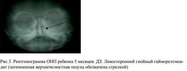
characterized by thickening of the sinus mucosa, the presence of exudate in it, which determines the form of the disease (purulent, catarrhal, serous). During the catarrhal process, a parietal darkening is noted in the sinus; an increase in edema can lead to a uniform total darkening. In the exudative form of sinusitis, darkening is detected in the lower sections with an upper horizontal level (Fig. 9). This level does not reach the bony walls of the sinus with severe swelling of its mucous membrane.
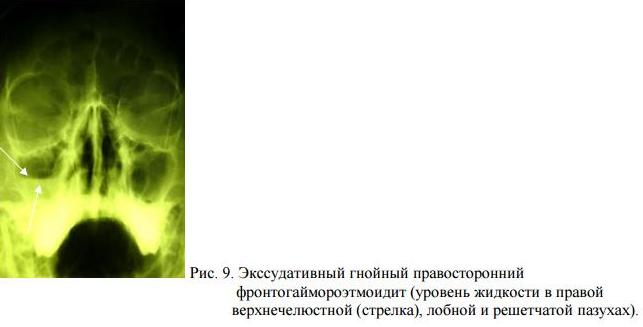
The intensity of the darkening can help to differentiate the nature of the exudate: medium intensity corresponds to a purulent process, low intensity to a serous one. When a large amount of exudate accumulates, the level is not determined - the darkening is total homogeneous. This may indicate the formation of a sinus empyema. Total homogeneous darkening of the sinus of low and medium intensity may correspond to severe swelling of the mucous membrane.
To correctly interpret the X-ray picture, it is important to know the clinical examination data: the presence of purulent secretion in the nasal cavity, body hyperthermia, and the severity of pain. The pain syndrome may be more pronounced with swelling in the sinus than with its empyema, when compression and death of nerve endings occurs. In such difficult cases, they resort to computed tomography
or
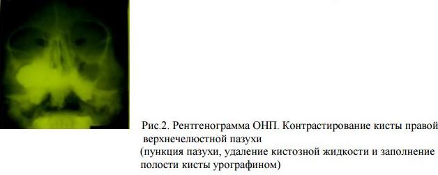
diagnostic puncture of the sinuses
.
Drainage of exudate is radiologically manifested by the restoration of transparency, starting from the superomedial angle of the maxillary sinus and the upper part of the frontal and sphenoid sinuses. After resorption or removal of exudate, thickening of the mucous membrane is determined in the form of residual wall deposits that persist for several weeks. This is especially true for the maxillary sinuses, in which the fluid level may be due to the injected medication during their previous drainage.
In everyday clinical practice, it is advisable to carry out control X-ray examinations immediately after the end of treatment only to determine the condition of the frontal sinuses.
Chronic sinusitis
has no characteristic manifestations, but more often there are productive forms in the form of rounded darkenings emanating from different walls of the sinuses, corresponding to polyps, cysts (Fig. 10 a), granulomas. The latter may be of odontogenic (dental) nature.
Large cysts can cause a total darkening of the sinus, simulating its empyema. Mucocele of the sinus should be distinguished from a cyst
, which does not have its own membrane and leads to stretching of the walls of the sinus due to obliteration of its natural anastomosis (Fig. 10 b).
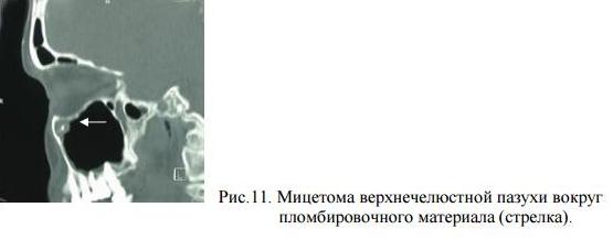
When the sinus is stretched with air as a result of the valve mechanism, stretching and thinning of the walls are also noted, but the pneumatization of the sinus is increased. When foreign bodies (filling material) get into the sinus, a fungal body may form around them - mycetoma
, which has a rounded outline on an x-ray (Fig. 11).
How the study is performed
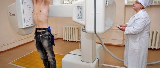
X-rays of the nasal bones are performed in frontal, nasomental or lateral projections, depending on the preliminary diagnosis. The examination can also be performed in a lying position if the doctor makes such a decision based on the general condition of the patient.
The irradiation itself lasts a matter of seconds, and the patient does not experience any pain.
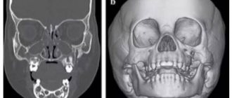
If there is a suspicion of a displaced bone fracture, the doctor prescribes x-rays in the frontal and nasomental projections. Left and right lateral images allow you to see the asymmetry of the damage. The X-ray picture of the transverse displacement of bone fragments is clearly visible in the axial projection. To diagnose comminuted fractures, it is necessary to obtain images in several projections at once.
Do not delay in seeing a doctor. Entrust your precious health to qualified specialists of the Doctor Nearby clinic network. Modern X-ray equipment and high professionalism of diagnosticians guarantee absolute accuracy of examination results.
About the x-ray procedure of the sinuses
When performing the examination in the nasochin position, the subject is in an upright position. The tip of his nose and chin are pressed tightly against the cassette, perpendicular to which the central beam generated by the X-ray machine is directed. The light stream passes in the sagittal projection, at the level of the outer edges of the eye sockets. The resulting image shows the pyramids and the base of the skull, along with the paranasal sinuses. When irradiated in the chin projection, the patient must sit. The chin touches the cassette.
Nasofrontal placement requires the patient to be in a standing or sitting position. The tip of his nose and forehead should touch the cassette. From this position it is possible to obtain a reliable image reflecting the state of the frontal sinus and ethmoidal labyrinth.
When performing radiography of the sinuses in the lateral projection, the patient's head is positioned so that the sagittal plane is parallel to the cassette. From this position, the radiologist is able to take a picture showing the condition of the walls of the frontal sinuses. This study has great diagnostic value in differentiating a pathological process localized in the frontal sinus from inflammation of the posterior cells of the ethmoidal labyrinth.
The affected areas (with neoplasms, cysts, accumulations of pus) are darkened in the image. Based on this, the radiologist interprets and writes a conclusion, which the ENT doctor uses as a basis when prescribing and conducting therapeutic or surgical treatment.