Modern diagnostic methods
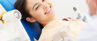
Effective treatment is impossible without correct diagnosis.
Such procedures allow you to determine an accurate diagnosis and find the best option for eliminating the disease. At the same time, modern medicine does not stand still. Every year, new analysis methods appear that make it possible to determine an accurate diagnosis. Thus, one of the most effective options is the use of digital equipment that provides panoramic images.
For this reason, such methods have almost completely replaced conventional computer diagnostics. OPTG of the dentition is especially popular. This analysis is deservedly considered the most effective. In this case, the inspection is carried out using the most modern equipment, which provides an accurate result!
Content
- What is OPTG in dentistry?
- Main types of procedure
- Diagnostic procedure
- Features of the procedure
- Main areas of use
- Children's examinations
- Equipment Features
- Indications and contraindications
- The advantages of an orthopantomogram and what is the difference between it and CT
- How is a dental x-ray done?
- Decoding the received data
- Why do dental x-rays take place?
- How often can children’s teeth be x-rayed without harming their health?
- Orthopantomogram or CT?
- Where can I do it?
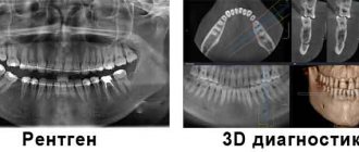
OPTG or CT: which is better / what is the difference?
Computed tomography differs from an orthopantomogram in that the output is a three-dimensional image. It can be rotated and laid out into layers, obtaining the most detailed information about the state of the structure of the area under study.
The radiation dose for both types does not exceed SanPiN norms, but there are differences. With a pantomographic examination, the patient receives 20-30 microsieverts, and with a CT scan - 60-70.
A computer image is considered more informative due to the absence of distortions inherent in a two-dimensional image. But it is prohibited to do it during pregnancy. Women who are expecting a child will be offered a pantomogram in the 2nd and 3rd trimesters.
It is up to the dentist to decide whether an orthopantomogram or a CT scan is better in a particular case based on the goals of treatment and the characteristics of the patient.
What is OPTG in dentistry?
Similar methods are used in various medical fields. For example, this diagnosis is carried out before the appointment of therapeutic, surgical and orthopedic procedures.
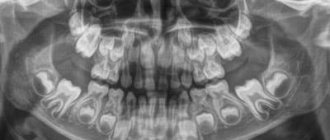
An OPTG image of teeth is prescribed in cases where conventional X-ray analysis is not enough. It gives the doctor a circular image of the oral cavity, which is unfolded in a plane. Such images show not only teeth and crowns, but also roots, canals and tissues.
Thus, such a procedure allows us to obtain maximum information about the condition of the dentofacial region. In some cases, making a correct diagnosis is only possible after obtaining a survey image.
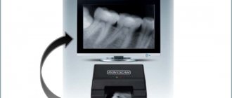
In this case, the image is automatically transferred to the computer in digital format. The use of special programs allows the doctor, if necessary, to enlarge individual parts of the image or set a certain viewing angle.
Orthopantogram of the dentition: what is it and how is it done?
28.05.2020
Orthopantogram of the dentition: what is it and how is it done?
- The essence of the OPTG method
- Indications and contraindications for the dental orthopantomogram procedure
- Advantages and disadvantages of the panoramic dental photograph method
- X-ray OPTG in modern dentistry
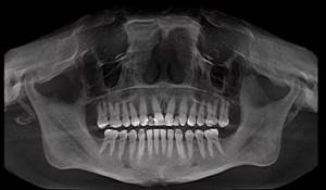
Diagnostic imaging is an integral part of modern dentistry. It helps doctors timely identify oral problems, plan dental treatment and monitor its effectiveness. The most popular method in this area is a dental orthopantomogram. This is a panoramic x-ray of the upper and lower jaw, showing all the teeth at once, including those that have not yet erupted. The OPTG also gives the doctor an idea of the condition of the jaw bone and temporomandibular joint.
The essence of the OPTG method
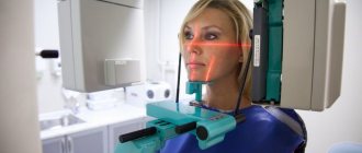
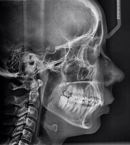
Sometimes an external examination of the oral cavity is not enough for a doctor to make a diagnosis or choose a treatment method. It is in such cases that an orthopantomogram is prescribed; you will find out what it is below. OPTG is an x-ray of the lower part of the face. Like any x-ray test, it uses ionizing radiation to create images of the body's internal structures. During the procedure, the patient places their chin on a small rest in front of the X-ray machine and gently bites down on a sterile mouthpiece. This ensures you remain still while shooting. The procedure is performed very quickly, the patient does not feel anything.
The standard method involves shooting directly on film. When conducting digital OPTG, information is processed on a computer.
Indications and contraindications for the dental orthopantomogram procedure
The procedure allows the doctor to assess the condition of all the patient’s teeth, determine their number, position and degree of eruption. Dental orthopathograms are also used to plan orthodontic treatment and examine the jawbone.
Additionally, orthopantomography reveals:
- caries or pulpitis;
- dental injuries and jaw fractures;
- sinusitis; periodontitis, periapical abscess;
- tumors, radicular cysts;
- disease, fracture or dislocation of the temporomandibular joint;
- foreign bodies;
- sialolithiasis (salivary duct stones).
Sometimes dentists use this method to diagnose children. In this case, OPTG of the dentition is used to monitor the growth and development of children's teeth (determining their location, shape, angle, etc.).
The only contraindication to the procedure is pregnancy. In this case, the doctor decides how much the benefits of the diagnosis outweigh the possible risks.
Advantages and disadvantages of the panoramic dental photograph method
A panoramic photograph of teeth in Kursk makes it possible to collect detailed information about the condition of the patient’s oral cavity. Compared to other types of dental diagnostics, an orthogram has a number of advantages:
- wide coverage of facial bones and teeth;
- low radiation dose;
- convenience and speed of examination;
- the ability to examine patients who are prohibited from opening their mouths;
- quick results;
- minimal number of contraindications;
- painlessness.
The disadvantages include the fact that the OPTG x-ray does not show small anatomical details, as well as the overlapping of teeth images on the photograph.
X-ray OPTG in modern dentistry
Orthogram is a highly accurate diagnostic method widely used in modern dental clinics. The study involves the use of a low dose of ionizing radiation. A digital orthopantomogram is used more often (how this study is done is described above). The procedure allows you to obtain a detailed image of the dental structures. Also, the Le-Dent clinic provides dental prosthetics services.
Main types of procedure
At the moment, there are three options for carrying out this procedure. Their varieties depend on the models of equipment used. Thus, a digital orthopantomogram is used for diagnosis. It is more expensive, but does not require expenses for consumables and photo laboratory services. At the same time, it provides high image quality.
In addition, it is possible to use film equipment. In this case, the level of radiation will be significantly higher. In this case, the accuracy of the result will be slightly lower, and the procedure itself will take longer.
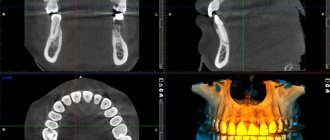
Another popular option is a 3D orthopantomogram. For this diagnosis, a special computed tomograph is used, which allows you to examine the tooth from all sides.
In this case, the image consists of a number of projections that provide a three-dimensional model of the desired area. This method is considered the most effective.
Digital or film orthopantomogram: what to choose?
Digital equipment today has practically replaced film analogues from practice. Although they cost clinics more, they do not require the costs of consumables and darkroom equipment.
In addition, film devices are characterized by a higher degree of radiation.
A panoramic image taken digitally is instantly transferred to the doctor’s computer and entered into the patient’s electronic record. If desired, the latter can receive the image on disk or in printed form.
Finally, the image quality with digital equipment is much higher than with film equipment. This means that the specialist receives better information for further work.
Diagnostic procedure
Many patients who encounter this procedure are interested in the question - what is an orthopantomogram and how is it done? It is worth noting that such diagnostics do not require complex and lengthy preparation. Moreover, the examination takes only a couple of minutes, after which the doctor has a ready-made image in his hands. The procedure itself consists of the following steps:
- The patient removes all metal jewelry and objects - chains, earrings, piercings, etc. Their presence may distort the results.
- The patient stands in front of the device and puts on a protective apron.
- After this, you need to hold the plastic holder between your teeth.
- The X-ray emitter then begins to rotate around the patient's head until a complete image is obtained.
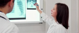
The finished image is automatically transferred to the doctor’s computer, after which the specialist interprets it. If necessary, the photo can be printed or sent to another medium.
Orthopantomogram: indications for performance
- before starting dental treatment or prosthetics – to diagnose the condition of all teeth at once in one image,
- in case of teething pathology,
- when diagnosing periodontitis,
- for diagnosing oncology of the bone tissue of the jaws,
- for the diagnosis of jaw fractures.
You should not hope that a panoramic image will allow you to evaluate specialized issues - the quality of bone tissue before implantation or help in planning orthodontic treatment, and it is not intended to search for small objects (perforations, root cracks, etc.). For all this, you will need a computed tomography, and specifically to correct the bite, the teleradiography method.
In addition, although an orthopantomogram shows the maxillary sinuses and temporomandibular joints, diagnosing joint diseases or the presence of chronic sinusitis using this type of image also does not seem to be a very good idea, because The projection of the image does not allow these formations to be clearly seen. Thus, the value of a panoramic image lies solely in the fact that it shows a picture of the condition of all teeth at once, as well as the bone tissue of the jaws.
Features of the procedure
Such a study can be called basic, since it allows us to assess the condition of the dentofacial region as a whole. For this reason, it often makes it possible to detect diseases that have not yet manifested themselves with pronounced symptoms, but could lead to serious consequences.
Many patients are interested in the question: what does an orthopantomogram of teeth show? It gives a complete picture of the condition of the oral cavity. So, the picture shows:
- jaws;
- maxillary sinuses;
- joints;
- roots;
- teeth, crowns, fillings;
- fabrics;
- bones.
For this reason, this diagnosis makes it possible to detect a disease that is associated with the dentoalveolar region. Therefore, this procedure is universal and can be used to detect any problem. At the same time, it guarantees the most accurate result. These features explain the high popularity of OPTG, which is only growing every year.
Main areas of use
There are a number of diseases for which the appointment of such diagnostics is the optimal or even the only solution. In addition, it can be used in various interventions, operations and procedures aimed at recovery. If patients are wondering why OPTG is needed, it is worth checking out the following list:
- Allows you to detect carious lesions, neoplasms and cysts.
- Makes it possible to identify cases of periodontal disease - this disease has no noticeable symptoms in the early stages.
- Helps detect bone diseases.
- Simplifies orthodontic correction, implantation and prosthetics.
OPTG also allows you to control the quality of operations performed, for example, installing a filling in a root canal. Thus, such diagnostics are multifunctional and help in carrying out a number of procedures. In addition, with the help of such images, a specialist can determine the density and height of the tooth, which is important when making prosthetics and installing fillings.
Children's examinations
The level of radiation exposure when creating an image is only 10-40 μSv. Thus, you can undergo the procedure up to a hundred times a year without harm to your health. It is also worth noting that this process is safe for children.
This level of radiation cannot harm the body. For this reason, OPTG is often used to detect permanent tooth buds. In addition, the method allows you to notice possible developmental anomalies in time. Indeed, in this case, the full row of teeth, including the roots, is visible. This uses an exposure mode that reduces the level of radiation exposure.
Indications and contraindications
A number of factors may be the reason for conducting a survey. These include the occurrence of inflammation, pain and swelling of the gums. In addition, the procedure is performed before implantation, tooth extraction or filling. Another prerequisite for OPTG is the occurrence of a cyst. The method has also shown high efficiency in diagnosing infectious diseases.
It is believed that there are no specific contraindications for the examination. However, this method is not recommended during the first trimester of pregnancy, or when feeding the baby.
The advantages of an orthopantomogram and what is the difference between it and CT
This procedure has a number of undeniable advantages. These include high image clarity and low radiation levels. In addition, OPTG allows you to zoom in on the image for a detailed inspection of individual areas. The information received is archived and can be reused if necessary. Another advantage is the speed of the process and its safety for the body.
For this reason, computed tomography is inferior to OPG in a number of indicators. In the first case, a separate area of the dentoalveolar region is examined. For this reason, problems such as cysts, granulomas and impacted teeth are difficult to identify. The difference between an orthopantomogram and a computed tomography scan is that the first option provides extensive information about the patient's condition. Meanwhile, who has worse accuracy and may not detect a number of important symptoms.
Sign up for a study by phone
+7 (812) 332-52-54
How is it carried out?
Before the examination begins, the person removes all metal objects: jewelry, removable dentures, hearing aids. He puts on a protective apron and stands or sits inside the frame of the device. The chin is placed on a special holder, the plastic mouthpiece is clamped with the teeth, and the hands hold the handles of the device. This is done to maximize stabilization of the head during the procedure.
The emitter with the cassette rotates around the person for 10-30 seconds. The result will be recorded on disk, film or paper. The examination is painless and takes 10-15 minutes.
Sign up for the study