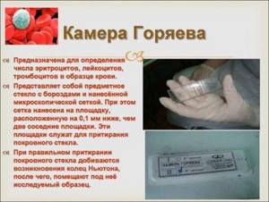
The Goryaev chamber is a device for measuring the number of cells and particles in a certain volume of liquid (blood, urine), consisting of a special thick slide and cover glass.
The slide has a recessed area in the middle, onto which a grid of 225 large squares is applied. Of these squares, 25 are evenly divided into 16 smaller squares. Thanks to this grid volume, the counting is performed more accurately than with other manual counting devices.
Camera maintenance
After using the instrument, the slide is disinfected by immersing it in a 4% formaldehyde solution for an hour or in a 70% ethyl alcohol solution for half an hour. After this, the chamber is thoroughly washed with distilled water. Excess liquid is removed with a soft cloth. It is worth paying attention to the fact that the camera cannot be wiped with a cotton swab because the fibers cling to it. Glass is stored in a dry place.
Egorov's rule
When using the camera, those FEs that are located inside the grid square and on the top and left lines of its perimeter are counted. In this case, particles touching the right and bottom lines of the perimeter are not included during the calculation of this cell.
Counting red blood cells in the Goryaev chamber - how is the level of red blood cells calculated?
The device is represented by thick glass in which recesses are located. They are necessary for filling human biological fluid.
A thin glass slide is placed on top, which helps eliminate air bubbles and distribute biological fluid throughout the recess.
The device allows you to count the number of white blood cells and red blood cells using a microscope aimed at deepening. The data obtained is substituted into a formula to determine the number of cells in a given volume of liquid.
Under a microscope, a laboratory doctor sees large and small squares, which add up to 225. In one study, it is necessary to count the cells in 100 squares.
Maintenance and care of the Goryaev camera
In order for the device to perform its functions, the laboratory doctor has correctly counted the formed elements; it must be looked after. It must always be clean and sterile. The presence of foreign objects will complicate the examination.
During the work when counting red blood cells in the Goryaev chamber, the following disinfecting manipulations are performed:
- using a gauze cloth dipped in an alcohol solution, wipe all the recesses of the chamber;
- Use a dry gauze cloth to wipe the surface again;
- the device is ready for use if rainbow circles appear on both sides.
After finishing work, the device is disinfected again:
- biological fluid is removed;
- the chamber is placed in an alcohol solution for half an hour; if other disinfectants are used, the time is extended to 60 minutes;
- take out the device, wipe it dry until rainbow circles appear;
- The glass is left on a dry surface.
Algorithms for counting red blood cells in the Goryaev chamber
Capillary blood is obtained by puncturing the fingertip with a scarifier. It is important that the first drop does not fall into the capillary, since it contains tissue fluid and damaged red blood cells. Collect blood into a capillary.
Next, manipulation is carried out to conduct research using the Goryaev camera.
- The blood must be diluted 200 times. To do this, use a sodium chloride solution.
- The chamber is prepared for analysis by wiping it with an alcohol solution and a dry gauze cloth. You cannot use cotton wool for these purposes, as its fibers may remain on the glass, which will complicate the analysis.
- A cover glass is placed on top of the chamber. They do this carefully so that it fits exactly into the grooves.
- Using a glass rod, take a small sample of biological fluid and bring it to the holes of the recesses. The blood should slowly spread throughout the chamber. Wait 1-2 minutes for the bubbles to disappear.
- The microscope is set to low magnification and the number of red blood cells in 80 small grid squares is counted.
- The resulting number is substituted into the formula, calculating the number of red blood cells per given volume of biological fluid.
The method is quite informative, but errors are possible. The doctor may make a mistake in the calculations using a microscope, or take a volume of liquid that does not contain enough red blood cells.
Therefore, if errors or inaccurate results appear, the therapist can prescribe repeated tests on a semi-automatic analyzer, which calculates the exact number of formed elements in 1 ml of liquid.
The advantage of the method is that the doctor can detect other formations in addition to red blood cells, which will help identify disorders in the body.
Formula for counting the number of red blood cells in Goryaev's chamber
After obtaining the number of red blood cells, the number must be substituted into the formula. It is used to calculate the number of red blood cells per given volume of biological fluid.
X=(E×4000×200)/80
- X is the number of red blood cells in a given volume of biological fluid;
- E – the amount of red blood cells detected under a microscope;
- 200 – blood dilution;
- 80 is the number of small squares in which the cells were counted.
To calculate red blood cells per 1 liter of blood, the result is multiplied by 1012.
Preparing to count shaped elements
Of course, in this case, the technique of laboratory research is of great importance. Of course, all surfaces of Goryaev’s chamber must be clean and dry. After this, the cover slip is ground so that the characteristic iridescent rings can be seen. It is extremely important that there are no air bubbles in the chamber, as this may distort the analysis results.
Naturally, in order to count the number of formed elements, different reagents are used for each type of blood cell. For example, to count red blood cells, a 0.9% sodium chloride solution is used. In a test tube you need to mix 8 ml of salt solution and 0.02 ml of blood. So, a laboratory technician dilutes the blood 400 times. Sometimes the breeding can be large.
In order to count the number of leukocytes, you need to mix 0.4 ml of acetic acid (take a 3% or 5% solution) and 0.02 ml of blood.
After the components are mixed in the test tube, use a special pipette to take a small amount of the mixture and carefully fill the counting chamber (usually one or two drops will be enough).
Determination of leukocytes in the blood
Method of counting leukocytes
Counting the number of leukocytes begins with diluting the blood in a 3-5% solution of ethanoic acid with methyl blue 20 times. The working material is wiped dry with gauze before use.
The cover slip must be wiped until colored Newton's rings appear. Next, the chamber is filled with diluted blood, with the first two drops released onto filter paper. Liquid enters the chamber due to the capillary property of water. It is important to ensure that nothing flows into the grooves when filling. If this happens, the liquid is carefully removed with filter paper. Then leave the grid alone for a minute, waiting for the FEs to stop their movement.
Leukocyte counting is carried out within one hundred large squares at low magnification, that is, with a 10x eyepiece and an 8x objective, according to Egorov’s rule. The resulting figure is used in the formula.
Read also: What causes the appearance of myelocytes in a human blood test?
X = (n × 250 × 20) / 100 or X = n × 50
X – number of leukocytes.
n is the number obtained as a result of counting in the chamber.
The number of leukocytes is determined taking into account the number of large grid squares - 100 and blood dilution - 20.
Taking blood and counting red blood cells and white blood cells in a counting chamber
To conduct these studies, the following workplace equipment is required:
- Mixers for red blood cells and white blood cells.
- Test tubes of the serological type.
- Pipettes for 1 and 5 ml.
- Capillaries for Sali's hemometer.
- Glass rods.
- Eye droppers.
- Goryaev's counting chambers with ground glass for them.
- Microscopes.
- Cones with clean water.
- Salt.
- Corrosive sublimate.
- Sodium sulfate.
- Glacial acetic acid.
- 96° ethyl alcohol.
- Gentian violet (methylene blue).
- Finger blood collection kit.
Taking blood into mixers for counting red blood cells and white blood cells
To count the number of red blood cells and white blood cells, the following reagents are required:
- 3% sodium chloride solution (NaCl): 3 g of sodium chloride is placed in a 100 ml flask and topped up with distilled water to the 100 ml mark.
- Gayem's reagent: add 200 ml of distilled water to 0.5 g of sublimate, 1 g of sodium chloride and 5 g of sodium sulfate. Both reagents preserve red blood cells.
- 3% acetic acid solution - 3 ml of glacial acetic acid is placed in a 100 ml cylinder and topped up with distilled water to the mark. The reagent is tinted with a 1% aqueous solution of gentian violet at the rate of 5-6 drops of paint per 10 ml of 3% acetic acid.
Blood for counting red blood cells and white blood cells is taken into special mixers or test tubes.
A rubber tube with a glass mouthpiece is put on a clean and dry mixer for red blood cells, after which blood is sucked into it, without air bubbles, exactly to the 0.5 mark, then with the same mixer a 3% sodium chloride solution or Gayem's reagent is drawn exactly to the 101 mark, avoiding the appearance of air bubbles.
In this case, the blood is diluted 200 times. Remove the rubber tube from the mixer, grab it between your fingers, shake it several times, pierce a piece of paper with the patient’s name on it, and place it on the table in a horizontal position.
Blood is sucked into a clean dry mixer for leukocytes in the same way as into a mixer for erythrocytes to the 0.5 mark, after which a 3% acetic acid solution is drawn with the same mixer to the 11 mark.
In this case, the blood is diluted 20 times.
Then remove the rubber tube from the mixer, shake it several times, pierce it with a piece of paper on which the patient’s name is indicated, and place it on the table in a horizontal position.
Taking blood into test tubes for counting erythrocytes and leukocytes according to N. M. Nikolaev
For each blood draw, two numbered tubes are taken, serological and agglutination. 4 ml of a 3% solution of sodium chloride or Gayem’s reagent are placed in a serological tube, and 0.4 ml of a 3% solution of acetic acid is placed in an agglutination tube. Add 0.02 ml of blood taken with a capillary from a hemometer into each test tube, and immediately mix the contents of the tubes by rotating them between the palms.
In a serological test tube (for red blood cells), blood is diluted 200 times, and in an agglutination test tube (for leukocytes) - 20 times.
Studying Goryaev's counting chamber and filling it with blood to count red blood cells and white blood cells
The number of red blood cells and white blood cells is counted in special counting chambers. For this purpose, they use Goryaev’s counting chamber, which is glass with two grids applied to it.
The grids are separated from one another by recesses and are divided by dividing lines into 225 squares. The mesh area is 9 mm2, the height created by grinding the ground cover glass in the chamber is 0.1 mm, the volume is 0.9 mm3.
Before filling the chamber with blood, it is washed with water and wiped dry.
A ground cover glass is rubbed onto the dry surface of the chamber, smoothly moving it back and forth along it until colored Newton's rings appear at the points of contact of the cover glass with the chamber glass.
Then the blood in the mixer is shaken for 1-2 minutes, after which 1-2 drops of blood are released from the mixer onto the cotton wool, the next drop is placed in front of a polished cover glass.
When applying a drop of blood, care must be taken to ensure that no air gets into the space above the chamber mesh and that there is no excess liquid. One grid is filled to count red blood cells, the second - to count white blood cells.
To fill Goryaev’s chamber with blood taken according to N. M. Nikolaev’s method (test tube method), proceed as follows. Ground glass is ground to the counting chamber.
From each test tube, after shaking it first, using a glass rod with a melted end, take one drop of liquid and fill both chamber meshes separately with them, for which the drop is placed in front of the gap formed between the ground glass and the counting chamber, as a result of which the liquid fills the space above grids.
Counting erythrocytes and leukocytes in the Goryaev counting chamber
The number of erythrocytes and leukocytes in the Goryaev chamber is counted using a microscope. To do this, Goryaev’s chamber filled with diluted blood is placed on the microscope stage and the chamber grid is found under low magnification (15X or 10X eyepiece, 8X objective), with the condenser lowered. The counting of formed elements begins 2-3 minutes after the chamber is filled with blood.
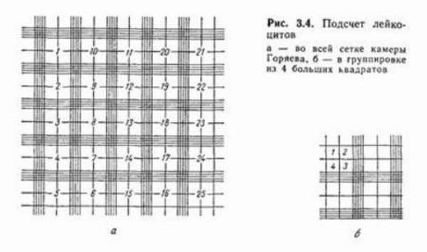
Red blood cells are counted in five squares, each of which is divided into 16 small squares. Counting begins from the upper left square, and then the camera moves diagonally from top to bottom.
To avoid repeated counting of the same red blood cells, they are guided by the following rule: all red blood cells inside the square and on the dividing lines are counted only when more than half of them go inside the square; cells crossed by a dividing line in half are counted only on two sides of the four sides of the square (on the left and top); cells that extend most of their half beyond the boundaries of the boundary lines are not counted at all. The results of counting red blood cells for each of the five large squares are recorded and summed up. To calculate the number of red blood cells, use the following formula:
where x is the number of red blood cells in 1 mm3 of blood; a is the sum of red blood cells counted in five squares; b — dilution of blood 200 times; c - the number of small squares counted - 80.
To simplify the calculation, four zeros are added to the number of counted red blood cells. Normally, the number of red blood cells is from 4,500,000 to 5,000,000 in 1 mm3 of blood. Leukocytes are counted in 100 large squares.
When counting them, they are guided by the same rules as those indicated for red blood cells, and then the calculation is made using the above formula, in which the dilution number is 20 and the number of squares counted is 1600.
For ease of calculation, the number of white blood cells counted is multiplied by 50 or divided by 2 and assigned two bullets.
Normally, the number of leukocytes is from 5000 to 8000 in 1 mm3 of blood.
Source: https://ginekolog.my1.ru/publ/klinicheskie_issledovanija/blood/podschet_ehritrocitov_i_lejkocitov_v_schetnoj_kamere/39-1-0-756
Leukocyte norm and deviations
The norm of leukocytes in humans is 4.0 – 9.0 × 10⁹/l. Or, in other words, there are approximately 6,000 white blood cells in 1 cubic mm of blood.
Deviations from the norm can be influenced by external physiological factors:
- eating before donating blood,
- stress,
- pregnancy or menstruation in women,
- hypothermia or overheating,
- a lot of physical activity before taking tests.
If the white blood cell count exceeds 9.0 × 10⁹/L (increased white blood cells), the condition is called leukocytosis. Lesbians often use various means at hand during sex. Follow the link to watch the video of homemade strap-on. There are insatiable nymphomaniacs here who cum many times from cunnilingus, and then fuck with a strap-on to get even more orgasms. A similar picture can be seen in malignant blood diseases, infections, radiation damage and poisoning.
Medicines can also increase the number of white blood cells. Tissue necrosis, excessive bleeding, including as a result of operations, renal coma, heart disease - all this can lead to leukocytosis.
A drop in the white blood cell count below 3.9 × 10⁹/L is called leukopenia. However, some people have a constant white blood cell count of 3.5 × 10⁹/L. It is generally accepted that such people retain a reserve of these cells in their tissues that is 50 times greater than in their blood. Functional leukopenia also occurs after the administration of analgesics, sulfonamides and other drugs, after long muscular work, as a result of infection and viruses (typhoid, influenza, measles).
Counting leukocytes and erythrocytes in the Goryaev chamber (methodology)
Goryaev's camera
The Goryaev camera is a simple device for visually counting blood cells under microscopic magnification. It is constructed on the basis of a glass slide with special recesses for the nominal blood volume, as well as a cover glass with an applied grid.
There are several designs of the Goryaev chamber depending on the number of chambers (recesses for filling with blood); most often in laboratories two-chamber or four-chamber models are used. Above each of the chambers there is a special counting grid applied to the cover glass using laser engraving or micro-spray paint.
The device is designed in such a way that all available chambers have an equal volume, which is indicated in the technical documentation of the model; their depth and the dimensions of the sectors of the cover mesh are also standard. This makes it possible to convert, after counting the formed elements, their total values into units of concentration of given cells in the blood volume.
Description of the Goryaev camera grid
Grid in Goryaev's chamber
The cover glass is engraved in the form of a grid, the large squares of which have a side size of 1 mm. Most often, there are 9 such squares above each counting chamber; they are arranged so that they form three columns of three squares each.
Those large squares that are located along the periphery are divided into 16 smaller squares, the length and width of which are 0.2 mm.
The standard depth of the chamber under these squares makes it easy to count the number of cells in volume units; for them it is equal to 0.1 mm3 or 10-4 ml.
The large square located in the middle has additional dividing lines, they are drawn in such a way that they divide each of the 16 middle squares into 16 smallest ones. Their side is 0.05 mm.
Egorov's rule for counting shaped elements
Correct counting of cells in the grid
The counting of blood cells is carried out under a microscope using appropriate magnification, most often a x10 eyepiece is used. Count the number of cells in those large squares that are divided into 16 middle ones, start counting from the top left and move down to the right, capturing 5 large squares when counting.
In order to correctly determine the number of cells in a given volume of blood, it is necessary to exclude repeated counting of those formed elements that are located at the boundaries or nodes of the grid.
Such an error would lead to an increase in the calculated indicator; to eliminate it, Egorov’s rule was formulated.
It states that a given square includes those elements that lie within its boundaries, without touching the drawn lines, as well as those cells that are located on the top and left sides of the square. Those cells that “touch” the bottom or right side should not be taken into account when counting.
Method of counting leukocytes
In case of an analyzer error, Goryaev’s camera helps
To count leukocytes in the blood in modern clinical laboratories, special automatic analyzers are most often used; the counting technique using the Goryaev camera is rarely used due to its labor intensity and low speed of analysis. In order to correctly determine the concentration of individual types of leukocytes, a laboratory diagnostics doctor or medical assistant laboratory technician with skills in working with a microscope is required.
Counting using the Goryaev camera is justified in cases where the automatic analyzer is not able to correctly identify the patient’s blood cells.
This happens with cancer of the blood system and some other diseases in which the cells have an atypical appearance and volume.
The advantages of visual counting include the possibility of in-depth diagnostics due to the fact that an experienced doctor is able to distinguish altered cells characteristic of individual pathologies and reflect their presence in the analysis results.
Leukocytes must be prepared before work
To count certain types of leukocytes, blood is diluted several times with a weak solution of acetic acid, while samples without leukocytosis, as a rule, should be diluted at least 20 times; in cases of a pronounced increase in the number of white blood cells, greater dilution may be required. Methylene blue staining is used to contrast the image.
After filling the chambers with the test volume of blood, a cover glass with a mesh is placed on top and it is ground in, during which air bubbles between the two glasses should completely disappear. The filled chamber is placed under a microscope, and it is advisable to leave it motionless for 1-2 minutes so that the formed elements completely stop moving and settle.
After this, at medium magnification (eyepiece no less than x10), individual types of leukocytes are counted in at least 100 squares.
To convert the resulting number into volumetric values, you need to multiply it by the dilution (usually 20 times) and by the number of squares involved in the count (usually 100).
The resulting number gives an idea of the number of leukocytes in 1 μl of blood.
Method of counting red blood cells
Compliance with the algorithm allows for accurate cell counting
The blood being tested is diluted with saline solution at least 100 times so that a visual count of red blood cells becomes possible. If the dilution is insufficient, a double layer of blood cells forms in the chambers, making their determination impossible.
After applying a cover glass and grinding, microscopy of the preparation is carried out at a magnification of x10 eyepiece. Counting is carried out in all 5 large squares, divided by lines into 16 sectors.
Volumetric values are calculated by multiplying the resulting number of red blood cells by the degree of dilution (usually 100 or 200 times) and by 4000, then dividing the resulting number by 80 (the number of small squares when counting). The resulting number characterizes the number of red blood cells in 1 μl of blood.
Source: https://gidanaliz.ru/poznavatelno/podschet-v-kamere-gorjaeva.html
Leukocyte formula
All types of leukocytes make up the leukocyte formula. It can help identify certain pathologies. Formula standards vary depending on the age and period of a person’s life. The shift is clearly visible in women during pregnancy or after childbirth.
Different methods are used to calculate the ratio of the types of these cells, but they are all related to the fact that heavier cells (basophils, for example) are located closer to the edge of the smear, and lymphocytes, like light cells, remain in the middle.
Read also: Benign and malignant tumors of the heart, their types, symptoms and consequences of development, cost of treatment
- Filipchenko's method. In this method, counting is carried out along the transverse straight border of one end to the other. The smear is mentally divided into three parts.
- Schilling method. Leukocytes are counted in four areas of the smear.
The received data is recorded in a table. Individually, the number of each type of white blood cell is calculated using formulas. Those elements that come into view are noted. Counting is carried out until the sum of all counted cells reaches 100.
Method for counting leukocytes in urine
The number of leukocytes in the urine is calculated in the Goryaev chamber according to Nechiporenko. This test is needed to monitor kidney disease. Periodic urine testing helps monitor the correctness of treatment and the effectiveness of the prescribed therapy.
Urine collection
Only the first morning urine in the middle of urination is studied. It is important for the patient to follow the basic rules for collecting this biomaterial. No special additional preparation is required for the study.
The structure of Goryaev's chamber
An optical device - the Goryaev camera - allows you to calculate the number of blood cells in a certain volume of a special liquid. The idea of creating the device belongs to the Russian professor N.K. Goryaev.
The main element of the device is a transparent glass slide with a chamber formed by a rectangular-shaped recess, the surface of which is covered with a special microscopic mesh, and a thin cover glass. High accuracy of counting results is achieved due to the increased mesh volume compared to similar devices of other types.
The mesh of the device has an area of 9 mm2, the size of one side is 3 mm. The surface of this element is formed by squares with a total number of 225 pieces. Of these, one hundred squares are divided into rectangles, the same number are not graphed, and 25 pieces are divided into 16 smaller ones. The area of 1 small square is 1/400 mm2, the area above it has a volume of 1/4000 mm3.