- Home >
- Directory >
- Is it possible to do a CT scan during pregnancy?
Computed tomography, like MRI, is an effective method of hardware diagnostics that allows us to identify many hidden pathologies. However, unlike MRI, computed tomography is contraindicated in pregnant women. Why and whether there are exceptions – we’ll tell you in this article!
Why is CT harmful?
The principle of CT scanning is the effect of X-ray radiation on the body. This radiation is practically harmless to an adult, if you do not exceed the permissible amount of research. However, exposure of a developing fetus to X-rays can result in embryonic death or congenital deformities.
Therefore, if it is possible to conduct an MRI instead of a CT scan, the doctor should choose the first option. CT scan is indicated only in cases where the life of the expectant mother is at stake.
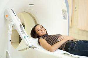
MRI in our center
Power 1.5 Tesla
High image quality
Study for patients up to 250 kg
Burn to disc for free
Is X-ray examination dangerous during pregnancy - MEDSI
Is X-ray examination dangerous during pregnancy?
X-ray examinations are carried out to determine the presence of any pathologies, tumors or diseases.
For this purpose, a directed beam of electromagnetic waves of a given length and frequency is used. It passes through human organs, the tissues of which reflect and absorb it in different ways. Thanks to this difference, various anomalies can be seen. The result is sent to the screen or recorded on a special film.
During a preventive examination, this examination is carried out in a special room. If the patient is injured, it can be used immediately in the emergency department, operating room or intensive care unit.
Effect of radiation on the fetus
X-ray radiation in general is not always beneficial for the body, since it destroys those cells that are in a state of constant division. This leads to the destruction or mutation of DNA chains.
There are not many such newly formed cells in the adult human body. But in the fetus in the early stages of development they are the basis. Therefore, such an examination is quite dangerous for him. Complications are most likely to occur when using x-rays during pregnancy in the first trimester (initial 12 weeks).
What problems can x-rays cause in early pregnancy?
Due to the gradual development of the fetus, the formation of future systems of the child’s body occurs in each week of its existence. Therefore, the exposure of the mother's body to large amounts of X-ray radiation at these stages can have various serious consequences:
- The first two weeks. Possible death of the embryo, miscarriage, ectopic pregnancy.
- Weeks three and four. Pathologies at an early stage of fetal development, miscarriage.
- Fifth-sixth week. Developmental disorders of a number of organs and systems: thyroid, thymus and gonads; immune, nervous, circulatory, endocrine systems.
- Seventh week. Damage to the liver, intestines. Metabolic disease.
- Eighth. Pathologies of the development of joints and limbs, the oral cavity.
- Ninth week. Damage to the respiratory and reproductive systems.
- Weeks ten and eleven. Problems associated with dental development. Heart disease.
- Twelfth week. Pathologies of the thyroid gland, immune disorders.
After this period, the effect of radiation on the fetus decreases, but it is still not recommended to do such a study until the end of pregnancy, unless absolutely necessary.
X-ray in early pregnancy
Doctors try not to prescribe x-rays to pregnant women, because even a minimal risk of harm from radiation always remains. It is especially great in the first twelve weeks.
The most dangerous types of tests for the fetus are:
- Abdominal x-ray
- X-ray of the pelvis and spine
- Mammography
- Fluorography
- CT scan
- Isotope scanning
The following types of x-ray examination are less dangerous:
- Chest (lungs, heart)
- Brain
- Limbs
Are there any non-hazardous types of examination?
The safest types of x-ray examination are:
- X-ray of teeth
- X-ray of the nose
In these cases, the impact occurs locally, and therefore the radiation dose is minimal.
The amount of radiation that a fetus can receive in two months is regulated by Sanitary Rules and Standards and should be no more than 1 millisievert (mSv).
There are other types of examinations that can be used instead of x-rays during pregnancy:
- MRI
- Visiography
- Ultrasound
Still, doctors try not to prescribe MRI in the first trimester of pregnancy, since statistical studies are insufficient to clarify its safety during this period.
What to do if you can’t do without an x-ray?
X-rays for pregnant women may be necessary in a situation where a disease or injury threatens the life and health of the mother and child, and it is impossible to use other diagnostic methods. And the harm from not using x-rays exceeds the potential harm from its use.
- If it is necessary to conduct an examination of an area that does not touch the pelvis, abdomen or spine, then they must be shielded with lead aprons and overlays.
- If in the early stages of pregnancy it is necessary to take an x-ray directly through the fetus, then the doctor may suggest terminating it to avoid mutations and miscarriage.
- A woman can refuse an abortion, but in this case she must understand the risk she is taking and the pathologies that may appear in the fetus.
In all these cases, after undergoing the study, it is recommended to go for an ultrasound to monitor the condition of the fetus and the appearance of certain pathologies.
If it is possible to postpone the use of x-rays until the last trimester or postpartum period, then it is necessary to do so.
At a very early stage, a woman may not be aware of pregnancy. Therefore, it is recommended to undergo additional examination before the x-ray.
Advantages of performing X-ray analyzes at MEDSI:
- 30 types of X-ray examinations
- The latest equipment with the ability to regulate radiation intensity
- Carrying out urgent examination in case of injury or other medical indications
- Technologies that are suitable for both adults and children
- Radiologists of high qualification categories, candidates of medical sciences
- Sign up for research and consultation by phone 8 (495) 7-800-500
- More than 20 diagnostic centers
Consequences of CT scan during pregnancy
What consequences can a CT scan during pregnancy lead to:
- Fetal death (especially in the early stages - 1-5 weeks)
- Malformations of internal organs: heart, liver, thyroid gland
- Deformities (eg, cleft palate and upper lip)
- Pathologies of the thyroid gland and, as a consequence, developmental delays
Take care of your health and the health of your child! Often an MRI is more revealing than a CT scan, and then it is not worth risking anyone's health. Sign up for an MRI and detect pathology without harm to the body!
What are the alternatives to CT?
The most common ways to assess health during pregnancy are ultrasound and MRI. In rare cases, conventional radiography can be used, but it is undesirable, just as computed tomography is contraindicated during pregnancy.
X-ray
This study is also unsolicited. Naturally, the radiation dose will be much less than a CT scan during pregnancy. But such a study will be prescribed only as a last resort, if there is no way to do without it.
MRI
The procedure is absolutely safe for the pregnant woman and the fetus, it has no consequences and will not affect the development of the child. However, the MRI procedure is no longer aimed at examining soft tissues, so it will not be possible to examine bone structures in detail. That is why in some cases you will have to think about whether a pregnant woman can have a CT scan.
When can a CT scan be done during pregnancy?
There are cases when an MRI cannot provide the necessary results, and the matter decides the issue of life and death of the expectant mother. In such situations, you cannot do without CT. What are these cases:
- Serious injury to the patient requiring prompt diagnosis
- Detection of acute infection, inflammation or tumor that may threaten a woman’s life
If a CT scan was performed before the patient found out about pregnancy, it is worth telling the gynecologist about this: you may have to undergo additional tests or think about terminating the pregnancy.
Is it possible to do a CT scan during pregnancy?

It is highly undesirable to perform a CT scan on pregnant women. This is especially true in the first trimester of pregnancy, when the vital organs of the fetus are formed.
The risk of developing pathology after a CT examination depends on the area of examination, the type of CT scanner that is planned to be used, and the stage of pregnancy.
- Survey area . Computed tomography of the head, neck, arms or legs is less dangerous for the fetus, since the source of X-rays is located at a distance from the fetal location, and the pregnant woman's stomach is additionally covered with a lead apron.
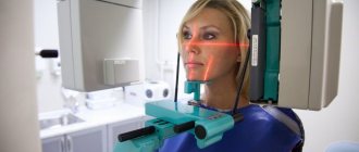
- View of a computed tomograph . Multispectral computed tomography allows for examination with a lower radiation dose to the patient compared to spiral CT. A dental computed tomograph gives such a small dose of radiation that pregnancy is often not a contraindication for examination with such a device. However, the capabilities of dental tomographs are limited.
- Gestational age . In the first days and weeks after conception, the embryo is most vulnerable to damaging environmental factors, including x-ray radiation. A number of practicing doctors insist on terminating a pregnancy if a CT scan was performed after conception. However, such categoricalness is not always justified.
The effect of X-ray radiation on the human fetus has not been studied, so there is no reliable data on how great the risk of congenital deformities after an X-ray examination or computed tomography is performed. The studies that were conducted on animals suggested exposure to much higher doses of radiation compared to those received by the patient during a CT scan. In this regard, these data cannot serve as a basis for referring the patient for an abortion.
It should also be remembered that in the first trimester of pregnancy the “all or nothing” rule applies . This means that if the harmful effects are strong enough to damage the fetus, a miscarriage occurs or the pregnancy ends. If the effect is minor, the pregnancy continues and the fetus continues to develop normally in the uterus.
MRI during pregnancy
MRI during pregnancy is not performed as often as ultrasound, but there are situations when only magnetic resonance imaging can help make a diagnosis. It is important that MRI will not harm either the mother or the child, which means that if you have a choice, give preference to magnetic resonance imaging.
Our experienced radiologists at the Elena Malysheva Diagnostic Center will do everything to quickly, efficiently and in the most comfortable atmosphere for the patient to identify possible pathologies.
Sign up for an MRI at the Elena Malysheva Diagnostic Center near the Baumanskaya metro station (see map) by phone or leave a request on the website.
Baumanskaya metro station (see map) 8
leave a request on the website
Indications for CT scanning

If there is no pregnancy or other contraindications, then a CT scan can be done for almost everyone. It does not have a serious negative impact on humans. When performing a CT scan with a contrast agent, doctors recommend drinking more water to remove it from the body as quickly as possible.
Computed tomography is a modern, convenient and very informative examination method and is used to diagnose a wide range of diseases. Thanks to it, pathologies can be identified at the most basic level. This is very important for diagnosing neoplasms, including malignant ones.
Exceptions to the rules

In some cases, a diagnostic method such as computed tomography is necessary. Such research may be needed to examine:
- cervical spine;
- skulls;
- bones of the hand, foot;
- chest;
- some joints.
Diagnostics in such a special condition can be prescribed to detect and study neoplasms, diseases of bones, and soft tissues. When the research is urgent, doctors take extreme measures - they prescribe CT scans for pregnant women.