X-rays during menstruation are performed in cases where other methods are not available. The study affects the endocrine, hematopoietic and reproductive systems. Therefore, its implementation is limited on critical days and their delay.
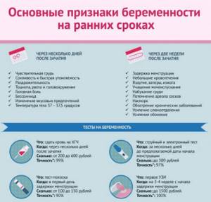
One of the first examinations prescribed when visiting a doctor is radiography. It is carried out to diagnose the musculoskeletal system, endocrine, excretory and reproductive systems. In which cases x-rays are not taken, and what conditions to observe in order to maintain reproductive health, we will discuss below.
In what cases is the examination carried out?
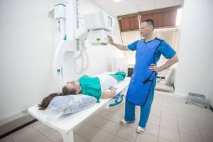
You can do x-rays or fluorography during menstruation. During this period, it is safe to examine the area of the head and neck, chest, and upper extremities. The pelvic organs are covered with a plate that does not transmit x-rays.
Ionizing radiation is directed horizontally and does not spread over long distances, so this method of protection is effective.
If necessary, even pregnant women can have their teeth taken. Examination of the pelvis, lower part of the spine, and hip joint during menstruation is carried out according to indications. For routine diagnostics, the image is taken in the last days of menstruation or after it. To understand the reason, you need to know how X-rays affect the reproductive organs:
- increase the intensity of bleeding;
- increase its duration;
- stimulate the production of sex hormones;
- increase the thickness of the endometrium;
- cause a delay in menstruation;
- stimulates the functioning of the ovaries.
The second factor influencing the course of the study is discharge. They make it difficult to visualize the uterus and nearby organs and distort the results of radiography.
Important! During menstruation, it is not recommended to conduct a contrast study - fluoroscopy.
Fluoroscopy
Critical days indirectly affect the accuracy of diagnosis, as they are accompanied by changes in intestinal motility and increased gas formation. The gas does not allow x-rays to pass through, covering the internal organs. This may lead to repeated imaging and increased radiation exposure.
To take an x-ray during menstruation, you need good reasons. These include emergency situations (fracture, organ perforation), acute surgical diseases (intestinal obstruction). Diagnostics may be required before surgery.
X-ray concept
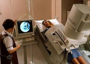
An x-ray is a diagnostic procedure that is prescribed for injuries, certain diseases and various lesions of the body. Radiography can be used not only as a primary diagnosis, but also as a full examination.
The impact is carried out by electromagnetic radiation. The rays pass through tissue, resulting in a clear image of the internal organs. The image is captured using special sensors and then presented in digital or film form.
This cannot but affect the organs, but the level of radiation is minimal. The procedure will not harm a healthy person - it is prescribed only according to indications when there is no risk of exceeding the radiation exposure limit . Sometimes the consequences appear after some time in the form of minor defects in tissues or organs located next to them.

Experts do not say unequivocally whether it is possible to take an x-ray during menstruation, but recommend, if possible, to refrain from the procedure. Each person’s body has its own characteristics, and sometimes it is difficult to predict how it will react to such an intervention.
Examination of the pelvic organs
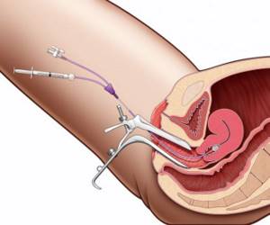
Hysterosalpingography
Contrast is also used for diagnosis. It is injected into the uterine cavity, fallopian tubes (hysterosalpingography) or into the abdominal cavity (pelviography). The examination helps to examine the contours and size of the ovaries and assess the patency of its tubes.
A bicontrast study is possible, when the drug is administered together with nitrogen, oxygen or carbon dioxide. The procedure is carried out after ovulation. When it is combined with critical days, the likelihood of infection and the development of complications increases.
The only indication for which contrast is used is infertility. Hysterosalpingography helps to exclude polyps, developmental anomalies, adhesions, and tumors.
The need for a removal procedure
In some situations, delaying tooth extraction is not advisable and even dangerous. Severe inflammation increases the likelihood of developing sinusitis, otitis, osteomyelitis, sepsis and other dangerous diseases.
Indications for removal on critical days:
- acute pain that is poorly relieved by medication;
- severe swelling, swelling of the cheek;
- purulent inflammation: periodontitis, cyst and other pathologies;
- toothache accompanied by fever;
- acute pulpitis;
- tooth root fracture;
- impacted “figure eight”, which causes severe pain and swelling.
Menstruation does not become an obstacle if a woman is planning a business trip or vacation in the coming days. It is better to solve the problem in advance than to suffer from pain, postponing treatment until you return.
Fluorography

The study is performed annually, and it often falls on the period of critical days. They are accompanied by a sharp change in hormonal levels, decreased immunity, and general malaise. The question of the need for examination is decided depending on how pronounced these factors are.
It is possible to do fluorography during a woman’s menstruation, since the x-ray load during the study is tens of times less than for the entire year from natural sources. If the reproductive and endocrine systems are normal, the procedure is performed any day. If there is a concomitant pathology (anemia), it can be postponed.
Exposure to rays is undesirable if a woman is planning a pregnancy. If the patient is sexually active and does not use protection, it is better to carry out fluorography during her menstrual period. This reduces the chance of pregnancy. Is X-ray diagnostics performed if there is a delay?
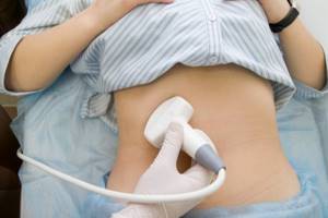
Delay is the absence of menstruation for 10 or more days after its expected date. In such cases, the study is carried out only after pregnancy has been excluded (examination by a gynecologist, express test, ultrasound diagnostics).
If menstruation is delayed for more than 1 week, there is a high risk of pregnancy. The effects of X-ray radiation on the fetus are known. These are abnormalities of organ development and chromosomal abnormalities. If the embryo is exposed to ionizing radiation before the gestation period, while it is in the fallopian tube, early miscarriage is likely. If pregnancy continues, it can proceed without complications.
Given these facts, women who suspect pregnancy need to inform their doctor. To clarify the diagnosis, other methods are used or the examination is postponed.
When the situation is urgent
Sometimes, for health reasons, you have to take an x-ray during menstruation, simply not paying attention to it. This situation can arise in the event of various injuries or serious illnesses, when it is very important to identify all damaged organs in a timely manner. In this case, even pregnancy and lactation are not absolute contraindications, since we are talking about a woman’s life.
The examination plan for infertility includes a type of x-ray examination such as hysterosalpingography or hysterosalpingoscopy. This manipulation is aimed at studying the reproductive organs at different stages of the menstrual cycle, including during menstruation.
In any case, an x-ray examination is prescribed by the attending physician, and he decides on the timing of its conduct, based on the data of a subjective examination.
How does X-ray radiation affect the menstrual cycle?
X-rays have the following effects on a woman’s body:
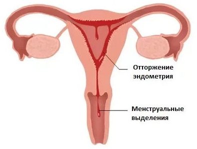
- accelerate endometrial detachment, which increases blood loss;
- disrupt the cycle;
- reduce the formation of blood cells.
More often than not, the examination leads to a delay. The regularity of menstruation changes not only due to radiation. This is largely due to the stress that occurs during a contrast study. Women suffering from infertility may become pregnant after exposure to ionizing radiation.
In healthy women, the examination does not cause adverse consequences. With anemia during menstruation, the symptoms of the disease may increase - palpitations, dizziness, weakness.
It is recommended to take an x-ray before menstruation, as the risk of impact on the reproductive organs is low. To conduct an examination, one condition must be met - to exclude pregnancy. What recommendations from gynecologists should be followed during the examination?
The issue of X-ray diagnostics is decided individually, depending on the characteristics of the woman and her reaction to previous examinations. If it is necessary to take photographs of the dental apparatus, hands, chest, they are taken without restrictions. The reproductive organs are protected with an apron.
The study can be postponed if the following symptoms are present:
- copious discharge;
- severe pain in the lower abdomen;
- headache and dizziness.
Gynecologists say that X-rays should not be taken during menstruation and for chronic diseases of the reproductive system. This statement is advisory in nature. The only contraindication for diagnosis using x-rays is pregnancy. Medical institutions require a statement indicating that the woman is not pregnant.

In case of urgent need, the study is carried out on any day of menstruation. To alleviate the condition, use antispasmodics - Drotaverine and painkillers - Paracetamol. They are taken as needed. When there is no pain, no appointment is required. In order to reduce gas formation, simethicone - "Espumizan" - is used. The day before the procedure, drink 2 capsules 3 times a day. On the morning of the examination, use 2 capsules once.
In a single study, the effect of radiation on the body is insignificant. The decision to conduct an x-ray is made by the doctor, depending on the underlying disease and general condition.
4. Interpretation of an x-ray image and what do the results depend on?
Interpretation of an x-ray image of the chest organs
Analysis and interpretation of the x-ray image is carried out by the attending physician to identify the cause of the disease or confirm the diagnosis. Here are the normal research indicators:
- The bones, including the ribs and spinal column, appear natural.
- The shape and size of the lungs and heart are satisfactory. Lung and cardiac tissue appear normal. No formations or growths were noticed within the lungs.
- The appearance of the vessels is normal.
- The location and shape of the diaphragm are satisfactory.
- No pathological accumulations of air and fluid were noted.
- The presence of foreign objects in the chest was not identified.
Let us list what refers to pathological indicators on x-rays:
- The presence of foci of inflammation caused by infectious diseases such as tuberculosis or pneumonia.
- The presence of swelling or swelling, which may be a consequence of heart failure.
- Detection of an increase in the size of the heart or accumulation of fluid around it, as well as an enlarged aorta and lymph nodes, pulmonary edema, etc.
- Bone fractures.
- Detection of foreign objects in the lungs, esophagus and respiratory tract.
What determines the results of a chest x-ray?
The quality of an x-ray depends on the following factors:
- a person’s ability to hold his breath for a while and not move;
- the presence of metal objects, the image of which is superimposed on the chest image;
- presence of obesity;
- scars that make it difficult to decipher the image.
Chest X-ray
is the process of obtaining an image of the chest and all its constituent elements - bones, internal organs - using x-rays. A chest x-ray allows a specialist to assess the condition of your internal organs, identify pathological and inflammatory processes, and also detect foreign objects in the chest area.
Recommendations from gynecologists
Despite the fact that modern X-ray equipment allows minimal impact on the body, specialists are obliged to warn the patient about the development of complications due to receiving a large dose of radiation. Therefore, during unscheduled X-ray examinations, it is recommended to cover the pelvic organs with a special protective apron .
In addition, there is no need to undergo the procedure if you feel unwell and have an exacerbation of inflammatory chronic diseases of the reproductive system .

If an x-ray is necessary for diagnostic purposes to identify the cause of infertility, then the patient is recommended to stop sexual relations from the beginning of menstruation until the day of the examination . It is usually done on days 7-11 of the menstrual cycle, since at this moment the thickness of the mucous epithelium is minimal and does not interfere with the passage of contrast into the fallopian tubes, thereby making it possible to obtain more informative and detailed images about the condition of these organs.