Computed tomography of the abdominal organs is a method of examining internal organs, often used in modern medicine. CT examination appeared in the diagnostic field relatively recently, in the 70s of the 20th century. It is based on the principle of the penetrating power of X-rays. The discovery of X-ray radiation occurred at the end of the 19th century, after which mathematical and technical research gradually led scientists to develop the possibility of scanning the human body with X-rays and converting the received information into tangible media. The first commercial tomography machines were developed in 1971. Since then, computed tomography techniques have improved every year.
What is computed tomography?
This is a diagnostic method in which the study of internal tissues and organs consists of creating many layer-by-layer X-ray images.
Using a computer, they are combined into a single 3D image - a tomogram. The doctor can study the structure of the organ both as a whole and in individual layers. Diagnostics takes 10-30 minutes, depending on the part of the body being examined. The study can be carried out with the introduction of a radiopaque substance (contrast) into a vein, orally or anally. This allows you to diagnose hollow organs, in particular the intestines, as well as any large vessels¹.
Video 1. Research method: computed tomography. Source: YouTube channel “TV Channel “Doctor”
Research limitations
Each diagnostic method has positive and negative aspects.
Disadvantages of magnetic resonance imaging (MRI):
- There is no possibility to conduct a comprehensive study of hollow organs. These include the gallbladder, urinary bladder, and lungs.
- There are restrictions on performing an MRI procedure on patients with metal objects in the body,
- To obtain accurate images, you should remain still and calm during the procedure.
Disadvantages of computed tomography (CT):
- CT scan is dangerous x-ray radiation,
- This procedure provides information only about the structure of organs and tissues, but not about their functioning,
- This type of diagnosis is contraindicated for pregnant and lactating women and children.
Brief history of creation
The idea of this method originated in 1959 in the USA. It was put forward by neurologist William Oldendorf, who worked on the problem of detailed diagnosis and study of brain structures. It was Oldendorf who created the CT scanner and patented it. However, it was not a full-fledged tomograph, but only a prototype.
The development of computer technology made it possible to obtain the first full-fledged tomograph only in 1969. This happened in London, and the device was created by US physicist Allan Cormack and British engineer Godfrey Hounsfield. The first study was carried out in 1971 - it was also a study of the brain.
Subsequently, devices were developed that had two radiation sources and the possibility of radiocontrast enhancement. The latest generation tomographs compose an image from many images in less than 1 second. This makes it possible, in particular, to obtain clear images of the heartbeat.
Is it dangerous to do a CT scan of the lungs after an x-ray?
Ionizing (X-ray) radiation is not beneficial for humans, but in excess amounts causes radiation syndrome and can become a “trigger” for the development of cancer in patients predisposed to it. According to the current “Radiation Safety Standards”, up to 30-50 mV of radiation per year is permissible, but we should not forget about the natural radiation background. A CT scan of the lungs (about 2.5 mSv) after an X-ray (about 0.1 mSv) is safe, and such precision diagnosis can save the patient's life.
However, to avoid additional radiation exposure, it is most advisable to immediately do a CT scan of the lungs without resorting to x-rays.
How does a tomograph work?
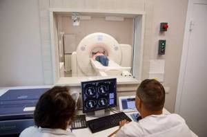
The main part of the apparatus is the gantry (or gantry), a ring frame with an X-ray tube inside. On average, its diameter is 70 cm, although there are larger tomographs - up to 90 cm in diameter. Inside the ring there is a table on which the patient sits motionless. The gantry allows the emitter to be rotated around it. The device can take pictures of the organ from different angles, giving the doctor a clear idea of its relief, density, and structure².
In modern devices, rotation occurs in a spiral (that’s why tomography is often called spiral). The rotation speed can be different - the higher it is, the more advanced the device and the better the image quality. For example, during a normal scan, the beam tube makes a full rotation in the gantry in 1-2 seconds, and when examining the heart - in 0.5 seconds. With each rotation, the system takes a picture of the organ. The beam of X-rays that the radiation tube emits can have different widths, depending on the area of the body being examined. If necessary, the frame can be tilted at an angle of up to 30°².
In a conventional X-ray machine, the rays passing through the patient's body are recorded on film. There is no film in the tomograph, and X-rays are received by detectors. They are also located in the gantry and are located opposite the beam tube. They transmit signals to a digital information conversion system that is connected to a computer. It remembers and combines the received images together.
7 differences between CT and conventional x-rays
1. Fluoroscopy gives a flat two-dimensional image of an organ, CT gives a three-dimensional, three-dimensional image.
2. In CT there is no effect of shadows from other organs and structures.
3. Resolution is 40-50 times higher, this gives incomparably higher clarity and information content.
4. The ability to examine the structure of an organ in sections up to 1 mm thick, which makes it possible to identify the smallest neoplasms and structural changes.
5. It is possible to diagnose any organs, bones, vessels and tissues that are inaccessible to conventional X-ray machines.
6. During an X-ray, the patient receives an average of 0.1 mSv of radiation, during a CT scan - from 1 to 10 mSv, depending on the area being examined. However, this radiation exposure is within the annual norm.
7. The price of tomography is higher, but this is compensated by the fact that the doctor receives the most objective and detailed information. In some cases, it can save the patient's life.
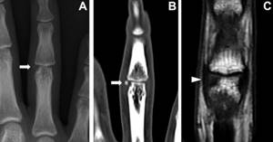
Comparison of X-ray, CT and MRI of the ring finger. X-ray (A), CT (B) and MRI (C). Photo: Case Reports in Plastic Surgery and Hand Surgery / ResearchGate (Creative Commons Attribution-NonCommercial 4.0 International license)
Contraindications for MRI and CT
There are several contraindications for CT scanning:
- pregnancy - the effect of x-rays negatively affects the fetus,
- the examination area is in plaster,
- Frequently performing similar procedures,
- lactation period,
- renal failure.
Before undergoing an MRI, you should pay attention to a number of contraindications, in the presence of which diagnosis is impossible.
- These are various electronic devices and metal implants in the studied areas of the body.
- Excess weight. The large size of the patient will not allow him to fit inside the MRI machine.
What does a CT scan show?
The device builds a three-dimensional image of almost any area. You can examine tissues and structures in increments of just 1 mm. On the tomogram you can see¹:
- benign and malignant neoplasms, including at the initial stage;
- metastases to organs;
- congenital/acquired anomalies in the development of organs and blood vessels;
- diseases of the lymphatic system;
- infectious foci;
- pneumonia, tuberculosis;
- damage/defects of bones, joints;
- vascular thrombi;
- coronary heart disease;
- abscesses, hematomas, aneurysms, internal bleeding;
- internal organ injuries;
- condition of intervertebral discs;
- muscle dystrophy;
- consequences of heart attacks/stroke.
What organs can be examined using CT?
The method is effective in diagnosing pathologies of the lungs, heart, large vessels, brain, liver, pancreas, stomach, intestines, kidneys, bladder, bones, joints, spine, sinuses, and eye orbits.
Difference between MRI and CT
Computed tomography (CT) is the best way to visualize bone structures. Therefore, CT is good to use to identify bone injuries and various injuries.
MRI clearly shows soft tissues: muscles, blood vessels, cartilage, spinal cord and brain. Therefore, MRI is better used to identify tumors and pathologies in neurology, neurosurgery, and endocrinology.
Under the influence of X-ray radiation, a computer tomograph (CT) produces a series of images of the object under study.
A magnetic resonance imaging (MRI) scanner operates based on the magnetic field in which the patient is placed.
MRI helps to obtain more accurate results in the following cases:
- the body’s response to the injected contrast agent was detected during computed tomography,
- you should check the condition of the brain, the condition of soft tissues,
- musculoskeletal diseases in children,
- it is necessary to check the condition of the pituitary gland, the condition of nerve cells in the brain,
- for damage to cartilage, joints,
- for suspected cancer.
CT is effective for:
- any mechanical damage, head injuries,
- damage to bones, their deformation under various influences,
- examination of blood vessels, heart,
- suspicion of the development of purulent diseases - sinusitis, otitis media,
- pathologies in the abdominal cavity,
- respiratory problems,
- suspicions of cancer, changes in the chest and its organs.
MRI is a harmless diagnostic method and does not irradiate the body, as with computed tomography. It is a good replacement if the body is intolerant to the contrast agent that is administered for CT scanning.
CT has a more intense effect on the body.
It is impossible to compare these two procedures, since they are completely different. Their main differences are contraindications, indications, and method of exposure. Therefore, the doctor himself makes a decision in favor of choosing one or another research method.
Types of computed tomography
CT head
A neurosurgeon, neurologist, traumatologist, surgeon, oncologist, maxillofacial surgeon, or ophthalmologist can refer you for this examination. The method allows you to identify circulatory disorders (malformations, thrombosis, artery blockage). They often cause headaches and serious pathologies, in particular hemorrhagic and ischemic stroke. In case of injury, the doctor will see hemorrhages, hematomas, tissue integrity violations, and foreign objects.
CT scan of the brain with intravenous contrast enhancement. Photo: Department of Radiology, Uppsala University Hospital. Uploaded by Mikael Häggström / Wikipedia (CC0)
It is possible to perform tomography of the entire head or individual areas:
- skull base;
- sinuses;
- eye orbits;
- facial bones;
- jaws;
- temporal bones.
You can see tumors that put pressure on nerves/vessels, and the images clearly show metastases of malignant tumors. A CT scan is recommended if the patient is suspected of having meningitis or encephalitis. The method is effective in identifying the causes of salivary gland dysfunction.
The examination is carried out during the removal of various tumors, cysts, operations for skull injuries, chemotherapy, plastic surgery (rhinoplasty, septoplasty).
Which is better - CT or MRI?
Magnetic resonance imaging is another modern diagnostic method. As with CT, a multilayer image is formed, from which the organ can be studied in various projections. But MRI does not use x-rays. Instead of X-rays, magnetic particles are used. CT shows well pathologies associated with hard tissues and hollow organs, and MRI shows pathologies associated with soft tissues. MRI is suitable for examining muscles, nerves, spinal cord, brain, and lymph nodes. This means that the tasks of these types of diagnostics are different. And the doctor prescribes one or another examination depending on the indications.
CT scan of the chest
Allows you to assess the condition of the lungs, blood vessels, soft tissues, sternum bones, and ribs. The doctor additionally asks the patient to hold his breath while inhaling/exhaling during the examination. Diagnostics can be prescribed by a therapist, surgeon, pulmonologist, oncologist to confirm/refute:
- benign or malignant tumor in the respiratory system;
- infection in the lungs (tuberculosis, pneumonia);
- presence of a foreign body;
- COPD;
- various inflammations.
A chest tomogram can provide the doctor with useful information when it is necessary to clarify the location and extent of the pathological process and assess damage to the mediastinal organs.
Is it possible to do a CT scan during lactation?
Breastfeeding is not considered a contraindication. However, after the procedure, the woman must stop breastfeeding for 24 hours.
CT scan of the pelvic organs
The doctor can obtain detailed information about the condition of the bladder and urinary tract. Examine the rectum and the vessels that supply it. This study is prescribed for men with pathologies of the prostate gland, seminal vesicles, and for women with pathologies of the uterus and ovaries. Tomography reveals:
- pathologies of bone structures in the pelvis, lumbar, sacral spine;
- suppuration, accumulation of fluid, blood;
- fractures, dislocations in case of injury;
- bladder stones;
- cysts, polyps of any size;
- inflammatory processes.
This is the main way to detect tumors and metastases. It allows you to monitor the patient’s condition after operations and injuries to the pelvic organs. The peculiarity is that if the presence of tumors is suspected, this examination is most often carried out with contrast³.
Abdominal CT scan
A referral for this study can be issued by a gastroenterologist, surgeon, oncologist, endocrinologist, gynecologist, nephrologist. Doctors can check the intestines, kidneys, adrenal glands, bladder, liver, spleen, gallbladder, pancreas, blood vessels, and lymph nodes.
This is the main method of investigating the cause of pain of unknown origin. The examination allows you to clarify the localization of inflammation, cysts, and abscesses. If an organ develops incorrectly, is damaged, or is compressed by a neoplasm, all this will be visible on the tomogram. CT is prescribed for internal bleeding, acute conditions, injuries, and suspected foreign body.
Is it dangerous to have a CT scan?
According to various sources and recommendations, a person can be exposed to no more than 20-50 mSv per year during research. The critical level is 150 mSv per year. To understand: with a chest CT scan without contrast, the body receives a load of 1.7-3.5 mSv, with contrast - 7-8 mSv. With CT scan of the brain - 0.9-2 mSv and 5-7 mSv, respectively. With CT scan of the abdominal cavity - 3.5-5.6 mSv and 14-15 mSv.
CT scan of the larynx
It is carried out in clinical cases when it is necessary to diagnose the cause of problems with the thyroid gland, esophagus, and soft tissues of the neck. They can be associated with tumors, cysts, metastases, compression of the esophagus, enlarged lymph nodes, various injuries, and edema. The patient may complain of discomfort or sore throat. He may have difficulty breathing, swallowing, or speaking.
During the examination, the device takes pictures of organs and tissues located in the neck, oral cavity, nasopharynx, retropharyngeal space, larynx, trachea. At a certain point, the doctor may ask the patient to reproduce certain sounds.
Kidney CT and Urology CT
The most accurate way to diagnose stones in the kidneys and ureters, urinary tract, polycystic disease, adenoma, abscess. An endocrinologist can identify various pathologies of the adrenal glands, in particular, insufficient/excessive production of hormones. Hemoblastosis, lymphogranulomatosis and other diseases of the lymphatic system can be determined.
Basically, tomography is performed to examine the kidneys. The photographs will show in detail their structure and deviations from the norm. A nephrologist or urologist will be able to obtain information about the causes of urinary retention and the presence of blood in it.
CT scan of the spine
It is used for various injuries, fractures, bruises, dislocations, and vertebral displacements. With its help, osteochondrosis, spondylosis, scoliosis, osteomyelitis, kyphosis, rheumatism are diagnosed, and bone tissue pathologies are identified.
You can examine the entire spine or just one of its sections - cervical, thoracic, lumbosacral, coccyx. You will get a detailed image of the spinal column and all its structures, including joints, cartilage, and blood vessels. Bone growths, stenoses, and tumors will be visible. A CT scan with contrast will help determine the cause of deterioration in the blood supply to the spine.
Vascular CT (angiography)
In this case, a CT scan with contrast is performed. The method allows you to identify congenital or acquired deviations from the norm in the vascular bed - arteries, veins in any part of the body. The doctor can determine the cause of poor blood supply to organs and tissues (stenosis, thrombosis). The walls of blood vessels can be damaged, and cholesterol plaques can be deposited on them. Often the vessels are compressed by various tumors. Aneurysms, dissections, and malformations form. Using tomography, the doctor analyzes the condition of blood vessels during ischemia, atherosclerosis, after a stroke, or injury.
CT with contrast
A radiopaque contrast agent is used to examine not only the vascular bed, but also the lymphatic system and abdominal organs3. The substance contains iodine - its peculiarity is that healthy and pathological tissues absorb it differently. When diagnosing blood vessels, it is injected into a vein - it spreads throughout the vascular system. In the photographs, different parts of it will be colored differently.
Hepatocellular carcinoma on CT images: A - without contrast, B - with contrast. Photo: Zhenyu Pan, Guozi Yang, Tingting Yuan, Lihua Dong, Lihua Dong / Wikipedia (CC BY 4.0)
Tomography with contrast will show pathological changes in the vascular bed, the presence of stenoses, blood clots, thinning, narrowing, blockages, malformations, and traumatic injuries. The method is also effective in detecting malignant neoplasms: new vessels are more actively formed around the tumor.
Is it harmful to use contrast?
No, modern iodine-containing preparations are not toxic, do not have any effect on the circulatory system or organs, and are quickly eliminated with fluid. The exception is patients with an allergy to iodine - the procedure is contraindicated for them.
Advantages
The MRI (magnetic resonance imaging) diagnostic method has the following positive aspects:
- The obtained results of the MRI study are characterized by high accuracy,
- MRI is the most accurate method for diagnosing diseases of the nervous system,
- MRI accurately determines the presence of hernias in the spine,
- MRI is not dangerous for pregnant women and children,
- There are no restrictions on the number of MRI procedures performed,
- There is no pain during the MRI procedure,
- Information based on the results of an MRI examination is provided in the form of a three-dimensional image,
- Information can be saved to any electronic media or computer,
- It is impossible to make mistakes in the results of an MRI study,
- There is no X-ray exposure.
Carrying out CT (computed tomography) is characterized by the following positive aspects:
- The result of a CT scan is a three-dimensional image,
- Images of bones are obtained with high accuracy,
- The CT scan procedure is absolutely painless,
- The duration of the CT scan is approximately a couple of minutes,
- The information obtained as a result of CT examination is simple and understandable,
- The radiation dose is much less than with an X-ray machine,
- There are no restrictions on undergoing the procedure for people who have metal or electrical devices in their bodies,
- CT scan provides accurate information about the presence of internal bleeding and tumors in the patient,
- The cost of a computed tomography (CT) scan is much less than an MRI.
Preparation
Most often, the study does not require special preparation. However, in any case, it is carried out on an empty stomach.
- Before a CT scan of the kidneys, abdomen, and pelvis, doctors recommend drinking more fluids and 2-3 days before stopping eating foods that increase the formation of gases in the intestines. The day before 21:00 you need to have a light dinner, and then you can only drink liquids. After dinner, take enterosorbent to reduce the risk of gas formation. Empty your bowels naturally in the morning. Enema is used only in extreme cases. If you are constipated, it is better to take a laxative. Half an hour before the CT scan, you can take 2 antispasmodic tablets to relax the intestinal muscles.
- A CT scan of the kidneys and bladder will require some simple preparation. Your doctor will recommend filling your bladder, but do not overfill it. An hour before the examination, you need to urinate and drink 2 glasses of water. Next, go to the toilet after the CT scan.
- Before a CT scan with contrast, it is necessary to exclude contraindications from the kidneys, since it is this organ that removes the radiopaque substance from the body.
How many CT scans can be done per year and how often?
There should be 6-12 months between scheduled studies, depending on the radiation exposure.
If a clinical case requires frequent tomography, this period can be reduced to 2-12 weeks, but taking into account the total annual radiation dose. Using a 3D image taken by a tomograph, the doctor can study the structure of the organ both as a whole and in individual layers. Photo: US Navy photo/DVIDS
The process of preparing for a CT scan of the abdominal cavity
For this type of examination, doctors put forward a number of special requirements regarding nutrition, since during the process the organs of the digestive tract are scanned.

Already a few days before the planned tomography, you need to completely exclude from your diet foods that increase gas formation in the intestines:
- bread, buns, muffins;
- dairy and fermented milk products;
- carbonated drinks;
- foods rich in coarse fiber.
In addition, alcohol is prohibited.
On the day of a computed tomography scan of the abdominal organs, you should not eat or drink at least 3-4 hours before the procedure, so it is recommended to schedule it in the morning or first half of the day so that the patient does not have to go hungry all day.
Failure to comply with these rules can distort the results of the study - shadows and spots that are not related to the presence of diseases may appear in the images.
The night before, a cleansing enema or laxative is recommended to cleanse the intestines. At this time, you should drink plenty of still mineral water.
When planning an examination with contrast, you must first take a creatinine test.
If you are taking medications, you should inform your doctor about this. In some cases, the doctor prescribes the use of drugs that block x-rays - they, in some way, act as a contrast agent if the contrast itself is not planned during a CT scan.
If you have one, you need to take with you to the procedure:
- doctor's referral;
- extract from the medical record;
- results of previously performed examinations and tests (these may be prescribed by a doctor in preparation for a CT scan, or as part of a general diagnosis of the body).
How is a CT scan done?
Tomography without contrast takes place without any discomfort. The patient will be asked to remove metal jewelry and outer clothing. He lies on his back or side on the tomograph table. The table automatically moves inside the gantry ring. Next, the most difficult thing is to lie still for 10-30 minutes. The doctor, who is in the next room, may ask you to hold your breath while inhaling/exhaling.
Tomography with contrast also does not cause discomfort. The substance is painlessly injected into a vein using automatic equipment that accurately doses the drug. In the first minute, some patients feel a surge of warmth, then this sensation passes. After the examination there are no restrictions. Doctors recommend drinking more fluids for 1-2 days to speed up the process of removing contrast from the body¹.
Preparing for a CT scan. Photo: myoceanstudio / freepik.com
Study of the results of CT scan of the abdominal cavity
Best materials of the month
- Coronaviruses: SARS-CoV-2 (COVID-19)
- Antibiotics for the prevention and treatment of COVID-19: how effective are they?
- The most common "office" diseases
- Does vodka kill coronavirus?
- How to stay alive on our roads?
After the examination is completed, the patient is given photographs of the abdominal cavity, a digital media with saved tomography results, as well as a report from a radiologist. It is necessary to take into account the fact that the quality of the images directly depends on the sensitivity of the tomograph, on the patient’s thorough compliance with the preparation rules, and on whether he was able to remain motionless during the process.
If all the requirements have been met, in the image the doctor will be able to detect the location, size and number of foci of inflammation, stones, neoplasms, thrombosis, and other disorders, and the degree of their spread.
Why is MRI more expensive?
Maintaining equipment that runs on electricity is a costly item for any medical institution. The high price of the examination is explained by the desire to recoup the center’s costs of equipping the diagnostic room and ensuring that the procedure is informative. Before starting the study, you need to properly equip the room, install modern computers with the necessary software, purchase tools for contrast and other related medical procedures.
A number of objective factors are taken into account that influence the cost of the session:
- Type of equipment. The use of open tomographs with low magnetic field generation is cheaper than the use of high-power machines, but the quality of the images is much worse.
- Workload of the medical facility. Clinics located in the city center are busier and are forced to carry out equipment maintenance and change equipment more often due to increased use. The warranty service for any MR device is no more than 1 year, then organizations monitor the serviceability of the equipment themselves, which is why prices in large medical centers are higher.
- Hire and train qualified radiologists, technicians and laboratory assistants.
- Own production or purchase of expensive contrast agents.
CT scans also use externally similar equipment, but the sessions are noticeably cheaper.
This is due to the fact that the operation of a computer scanner does not require as much electricity as the launch and maintenance of a multi-ton magnet in MRI equipment. The price is also affected by the duration of the procedure. The CT session time is no more than 15 minutes, while the MRI procedure begins with this figure and can last up to 1.5-2 hours (depending on the volume of the body area, contrast and complexity of the disease).
Where is tomography performed?
Regardless of which scanning method is chosen and prescribed, diagnostics require a specialized clinic equipped with modern tomographs. Only verified clinics are registered on our service, which are distributed according to ratings, equipped with descriptions, prices and reviews. To find the right medical facility near your home, use the filtering system on the website. Set your own search criteria for a center in the area you are looking for or at the nearest metro station, study the offers and choose the best ones at your discretion. Call the service administrators at the number at the top of the screen and sign up for the procedure with discounts.