To monitor the course of pregnancy, laboratory and instrumental tests are carried out. Mandatory diagnostic methods include ultrasound examination (US). What is the diagnostic value of this method, how often and when should ultrasound be performed?
Ultrasound allows you to obtain images of internal organs and visualize them. In this case, the body is not exposed to harmful radiation, as during fluoroscopy, and pregnant women can undergo this procedure without fear. Ultrasound is a screening diagnostic method, that is, identifying abnormalities that do not manifest symptoms. The research allows:
- identify intrauterine pathologies;
- assess the dynamics of fetal development, the condition of internal organs;
- determine the duration of pregnancy;
- find out the gender of the unborn child;
- diagnose ectopic pregnancy;
- monitor the effectiveness of therapy for complications.
When to do a fetal ultrasound during pregnancy:
The first ultrasound during pregnancy is at 5 - 8 weeks. At such early stages, ultrasound helps to confirm with 100% certainty the very fact of pregnancy, as well as to find out the place of implantation of the fertilized egg (an ectopic pregnancy is diagnosed). In addition, the viability of the embryo is determined by the number and rhythm of the heartbeat. Ultrasound of the fetus at 10 - 14 weeks (1st trimester) Routine ultrasound to determine the date of birth and the nature of the pregnancy. And most importantly, it is already possible to determine fetal malformations (hydrocephalus, Down syndrome and other chromosomal diseases). 20 - 24 weeks of pregnancy (2nd trimester) It is during these periods that all internal organs of the fetus are already fully formed, which makes ultrasound at this stage of pregnancy especially important, because Some developmental disorders of certain organs that can lead to fetal loss can be treated in utero. This includes carefully analyzing the condition of the placenta and amniotic fluid. 30 - 34 weeks of pregnancy (3rd trimester) In addition to a thorough examination of the child’s internal organs and motor activity, the condition of the placenta, amniotic fluid and uterus is also assessed.
Why is this necessary?
How to do it right
Experts tell us how an expectant mother should eat properly, what myths about pregnancy should not be believed, and how to quickly get ready for the maternity hospital.
Getting up early and taking tests on an empty stomach, sitting in queues, ultrasound examinations, examinations and measurements seem to many women to be useless procedures that take too much time and effort.
If they feel normal, some expectant mothers try to avoid even planned examinations
.
But all the data that is collected during pregnancy is needed by the doctor for only one purpose - so that your pregnancy goes well and you give birth to a healthy baby. Including those without genetic abnormalities. Take your trips to the antenatal clinic not as a severe necessity, but as a manifestation of your care
for your unborn baby, which begins even before his birth. After all, many hidden problems with your or his health can only be detected by test results.
The condition of which organs is monitored by ultrasound during pregnancy?
The uterus is where the embryo develops and the fetus is born; it is thanks to the peculiarities of its muscles that the fetus is expelled from the mother’s womb. Therefore, diagnosing her condition is very important. Particular attention is paid to:
- Uterus size
- The condition of its walls (tone, thickness, presence/absence of myomatous nodes)
The placenta not only protects the fetus from the harmful effects of the external environment, but is also a direct supplier of oxygen and nutrients to the developing body of the child. That is why it is so important to monitor its condition, degree of maturity and normal functioning. Umbilical cord – connects the placenta to the fetus. And a lot depends on the quality of this connection. A more specific target of the study is the umbilical cord vessels. Amniotic fluid - by its condition, quantity, structure, transparency, you can determine the condition of the placenta. Cervix - during pregnancy, its length changes, the internal and external pharynx should be closed, and closer to childbirth the surface of the cervix begins to smooth out. If any of these processes slows down or, on the contrary, accelerates, this requires special attention from the doctor leading the pregnancy.
Ultrasound during pregnancy 3D/4D, recording on disk, additional screen on the wall for the expectant mother and her family.
Over the past decade abroad and in the last two years in our country, a new method of ultrasound diagnostics has become increasingly popular among both patients and doctors - three-dimensional ultrasound, which significantly expands diagnostic capabilities, while remaining the same safe and reliable method. fetal imaging. Three-dimensional examination data provides additional information, especially for the diagnosis of certain fetal malformations: limbs, face, spinal column. The combination of qualified two-dimensional and three-dimensional ultrasound is a unique diagnostic method in obstetrics, ensuring the modern right of the patient to full ownership of medical information. In addition to medical purposes, three-dimensional ultrasound examination for the first time makes it possible to see the fetus, individual parts of the body, the face, and record all this on DVD, forming a video archive of the child even before his birth.
Ultrasound during pregnancy
Level I – Standard
Recommended indications . For everyone who wants to undergo an ultrasound in obstetrics during the screening period. Optimal for dynamic monitoring of pregnancy development and excluding hidden complications.
- Determination of the number of fruits.
- Determination of fetal presentation.
- Localization of the placenta, its thickness, structure (I – III).
- Determination of the amount of amniotic fluid (normal, oligohydramnios, polyhydramnios).
- Determination of gestational age based on the size of the fetal organs and their maturity.
- Detection of gross malformations of the fetus (II – III trimester of pregnancy).
- Diagnosis of fetal sex two-dimensional image of the fetus (II-III trimester of pregnancy)
- Doppler study (if indicated).
Level II – Fundamental
Recommended indications. It is recommended for all pregnant women between 18 and 27 weeks as a one-time study to exclude congenital malformations. At any stage - for patients with increased attention to the health of the child. Optimal for any complications during pregnancy, including complications in the pregnant woman’s health.
- All level I studies.
- Complete anatomical, morphological and functional assessment of the fetus’s condition, identifying the vast majority of congenital malformations.
- Demonstration on the screen of the structures of the face, brain, skeleton, and some internal organs. Video recording in two-dimensional mode of all organs and parts of the fetal body in standard projections (without recording audio comments).
- If necessary, calculate the weight and height of the fetus.
- Doppler study of blood flow velocity in the vessels of the umbilical cord (according to indications).
Level III – Expert
Recommended indications. Suspicions of any malformations or diseases in the fetus based on the results of various studies and tests. Presence of children with congenital diseases in the family. Pregnant women after IVF or other reproductive technologies. Multiple pregnancies. Any complications during pregnancy. For patients with increased attention to the health of the child. For those who want to have complete audiovisual information about ultrasound results, which are adapted to the understanding of people without medical education. The expert level at the present stage represents the most advanced technique for ultrasound examination of the fetus. The methodological techniques used are carried out by specialists only of the highest qualifications and make it possible to exclude most known diseases and malformations of the fetus, including minor ones (up to 95%). This study includes unique methods for studying the brain, heart, lungs, gastrointestinal tract, kidneys, face, musculoskeletal system of the fetus, etc., as well as color Doppler mapping of the vessels of the brain and heart of the fetus to identify the most insignificant deviations.
All types of three-dimensional ultrasound obstetric examination include:
- Mandatory medical report on the condition of the fetus.
- Visual support of the examination for the patient and, if desired, accompanying persons.
- Recording fragments of fetal examination on DVD in 3D mode.
Doctors who perform fetal ultrasound examinations in Medical:
Chugunova Svetlana Alekseevna - doctor of prenatal antenatal diagnostics in N. Novgorod. FMF certificate. The highest category. Work experience over 20 years. Leading specialist of the Regional Diagnostic Center. Member of the board of ultrasound experts in Nizhny Novgorod. Ultrasound of pregnant women with 2D, 3D, 4D sensor, ultrasound of fetal vessels.
Gribova Katerina Viktorovna – obstetrician-gynecologist, ultrasound specialist. Work experience more than 10 years. FMF Certificate. All types of pregnancy ultrasound, fetal screening ultrasound, pelvic ultrasound.
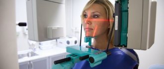
How is an ultrasound performed during pregnancy?
- Painless
- Relatively fast (20-25 minutes)
- Safe for both mother and child (ultrasound is rightfully considered the most gentle and safe diagnostic method)
- Intravaginally (for less than 12 weeks)
- Abdominal (more than 12 weeks).
Normally, fetal ultrasound is performed 3 times during the entire pregnancy (confirmation of pregnancy in the early stages using ultrasound and ultrasound before birth are not mandatory). However, if any abnormalities have been identified, the doctor may prescribe additional sessions to monitor the situation. Duplex scanning during pregnancy is prescribed for:
- More thorough diagnostics of cardiac blood flow if pathology of the development of the fetal cardiovascular system is suspected.
- Diagnosis of the condition of large blood vessels and arteries: aorta, umbilical artery, inferior genital vein, arcuate arteries of the uterus and middle cerebral artery of the fetus.
A special feature of Doppler is that monitoring of blood flow occurs in real time.
You can sign up for a fetal ultrasound at the Profmedical Examination Plus clinic by calling the numbers listed on the website.
First trimester
The first visit to the antenatal clinic takes place at 7-8 weeks
of pregnancy.
It is around this time that most expectant mothers find out that they are pregnant. An obstetrician-gynecologist will measure your weight, height, blood pressure and pelvic size. You can also consult with your doctor about taking vitamin and mineral supplements. But you will leave the antenatal clinic for a reason, but with a whole bunch of referrals for tests and consultations with specialists. In two weeks you will have to pass: - Urinalysis
.
The morning urine sample is collected on an empty stomach. Based on the results of this analysis, kidney function and the bacterial “population” of the bladder are assessed. — Vaginal smear
for microscopic examination.
It will show whether there are inflammatory processes in the genital organs, obvious and hidden infections. - General blood analysis
.
Used in the morning and on an empty stomach. It shows the composition of the blood and allows you to track its changes over time. For example, such an important factor as the level of hemoglobin in the blood. This substance is the only transporter of oxygen in the blood, and the oxygen supply of the fetus depends on its quantity. — Analysis for blood group and Rh factor
.
Even if these indicators are tattooed on your forearm, the doctor leading your pregnancy is obliged to check them. Moreover, if your Rh factor is negative, the father of the unborn child will also be tested to find out whether you will have a Rh conflict (Rh factor incompatibility). — Blood test for HIV, hepatitis B and C and syphilis
.
Even if you are completely confident in yourself and your partner, you should treat the need to get checked again as an additional guarantee that everything is fine with you. — Blood test for TORCH infections
.
These include toxoplasma, mycoplasma, cytomegalovirus and herpes virus. They can remain in a woman’s body for years and not cause her any inconvenience, but they lead to developmental defects in the unborn child. If the test is positive, the doctor selects special treatment for the woman. — Blood test for sugar
.
Pregnancy creates increased stress on all of a woman’s organs. Including the pancreas. This analysis allows you to determine how effectively it works and to foresee all the risks associated with the risk of developing diabetes during pregnancy. — Blood test for coagulation
, or coagulogram.
This test allows you to determine how blood clots - whether there is a tendency to clot or bleed. During the same two weeks, you need to visit a therapist, endocrinologist, ophthalmologist and otolaryngologist and have an electrocardiogram done. A second visit to the doctor is expected at 10 weeks
.
You take a urine test again and prepare for the fact that before each visit to the doctor until the very birth you will come with a characteristic jar. At this meeting, the doctor will look at the results of your tests and the appointments of other specialists and make a conclusion about the state of your health. At 12 weeks
you must undergo the so-called
first screening
. It consists of a blood test, which reveals abnormalities in the child’s development, and an ultrasound examination, which, based on certain parameters, shows the risk of genetic abnormalities in the baby, for example, Down syndrome.