Preparation for ultrasound of the abdominal organs
Ultrasound examination of the abdominal organs includes checking the condition of the stomach, pancreas, esophagus, liver, and intestines. Indications for its implementation are the presence of a whitish coating on the tongue, pain in the abdomen, a bitter taste in the mouth, increased formation of gases in the intestines, assumptions about the presence of appendicitis, and previous inflammatory liver disease - hepatitis A, B or C.
On the eve of the examination, exclude from the child’s diet foods that contain a large amount of fiber - cabbage, raw vegetables and fruits, brown bread, beans, peanuts, peas and other legumes, as well as soda. Failure to comply with this rule threatens increased formation of gases in the intestines of a small patient, as a result of which diagnosis will be difficult, and the doctor may have difficulty obtaining a reliable and detailed image of the internal organs.
If the child suffers from bowel problems, in particular constipation, and has not had a bowel movement for one or two days, it is recommended to perform a cleansing with an enema before the test.
An ultrasound is performed on an empty stomach, so you should not feed or drink the baby, or give him sweets, chewing gum or medications. Since this recommendation cannot always be followed, for example with a newborn, it is best to discuss it with your doctor on an individual basis.
Rules for preparing for diagnostic studies
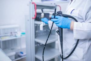
ESOPHAGOGASTRODUODENOSCOPY
Patient preparation
Preparation for the FGDS begins three days before the date of its conduct. Eliminate fatty, smoked foods, carbonated water, and foods that increase acidity levels from your diet.
- The last meal should be twelve hours before the diagnosis. At the same time, the menu should include only easily digestible foods. It could be porridge, boiled chicken meat.
- If the patient smokes, then at least three hours before the procedure he must give up cigarettes.
- Before the procedure, it is better not to take any medications except those that are absolutely necessary. This is in case you have to use some medications during FGDS.
- The preparation algorithm also involves the collection of anamnesis by the doctor. Important information for the doctor will be a message about what medications you are allergic to, what chronic and acute pathologies are present.
The patient must bring a diaper with him to the procedure. If the diagnosis was carried out earlier, then its results. The patient must put on shoe covers or replaceable shoes on his feet.
If you come to the procedure unprepared, the results will be uninformative.
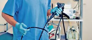
RECTOSIGMOIDOCOLONOSCOPY DIAGNOSTIC
Patient preparation
Colonoscopy is an excellent examination method that allows you to examine the entire colon. The main condition for an informative examination is proper preparation for intestinal colonoscopy. Since a visual examination is possible only if the intestines are clean, completely free from the presence of feces, preparation for the study begins 3 days in advance.
The diet is prescribed three days before the procedure, and if the patient suffers from constipation, the diet will need to be followed for 5 days. All products containing a large amount of plant fiber or contributing to the intense formation of gases in the intestines are completely excluded.
Preference is given to light dishes - fermented milk, low-fat broths. Most fruits and vegetables are excluded. It is undesirable to drink carbonated drinks and strong tea/coffee. Alcohol is excluded in any form. You can continue taking medications only after consulting your doctor; some of them are discontinued until the colonoscopy is performed.
Cleansing the intestines:
Fortrans
This medicine was specially developed to prepare patients for various types of examinations of the gastrointestinal tract. It is available in powder form, which the patient dilutes independently immediately before use. During this preparation you will need to drink a very large amount of water. The calculation is as follows: 1 liter for every 20 kg of a person’s weight.
The simplest algorithm for such preparation is to dilute the required dose of the drug in water and drink a glass of the prepared solution every hour, starting at 15:00 (the day before the study). When preparing for FCS in another way, you should drink half the dose immediately the day before, and the second 3-4 hours before the procedure. This is more difficult for patients because it is not easy to drink a large volume of liquid at once.
Duphalac
This is a mild laxative, available in syrup form. During preparation, the medicine is diluted in 2 liters of water, starting 1.5-2 hours after lunch. Within three hours you will need to drink the entire prepared solution. If you are worried about bloating, it is recommended to take Espumisan.
Lavacol
To dilute the drug, you only need a glass of warm water. The powder is dissolved immediately before use and drunk 3 hours after lunch. The medicine does not have a very pleasant taste, which can be improved by adding fruit syrup or honey to the prepared solution.
Important!
If, during the period when you are scheduled for a colonoscopy, you are taking medications containing warfarin (or other blood thinners), aspirin, diclofenac, indomethacin, ibuprofen and other anti-inflammatory drugs, as well as insulin, be sure to tell your doctor this before starting the preparation. to the procedure.
With the consent of the doctor, all of the above medications should be stopped the day before the examination.
When should you not have a colonoscopy? Contraindications for colonoscopy
Despite the fact that, in general, colonoscopy is a fairly low-traumatic and safe procedure, its implementation, in some cases, is associated with a high risk of complications and is therefore either prohibited or performed only if it is not possible to solve the problem in any other, even less traumatic way.
In particular, colonoscopy is contraindicated during pregnancy and is only allowed in cases where open bowel surgery is an alternative.
Preventive colonoscopy is also contraindicated during an exacerbation of Crohn's disease or ulcerative colitis, and during an attack of diverticulitis (in such cases, colonoscopy is postponed until the disease is in remission).
Attention!
Contact your doctor as soon as possible if you experience symptoms such as:
- Temperature above 38 C
- Stomach ache
- Severe nausea and vomiting
- Bloody discharge from the rectum
- Diarrhea with blood
- Severe weakness, dizziness, loss of consciousness.
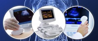
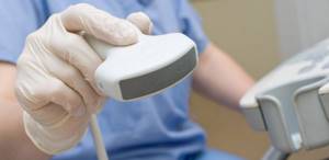
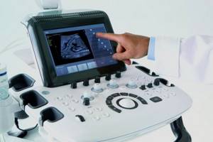
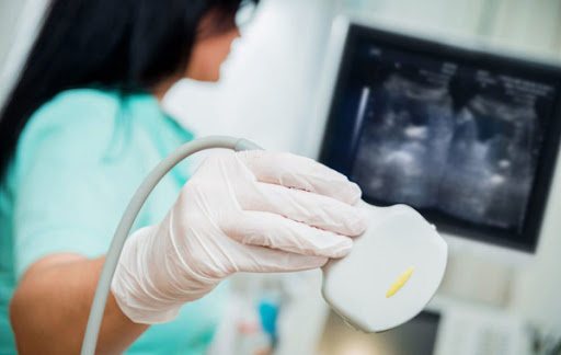
ULTRASONOGRAPHY
An abdominal ultrasound includes examination of the liver, pancreas, gallbladder and spleen.
Patient preparation
Doctors' instructions on how to prepare for an ultrasound of the liver and pancreas, stomach with spleen and gall bladder are identical:
- Follow a diet that excludes fatty and gas-forming foods for three days before the examination;
- Refuse food and water eight hours before the ultrasound;
- Take only medications approved by your doctor;
- Avoid chewing gum and smoking on the day of the test.
The diet before an abdominal ultrasound allows for foods that promote normal digestion: beef, chicken breast, white turkey meat, river fish, steamed or stewed vegetables, boiled eggs.
In preparation for an abdominal ultrasound, you can eat other dishes: soups, rice porridge with water, boiled potatoes, etc. The main thing is that the foods from your diet do not cause a feeling of heaviness and bloating, intestinal disorders and other problems.
It is recommended to maintain a healthy drinking regime - at least 1.5 liters of water per day, not counting tea and coffee. Preparation for an abdominal ultrasound examination begins 3 days before it.
What not to eat before an abdominal ultrasound
Preparation for an ultrasound of the abdominal organs includes the mandatory exclusion from the diet of foods that provoke an individual reaction - fermentation in the stomach, gases.
Gases produced by some products prevent ultrasonic waves from penetrating into the area being examined and seriously impair vision, distorting the projection of organs on the screen and in photographs.
Before an abdominal ultrasound, it is necessary to exclude the following foods:
- milk and any dairy products (soft cheese, cottage cheese, ice cream, etc.);
- carbonated drinks;
- alcohol;
- legumes;
- cabbage, radish, radish;
- pasta and flour products;
- all cereals except rice;
- fresh apples, pears, grapes;
- products containing artificial sweeteners.
Is it possible to eat before an abdominal ultrasound?
Eating before the study is allowed no later than 8 hours. If the diagnosis is scheduled for 18:00, you need to have time to eat before 10:00 am. Usually the examination is carried out in the morning, so you will be able to have breakfast only after the ultrasound.
Fish and meat are not suitable as dishes for dinner or breakfast before an ultrasound.
Many patients are interested in whether it is possible to drink coffee before an abdominal ultrasound. Alas, you will have to do without a morning cup of coffee on the day of the study. Tea, juice, and other coloring drinks are also not allowed.
Is it possible to drink before an abdominal ultrasound?
The answer to the question whether you can drink water before an abdominal ultrasound is negative. Complete refusal of food and any liquid, including tea, coffee, water, is a necessary measure. The stomach must be empty - otherwise there is a possibility of distortion of the image obtained using an ultrasound sensor.
The last intake of clean water is allowed 2 hours before the test. To obtain the most objective data on the functioning of the gallbladder and liver, it is advisable to refrain from drinking before the ultrasound for longer: for 8 hours.
Ultrasound of the kidneys and bladder
Patient preparation
Before an ultrasound scan of the kidneys and bladder, you must follow a diet. If you do not adhere to the established rules, the diagnostic results will be distorted.
Preparatory activities include two stages.
The first begins 3-4 days before the procedure, and the second - on the eve of the examination. The diet before an ultrasound involves excluding foods that can aggravate pathologies in the urinary system and increase gas formation. Patients with normal functioning of the digestive tract should remove baked goods, sweets, legumes, cabbage, fresh vegetables and fruits, fiber, fermented milk products, alcohol and carbonated drinks from their diet for 3 days. If there are problems with the functioning of the gastrointestinal tract, or stool is not daily, then you need to stick to the diet for a week. In your diet you need to minimize pickles, spicy foods, and fatty foods. The daily volume of fluid consumed should be reduced to one and a half liters.
On the eve of diagnosis, you should cleanse the intestines and take medications to eliminate bloating, if necessary. If you have daily bowel movements, there is no need to do an enema or use laxatives. It is enough to simply go to the toilet a few hours before the test. If you have problems with bowel movements, you should use symptomatic, quick-acting remedies for constipation (suppositories or enema).
This should be done not on the day of the ultrasound, but the night before in order to avoid increased peristalsis of the muscle organs and distortion of the indicators. It is recommended to take the last meal 8-10 hours before the procedure. If the digestive tract is examined along with the kidneys, then this interval should be longer. It is advisable to choose something easily digestible from food. The bladder must be full during diagnosis. To do this, just drink a few glasses of water in half an hour. If necessary, the doctor will suggest emptying your bladder, after which a repeat scan will be performed.
Recommendations for pregnant women
Before an ultrasound scan of the kidneys and bladder, expectant mothers are given the same preparation as all patients, with the exception of some points.
It is especially important for a pregnant woman to follow a diet that prevents flatulence. At the same time, you can take sorbents that remove gases from the intestines and carminatives, but you must first consult with your doctor about the safety of them. The peculiarity of preparing for an ultrasound of the kidneys and bladder during pregnancy is that the expectant mother should not use laxatives.
Even if the intestines are not functioning properly, and bowel movements are not daily, it is contraindicated to take fast-acting medications for constipation. They increase the tone of not only the digestive tract, but also the uterus, which is quite dangerous. Approved lactulose-based laxatives should also not be used, as they increase gas formation and may complicate diagnosis. Therefore, the expectant mother needs to adhere to the most gentle diet possible, eating food that does not cause flatulence.
Ultrasound of the prostate gland
To prepare for an ultrasound scan of the prostate gland in men, you need to fulfill simple requirements for 2-3 days before the examination.
Patient preparation
For the convenience of preparing for a prostate ultrasound, it is worth remembering a short reminder for the patient:
- You need to give up foods that cause gas and constipation: legumes, baked goods, bread, cabbage, apples, plums, dairy products.
- Take activated carbon, Espumisan or Filtrum for 3 days before the examination.
- If the digestive system is not functioning well, take Festal or Mezim with meals.
- You should stop eating 8 hours before the examination (it is also advisable not to overeat before this).
- Before the procedure, you need to cleanse the rectum; for this, it is recommended to take mild laxatives, microenemas or suppositories.
- You need to fill your bladder before the procedure. To do this, it is recommended to drink 1-1.5 liters of pure still water.
If you are taking any medications, you must inform your doctor. Their action may distort the test result.
To prevent feces from distorting the image, you need to cleanse the intestines:
- To do this, you can do an enema before an ultrasound of the prostate. To completely cleanse the intestines, it can be carried out 2 times: the night before and 2-3 hours before the procedure
- It is possible to use Microlax before an ultrasound of the prostate. This is a microenema with a ready-made solution, which works effectively and gives quick results.
- Glycerin suppositories are also suitable for cleansing the intestines. The suppository should be administered in the morning on the day of the procedure.
- You may take laxatives. Their use is recommended the day before the test.
You should also take care of hygiene rules. In the morning before going to the doctor, it is advisable to take a shower and thoroughly wash the perineal area. Sometimes it is recommended to shave the hair on the scrotum. This procedure is not required, but it will make the research easier.
Is it possible to eat?
Whether you can eat before a prostate ultrasound or whether the examination needs to be done on an empty stomach depends on the time for which it is scheduled. If the study is carried out early in the morning, then the last meal should be in the evening. If scanning is carried out during the day, you can eat and drink in the morning before the scan. Breakfast should be light and take place at least 6 hours in advance.
Is it possible to have sex?
You can have sex before the examination; this will not affect its accuracy or the parameters being examined.
Is it possible to play sports?
Playing sports did not affect the results of the study in any way. However, after them, more attention should be paid to personal hygiene.
What not to do before the procedure
Before a prostate ultrasound, you should not:
- drinking alcohol;
- drink strong tea and coffee;
- eat fatty or fried foods;
- eat immediately before scanning.
Ultrasound of the breast
When performing an ultrasound scan of the mammary glands, it is necessary to take into account the phase of the menstrual cycle. The menstrual cycle in women determines changes in hormonal levels and the functional state of the reproductive system and mammary glands. Therefore, most diagnostic procedures must be coordinated with certain periods.
Calculation of the optimal time for ultrasound is carried out according to schemes depending on the duration of the cycle:
- A cycle from 21 to 28 days - the study is best carried out from 5 to 12 days after menstruation.
- The duration of the cycle exceeds 28 days - the optimal time for research is from 7 to 14 days after menstruation.
- Irregular cycle - the optimal period for the study is determined by the attending physician based on the results of a blood test for estrogen levels, which is performed on the 3rd day after menstruation.
- During pregnancy, as well as during lactation, cyclic hormonal changes in the female body cease, so an ultrasound can be performed any day. The ultrasound result does not depend on the day of the examination.
- The breast ultrasound procedure during menopause and postmenopause does not depend on the day of the study
Patient preparation
To prevent unreliable results associated with functional or structural changes in the breast tissue, a woman must observe a number of restrictions on the eve of the study, usually 2-3 days before:
- Visiting places with high air temperature and humidity is excluded, these include bathhouses and saunas.
- Prolonged exposure to the sun is not recommended, as exposure to infrared and ultraviolet radiation leads to a local increase in temperature in the tissues.
- 2 days before the ultrasound, you cannot undergo physical procedures that involve exposure to certain physical factors, including heat, on the human body.
- On the eve of the study, you cannot undergo radiography, fluoroscopy, computed tomography or other diagnostic techniques that involve exposure to ionizing radiation on the body.
- The day before and on the day of the test, you should not drink alcohol, including drinks with low alcohol content.
- In the morning, it is necessary to limit excessive physical activity.
- If possible, emotional stress and stress should be eliminated.
- A woman who smokes should try not to smoke in the morning.
Before the study, a light breakfast and black tea are allowed. Drinking coffee or other stimulating drinks is not recommended.
The study can be carried out on a man against the background of the development of mastopathy, which is characterized by pathological enlargement of the mammary glands against the background of changes in hormonal levels.
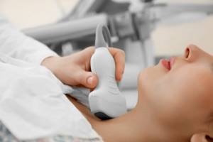
Ultrasound of the thyroid gland
Ultrasound of the thyroid gland does not require any special preparation measures. All you need is a towel that can be placed under your head. They then remove the remnants of the special conductive gel used during the examination.
If the thyroid gland has been examined previously, it is better to bring the results of such tests to the doctor. No other preparation is needed.
ELECTROCARDIOGRAPHY
Patient preparation
Patients who are prescribed an ECG must begin preparing for the examination the day before diagnosis with the following activities:
1. Complete peace of mind. On the eve of the ECG, it is necessary to completely protect yourself from stressful situations if possible. Experiences and high anxiety can distort the results of a cardiogram.
2. Full sleep. Before the study, be sure to get enough sleep. In a tired body, the heart functions in an enhanced mode.
3. Avoid alcohol. It is strictly forbidden to drink alcohol before an ECG. Alcohol causes the blood to thicken, causing the heart to work much harder.
4. Proper nutrition. You should not overload the body with too heavy dinner and breakfast before the ECG. It is advisable to exclude fatty and heavy foods from the diet. Otherwise, the body will devote all its reserves to digesting food, and the heart will be forced to work “for wear and tear.”
5. Refusal of physical activity. In order for the cardiogram to show reliable results, doctors advise not to exercise on the eve of the ECG, and to do without exercise or jogging in the morning.
6. Elimination of thermal procedures. It is not recommended to visit saunas or steam baths before taking an ECG. Doctors advise against taking a bath. Any thermal procedures can increase blood circulation and heart activity.
7. Taking medications. The day before the examination, it is necessary to completely avoid taking medications, except those that are prescribed on a regular basis. Be sure to notify your doctor about this.
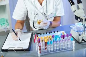
BIOCHEMICAL BLOOD STUDY
Patient preparation
- Refrain from physical activity, drinking alcohol and taking medications.
- You should not eat after dinner, you should go to bed the night before at your usual time and get up no later than an hour before taking blood.
- You cannot drink juices, tea, coffee and other drinks. Use water for drinking.
- 1-2 days before the examination, you must adhere to a diet that excludes fatty and fried foods from your diet. If there was a feast the day before, it is necessary to reschedule the laboratory test for 1-2 days.
- Before donating blood, you need to avoid temperature changes, that is, baths and saunas.
Before a hormonal blood test in women of reproductive age, you should follow the recommendations of your doctor about the day of the menstrual cycle on which you need to donate blood, since the result of the analysis is influenced by physiological factors of the phase of the menstrual cycle.
Before donating blood, you need to calm down in order to avoid an unmotivated release of hormones into the blood and an increase in their levels.
It is not recommended to donate blood after using medications, especially when administered intramuscularly or intravenously. You should not donate blood after exposure to x-rays (“X-rays”), physiotherapeutic procedures, ultrasound, or ECG. Taking into account the daily rhythms of changes in blood parameters, it is advisable to take samples for repeated studies at the same time.
If you have difficulty stopping medications, be sure to tell your doctor about it.
It is very important that you follow these recommendations exactly because... Only in this case will the correct blood test results be obtained.
Blood test for hormones (T3, T4, TSH, AT-PO)
The levels of these hormones must be checked on an empty stomach. At least 8 hours must pass between the last meal and blood collection. Juice, tea, coffee (especially with sugar) are not allowed. You can drink water. Blood is donated on any day of the menstrual cycle. Immediately before drawing blood, the patient should be at rest for about 30 minutes.
2-3 days before collecting blood for analysis, you need to prevent taking iodine-containing drugs, iodine-131, technetium-99m. Stop taking thyroid hormones for 1 month (except on special instructions from the treating endocrinologist). It is recommended to limit physical activity and psycho-emotional stress. Subsequent monitoring should preferably be carried out using the same method and in the same laboratory.
For a general blood test, markers of viral hepatitis and other infections, no special preparation is required.
General urine analysis
For general analysis, it is preferable to use “morning” urine, which is collected in the bladder during the night; this reduces the natural daily fluctuations in urine parameters and thereby more objectively characterizes the parameters under study. The volume of urine for a complete study is 100-200 ml. Urine should be collected after a thorough toilet of the external genitalia in a dry, clean container, well washed from cleaning agents and disinfectants. Failure to comply with this rule may result in an increase in the number of red and white blood cells in the urine, which will make it difficult to make a correct diagnosis. You can use a soap solution (followed by washing with boiled water), 0.02% furatsilin solution, 0.02-0.1% potassium permanganate solution. For analysis, you can collect all the urine, but it may contain elements of inflammation of the urethra, external genitalia, etc. Therefore, as a rule, the first portion of urine is not used. The second (middle!) portion of urine is collected in a clean container, without touching the body with the bottle. The container with urine is tightly closed with a lid. A urine test is performed no later than 2 hours after receiving the material.
Urine that is stored longer may be contaminated with foreign bacterial flora. In this case, the urine pH will shift to higher values due to ammonia released into the urine by bacteria. Microorganisms consume glucose, so with glycosuria you can get negative or underestimated results. Bile pigments are destroyed by daylight. Storing urine leads to the destruction of red blood cells and other formed elements in it.
Before submitting urine for analysis, it is undesirable to use medicinal substances, because some of them (in particular, ascorbic acid, which is part of most complex vitamins) affect the results of biochemical studies of urine (protein, glucose, hemoglobin).
Transportation of urine should be carried out only at a positive temperature, otherwise precipitated salts can be interpreted as a manifestation of renal pathology, or will completely complicate the research process.
To quantitatively study the sugar content in daily urine (for diabetes mellitus, etc.), it is necessary to collect daily urine - i.e. all the urine in one day. In this case, the container with urine must be stored in a cool place (optimally in the refrigerator on the bottom shelf at 4-8ºC), preventing it from freezing. If there is a large amount of daily urine, it is permissible to transport only part of it to the laboratory. Previously, the patient measures the volume of urine as accurately as possible, records it in the doctor’s direction, and then, after thoroughly mixing, pours 50-100 ml of the total volume into a clean container, after which he delivers the urine to the laboratory along with the direction. In case of diabetes mellitus, it is also possible to determine sugar in urine collected at fixed (prescribed by a doctor) periods of time.
Study of glucosuric profile
Urine is collected at certain time intervals: I portion - from 9 to 14 o'clock, II - from 14 to 19 o'clock, III - from 19 to 23 o'clock, IV - from 23 to 6 o'clock in the morning, V - from 6 o'clock in the morning to 9 o'clock in the morning. Before analysis, urine samples should be stored in the refrigerator at 4ºC.
Urinalysis according to Nechiporenko
The morning portion of urine is examined in the middle of urination (“middle” portion of urine). 15-25 ml is enough. Storage and delivery to the laboratory - (see general laboratory urine test).
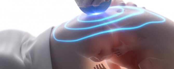
What screenings are prescribed during pregnancy?
Screening of pregnant women is a complex of examinations that can detect the presence of genetic abnormalities and developmental pathologies in the fetus. Screening consists of:
- Ultrasound;
- Blood test.
For unification and ease of interpretation of the results, uniform dates for ultrasound screening and blood donation during pregnancy were adopted. In this case, accuracy down to the day is very important, since computer programs operate in a specific time interval.
Today screening is indicated for certain categories of women:
- If the mother is over 35 years old or the father of the child is over 40.
- There are relatives in the family with genetic disorders.
- There is a history of miscarriages and frozen pregnancies.
- The woman took medications in the first trimester that were potentially dangerous to the fetus.
- A woman suffered from an infectious disease during pregnancy.
- A woman works in hazardous work.
At what stage of pregnancy is 1st ultrasound screening performed, and what is it?
The first screening during pregnancy is done between 11 and 13 weeks. Its decoding suggests the presence of such genetic developmental abnormalities as Down syndrome, Edwards syndrome, Patau syndrome, as well as preeclampsia and fetal death.
At week 12, 2 ultrasounds are performed, during which the following indicators are determined:
- KTP – coccygeal-parietal size (43-65 mm);
- BPR – biparietal size (17-24 mm);
- TVP - thickness of the collar space (1.6-1.7 mm);
- Length of the nasal bone (2-4.2 mm);
- Heart rate – heartbeat (140-160 beats/min), etc.
The next day, a blood test is done, which determines the following hormones:
- B-hCG – human chorionic gonadotropin;
- PAPP-A is plasma protein A.
It is worth noting that screening results only suggest the presence of abnormalities and require further examination.
At what week is the 2nd screening scheduled?
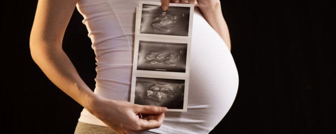
The timing of the second screening varies from 16 to 21 weeks. At 20 weeks of pregnancy, an ultrasound scan can already print out a photo in which the silhouette of the little man can already be guessed.
Ultrasound is used to determine:
- BDP - biparietal size (26–56 mm).
- DBL - length of the femur (13–38 mm).
- KDP - length of the humerus (13–36 mm).
- OG - head circumference (112–186 mm) and a number of other parameters.
A blood test shows the qualitative and quantitative content of the following hormones:
- B-hCG;
- Free estriol;
- AFP.
This examination suggests the presence of Meckel syndrome and pathologies in the development of the neural tube and intestines in the fetus.
Third ultrasound screening during pregnancy
3 Ultrasound screening during pregnancy does not require simultaneous blood donation. It aims to determine the degree of fetal development and helps the doctor decide whether to have a natural birth or whether a caesarean section is necessary.
Using ultrasound, the following parameters are determined:
- BDP - biparietal size (67–91 mm).
- DBC is the length of the femur (47–71 mm).
- KDP - length of the humerus (44–63 mm).
- OG - head circumference (238–336 mm).
The condition of the amniotic fluid, cervix and placenta is also determined. At this stage, they play a decisive role in determining the method of delivery and even the timing of the operation.
Sometimes you can hear the question, at what week is the 4th screening during pregnancy carried out? Screening 4 refers to an ultrasound scan immediately before childbirth, which is done for medical reasons.
At what stage does an ultrasound show pregnancy?
Most of all, women are interested in the question: will ultrasound determine pregnancy in the early stages? And if so, which ones exactly? Doctors at the Art Medic clinic claim that the fertilized egg is clearly visualized in the uterine cavity already 2.5-3 weeks after conception. Based on a normal 28-day cycle, this is the first week of a missed period. At this stage, it is already possible to determine the week of pregnancy and the very fact of its presence.
Which ultrasound most accurately shows the gestational age? At this stage, doctors resort to the transvaginal method of examination, as it provides better visualization. From about 8-10 weeks, all diagnostics are carried out “in the abdomen”.
Definition of ectopic pregnancy
From the point of view of doctors, it is most advisable to use ultrasound specifically to determine an ectopic pregnancy. This pathology can be detected as early as 5-6 weeks (the first week of delay). An ectopic pregnancy is characterized by the fact that the fertilized egg does not reach the uterus, but implants in the fallopian tube. If it is not detected in a timely manner, then as the fetus grows, it will rupture the tube. Without timely medical care, a woman will face death. Fortunately, modern medicine has all the means to not only save a pregnant woman, but also avoid removal of the fallopian tube, preserving the possibility of childbearing in the future.
Ectopic pregnancy is quite rare and, if detected at 5-6 weeks, can be terminated without irreparable harm to the woman’s fertility.
Definition of frozen pregnancy
A frozen pregnancy is one in which the fetus does not develop. Usually this condition ends in spontaneous miscarriage, but sometimes nature fails. The woman herself practically cannot feel that the pregnancy has stopped, especially if this is the first child.
The fetus can “freeze” at any stage, and the main criterion for this sad fact is the absence of a heartbeat. According to research, the heart of the embryo begins to beat already at the 5th obstetric week (3 weeks after conception), however, this can only be determined using ultrasound starting from the 6th week of pregnancy. Moreover, in the vast majority of cases, doctors recommend waiting until week 7, since imaging errors are possible before this period.
To monitor the situation, especially if there have already been cases of frozen pregnancy in the anamnesis, it is enough to undergo diagnostics at the 7th obstetric week, and then do scheduled screenings on time. An emergency ultrasound is performed according to the doctor’s indications in the presence of threatening symptoms.
Reviews from patients about ultrasound in medical
Our patients often leave reviews about pelvic ultrasound during pregnancy, which they underwent at the Art Medic clinic. You can view them in a special section of the medical center’s website.
Congratulations to the team on March 8th. My husband and I express our deep gratitude to all the doctors and personally to the director T.P. Kryachek. for good organization. A highly professional team has been selected here and every problem is treated with understanding. The legs carry themselves to such doctors. Happiness and prosperity!
05 March 2012
What kind of ultrasound is done during pregnancy?
Depending on the stage of pregnancy, ultrasound is performed transvaginally or transabdominally. In the first case, the sensor is inserted into the vagina, and in the second, imaging is carried out through the abdominal wall.
Usually, at any stage, doctors resort to the transabdominal technique so as not to provoke a miscarriage or premature birth. Only the first examination is performed transvaginally, during which the fact of pregnancy is actually established.
It is worth noting that both methods are absolutely safe for the fetus, and if women feel excellent, they can be used without restrictions.

From the point of view of better visualization, doctors distinguish between classic and three-dimensional ultrasound. The latter is also called 3D and even 4D. Three-dimensional images can only be obtained using special ultra-modern equipment. From a medical point of view, in the vast majority of cases there is no need to do a 3D ultrasound, however, it allows you to see the child in the smallest detail, right down to the facial expression in the later stages, which gives future parents a lot of positive emotions.
Is it possible to go through an entire pregnancy without an ultrasound?
There are often cases when a woman never visits an ultrasound diagnostic room during the entire pregnancy. Most often this is due to religious or other beliefs.
Doctors strongly recommend doing an ultrasound at least after pregnancy in order to prevent possible complications (bleeding, infection, etc.) in time. The examination is also indicated after an abortion or miscarriage. It is carried out three times - the first with an interval of 10 days, and then another after 2 months.
Can pregnant women have an ultrasound?
Medicine does not yet have data on the potential harm of ultrasound to the fetus. Moreover, this diagnostic method has been used for several decades, and there is no difference between babies born after ultrasound and babies whose mothers were not examined at all.
However, there are some nuances that should be considered before making an appointment with a doctor. At what week of pregnancy is the first ultrasound done? Usually this procedure is carried out no earlier than the 5th obstetric week, which is approximately the first week of missed menstruation. Before this period, it is almost impossible to see the fertilized egg.
At what stage of pregnancy can a repeat ultrasound be done? Any, if there are medical indications. Almost half of pregnant women undergo 10 or more sonographies over a 9-month period as prescribed by a doctor, and this does not affect the child in any way.