Indications
All women at risk should have a mammogram
- age over 39 - 40 years, according to statistics, most often it is during this period that pathological processes begin to develop;
- presence of cancer pathologies in close relatives;
- an abortion was performed and there are gynecological abnormalities;
- absence of children, late childbirth, refusal of breastfeeding;
- the onset of the menstrual cycle at an early age;
- obesity, etc.
Breast examination
must be carried out in the presence of alarming symptoms:
- the shape of the glands and nipples has changed;
- swelling and tenderness in the chest appeared;
- there are compactions, lumps, etc.;
- there were injuries in the sternum;
- disturbed by discharge from the nipples;
- lymph nodes in the armpit are enlarged;
- there are cracks, peeling on the skin and in the nipple area, the color of the integument has changed;
- in preparation for surgery.
All women are recommended to have a mammogram
, even in the absence of complaints. It is important not to miss the development of mastopathy, mastitis, cystic formations, fibroadenomas, oncopathologies, etc. A basic mammogram is necessary for subsequent examinations; subsequent results will be compared with this image. Starting from 39 to 40 years of age, a woman should be diagnosed annually.
Types of X-ray
X-ray mammography – viewing the structure of the breast using x-rays. For preventive purposes, it is performed for women after 40 years of age every 2 years, after 50 years of age - every year. Patients under 50 years of age, but at risk (primarily those whose close relatives have been diagnosed with carcinoma or sarcoma), are examined more often. Diagnostics is indicated for suspected tumors and other diseases. Information content is 90-95%. After additional tissue examination, false-positive results are detected in 25% of patients.
What does the methodology determine:
- infiltration (compaction) of different sizes and localization;
- calcifications – calcium deposits in the ducts and tissues of the gland;
- cystic formations;
- degree of patency of the milk ducts;
- detailed visualization of neoplasms.
Film X-ray mammography is a traditional diagnostic method. After Ro-rays pass through the breast tissue, an image is formed on film. This is the most accessible, cheapest, but also the least informative option, since the study is carried out on old-style devices. In addition, the radiation dose when performing classical mammography will be the highest compared to more advanced analogues.
Digital mammography is an examination performed on modern equipment using X-rays with minimal radiation exposure. The radiation dose is four times less than with traditional mammography. All information is transmitted in digital format, and not in black and white. It is possible to increase, detail, change the contrast, and clarity of the resulting image in 3D format. The doctor has the opportunity to work with images in electronic form; as a rule, there is no need for repeated examination.
Mammography with tomosynthesis is the use of X-rays to obtain a tomogram (layered sections with a three-dimensional image). The efficiency is close to 100%. Thanks to innovative automated equipment, diagnostics are painless and comfortable for the woman. Prescribed only if pathology is suspected; tomosynthesis is not performed as screening.
There are other types of mammography. They are not radiological, but are also informative.
Electrical impedance mammography is a technique based on the resistance of gland tissue to electrical impulses. The examination area is exposed to alternating current for half a minute. Healthy and altered tissues react to current differently, which determines the principle of obtaining a picture. The sensor displays the image on the monitor in real time. A complete breast examination takes 10-15 minutes. The technique is harmless for pregnant women and nursing mothers. It is recommended for young girls with symptoms of mastopathy.
MRI (magnetic resonance mammography) is a harmless way to diagnose breast diseases, based on the interaction of a magnetic field with biological fluids of the body. The examination does not use ionizing or radioactive radiation, so the method is absolutely safe. MRI is prescribed when mammography is insufficiently informative for the purpose of differential diagnosis.
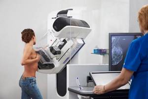
How is mammography performed?
Digital
The diagnosis is safe for health, painless and takes 5–10 minutes. The study is carried out in a separate room where a mammograph is installed. During the procedure, each breast is carefully placed on a special platform and gently pressed by scanning plates. Such fixation allows the tissues of the glands to be evenly positioned and fixed. The pressing force of the plates and the examination time depend on the density and size of the breast, and the presence of implants in it.
Pictures are taken in various projections: from bottom to top and at an angle. To obtain clear images, the woman may be asked to hold her breath for a minute. During the scanning process, the patient may feel slight movements of the device plates. There is no pain or discomfort; after the diagnosis is completed, the images are given to you.
When going for a consultation with a mammologist, it would be correct to capture data from previous examinations so that the doctor can analyze the dynamics of changes.
Advantages and disadvantages of the method
Breast X-ray is an accessible and informative method for diagnosing female diseases. Detects any changes - cysts, tumors, infiltration, inflammatory processes. The image visualizes small details, boundaries of formations, and tissue texture. It is used not only to detect pathology, but also as screening in women after 40 years.
The disadvantages of the method include radiation exposure to the body - receiving a dose of ionizing radiation. During the examination, some women experience discomfort and even pain. For girls of reproductive age, the technique is not very informative. It is difficult to diagnose women with small or excessively large breasts.
Where to do the research
High-quality diagnosis increases the chances of successful treatment. Take a study in Moscow
You can visit the Doctor Nearby CDC on Simonovsky Val.
Ultra-modern installations are installed here that allow you to regulate the radiation dose. done
for the purpose of screening control over the condition
of the mammary glands
or according to the indications of a mammologist. If you have complaints and alarming symptoms, you need to get diagnosed as soon as possible.
Digital
equipment and highly qualified doctors make it possible to detect pathological changes in the initial stages.
The advantage of the latest mammography systems is that there are no restrictions associated with breast size. Mammography
is performed on patients with implants, as well as women in a wheelchair.
Where to get an x-ray and how much does it cost?
Breast X-rays are performed both in public clinics and private diagnostic centers and hospitals.
If you need a regular mammogram, with a referral from a gynecologist, it can be done free of charge at a public medical institution (at your place of residence). Such programs are funded by the Ministry of Health with the aim of timely detection of breast cancer in women.
If it is necessary to carry out detailed diagnostics using expensive equipment, the patient is sent to a private clinic.
The cost of mammography depends on the x-ray method (digital, CT, tomosynthesis), as well as the area being examined.
Preparing for the study
Sign up for a mammogram
better taking into account the phase of the menstrual cycle, optimally in the period from 5 to 12 days.
This is due to the fact that the mammary glands
become less elastic and their structure can be seen. During menopause and menopause, a mammogram is done any day.
On the eve of diagnosis, you must stop using cosmetics, perfumes, lotions, etc. This is an important condition, since these products can distort the results of the examination. Immediately before the examination, remove clothing and jewelry that is located above the waist. If you have endoprostheses or silicone implants, you must inform your specialist about this.
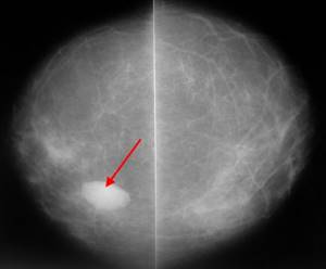
What does the radiologist see in the images?
In mastopathy with fibrous growths, heavy shadows are visible in the photo. The glandular tissue is enlarged, and isolated cysts are present.
Calcifications are detected during milk stagnation in lactating women. This is also a sign of the presence of a tumor. Therefore, an additional biopsy is performed, even if the image shows no signs of a neoplasm.
Fat involution is observed in women during menopause. On the mammogram there are no clear divisions between the structures of the gland. This is due to the replacement of glandular tissue with adipose tissue.
Signs of a benign tumor
A benign breast tumor is suspected based on the following signs:
- darkening in the image has a regular round shape;
- the boundaries of the tumor are clear;
- there is no reaction from surrounding tissues;
- in dynamics, non-infiltrative growth is observed (it moves apart healthy tissues, and does not penetrate them).
Signs of a malignant tumor
How to recognize breast cancer in mammography images:
- the shape of the tumor is irregular, asymmetrical;
- the edges are torn, the contours are blurred, the structure is heavy;
- a path stretches from the formation to the nipple;
- the presence of small calcifications in the milk ducts;
- there is no clear rim in the form of darkening around the tumor;
- increased proliferation of blood vessels;
- replacement of healthy connective tissue;
- thickening of the dermis in the area of the tumor;
- bulging or recessed nipple.
Questions and answers
Can a patient with implants have a mammogram?
The presence of implants and endoprostheses in the tissues is not a contraindication to diagnostics. Radiologist performing mammography
using special devices. The force of tissue compression by scanning plates is controlled, and there is absolutely no chance of damage or rupture of silicone implants.
On what days of the monthly cycle is it optimal to take a chest x-ray?
During the menstrual cycle, hormonal changes occur in a woman’s body, and the sensitivity and density of the glands changes. For your own comfort and to obtain excellent results, it is recommended to do
diagnostics from 5 to 12 days from the beginning of the cycle. During menopause, a mammogram is done any day.
What will a mammogram show?
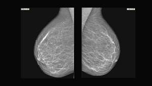
The patient receives 4 (four) photographs of the mammary glands. A radiologist is able to identify heterogeneous compactions, tumors, cysts, thickened areas, accumulations of calcifications and other pathological changes. If there are deviations from the norm, the woman is sent for an ultrasound of the mammary glands, biopsy, MRI or other additional studies prescribed by the doctor.
How is the image description protocol formulated?
The research protocol is carried out according to one of three options.
- The mammogram did not reveal any structural changes in the tissue.
- Most of the glandular tissue is without pathological changes. Minor destructive areas were identified. Recommendation: repeat x-ray in six months.
- The image revealed diffuse tissue changes and pockets of calcium accumulation (calcifications). Recommendation: biopsy to identify the pathological process.
If the image shows no signs of malignancy, it cannot be completely excluded. According to the findings of practicing oncologists, the tumor is clearly visible when it reaches a size of 1-1.5 cm.