Types and causes of thrombocytosis
Thrombocytosis is of two types:
- Primary thrombocytosis, in which the number of megakaryocytes in the bone marrow increases. This, in turn, leads to an increase in the number of platelets with a normal life expectancy, but impaired functions and abnormal structure. These platelets tend to be large, increasing the tendency for them to bleed and form clots that can block blood vessels. Bleeding occurs because platelet aggregation is disrupted and because most of them can be used to form blood clots. This condition can provoke serious complications: myocardial infarction, stroke, gastrointestinal bleeding. At the moment, the reasons why the division of megakaryocytes in the bone marrow is disrupted are unknown, but scientists have information that patients have a mutation in the V617F gene. Primary thrombocytosis is a myeloproliferative disorder. In such diseases, the hematopoietic function of the bone marrow is disrupted, which stimulates the formation of blood cells.
- Secondary (or reactive) thrombocytosis, in which platelets function normally, but the disease itself is formed due to one of the following abnormalities:
With secondary thrombocytosis, the likelihood of bleeding and clots is usually lower than with primary thrombocytosis.- Oncological diseases (most often ovarian, lung, stomach cancer);
Response to irritation of the bone marrow by certain substances that damaged tissues release during: fractures of large bones; infectious diseases (usually bacterial, less often viral, fungal and parasitic); serious and large-scale surgical operations: Removal of the spleen (splenectomy);
- Chronic or acute bleeding;
- Chronic inflammation;
- Taking certain medications: chemotherapy drugs and glucocorticosteroids;
- Recovery from thrombocytopenia (which was caused by alcohol or vitamin B12 deficiency).
Thrombocytosis
Thrombocytosis is a significant increase in the number of platelets in the blood, which disrupts the properties of the blood and increases the likelihood of thrombosis (blockage) of blood vessels. Platelets are cells that are responsible for blood clotting.
Thrombocytosis can be either an independent disease or a consequence of a number of diseases of the blood or any organs.
Primary thrombocythemia occurs most often in people over 60 years of age. The prognosis is favorable - the life expectancy of patients with primary thrombocytosis with proper observation and treatment is practically no different from that of healthy people.
Young children are more susceptible to secondary thrombocytosis. The platelet count usually returns to normal after recovery from the underlying illness.
Synonyms Russian
Essential thrombocythemia, primary thrombocythemia, secondary thrombocythemia, thrombocytophilia, chronic thrombocythemia, chronic megakaryocytic leukemia, idiopathic thrombocythemia.
English synonyms
Primary thrombocythemia, essential thrombocythemia, idiopathic thrombocythemia, primary thrombocytosis, essential thrombocytosis, secondary thrombocytosis, reactive thrombocytosis, secondary thrombocythemia.
Symptoms
Symptoms usually develop gradually and may be absent in the initial stages of the disease. The main manifestations of thrombocytosis are caused by two factors: the formation of blood clots in blood vessels and increased bleeding. With secondary thrombocythemia, the likelihood of these disorders is lower, since the number of platelets is lower than with primary thrombocythemia.
The main symptoms of thrombocytosis:
- headache,
- pain in the hands and feet, their numbness,
- weakness, irritability,
- visual impairment,
- bleeding gums,
- nosebleeds,
- blood in the stool.
General information about the disease
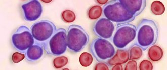
Platelets are small, colorless plates that do not contain a nucleus. They are formed in the bone marrow and are “fragments” of megakaryocytes - giant multinucleated cells. From the bone marrow, platelets enter the blood, and some of them are retained in the spleen. They exist for about 7-10 days and are then destroyed by liver and spleen cells. Platelets are responsible for blood clotting and stopping bleeding. Their normal amount in the blood is 150-450×109/l.
There are two types of thrombocytosis.
1. Primary thrombocytosis. In this case, an increased number of megakaryocytes is formed in the bone marrow, which increases the number of platelets that have a normal life expectancy, but an abnormal structure and impaired functions. Platelets are large, and there is an increased tendency to form clots that block blood vessels and cause bleeding. Bleeding occurs due to disruption of platelet adhesion, and also due to the fact that most of them can be used to form blood clots. This can lead to serious complications: stroke, myocardial infarction, gastrointestinal bleeding. The causes of impaired division of megakaryocytes in the bone marrow are not fully known, however, there is information about the presence of a mutation in the V617F gene in patients. Primary thrombocytosis refers to myeloproliferative diseases in which the hematopoietic function of the bone marrow is disrupted, which stimulates the formation of blood cells.
2. Secondary (reactive) thrombocytosis. With it, platelets function normally, and the cause of the disease itself is some other abnormality, one of the following.
- Oncological diseases, most often cancer of the stomach, lungs, ovaries. Tumor cells secrete biologically active substances that activate platelet production.
- Response to irritation of the bone marrow by substances that are released by damaged tissues during: infectious diseases, most often bacterial, less often parasitic, fungal and viral,
- fractures of large bones (femur, humerus, pelvic bones),
- extensive surgical operations.
Such thrombocytosis always does not last long and disappears when the patient’s condition normalizes.
- Splenectomy – removal of the spleen. In this case, thrombocytosis is associated with the entry into the blood of those platelets that are normally found in the spleen, as well as with a decrease in the amount of substances synthesized by the spleen and inhibiting the formation of platelets in the bone marrow.
- Acute or chronic bleeding. Acute occurs suddenly and is caused by trauma, surgery, chronic lasts a long time and can accompany a stomach or duodenal ulcer, or intestinal cancer. As a result of blood loss, iron deficiency anemia occurs, that is, a decrease in the amount of hemoglobin, red blood cells and iron included in their composition. The mechanism of development of thrombocytosis in response to iron deficiency has not been fully studied. Another factor is important in this case: during blood loss, the production of red blood cells in the bone marrow is activated. The process of more active division also involves megakaryocytes, that is, the number of platelets in the blood increases. In addition, thrombocytosis is a natural response of the body, which needs additional platelets to stop bleeding.
- Chronic inflammation (colitis - inflammation of the large intestine, vasculitis - inflammation of the walls of blood vessels, rheumatoid arthritis - inflammatory disease with joint damage), in which interleukin-6 is released - an active substance that stimulates the formation of thrombopoietin, which promotes the division of megakaryocytes and the formation of platelets.
- Taking medications: glucocorticosteroids (synthetic analogs of adrenal hormones), chemotherapy drugs (vincristine).
- Recovery from thrombocytopenia caused by vitamin B12 deficiency and alcohol. Thrombocytosis in this case occurs as a response to thrombocytopenia therapy.
The likelihood of clots and bleeding with secondary thrombocytosis is lower than with primary thrombocytosis.
Who is at risk?
- People over 60 years of age (for primary thrombocytosis).
- Children (for secondary thrombocytosis).
- Patients with iron deficiency anemia.
- Having undergone surgery, severe injuries.
- Suffering from cancer.
Diagnostics
Thrombocytosis is often asymptomatic. A doctor may suspect it during a routine checkup. An important point in diagnosis is to determine the type of thrombocytosis - primary or secondary. In the case of secondary thrombocytosis, the doctor may prescribe a number of additional studies necessary to determine its cause.
Symptoms of thrombocytosis
Typically, symptoms develop gradually. At the initial stages of the disease they may be completely absent. The main manifestations of thrombocytosis are provoked by two factors: increased bleeding or the occurrence of blood clots in the blood vessels. If thrombocythemia is secondary, then the likelihood of such disorders occurring is much lower: the number of platelets in this form of the disease is lower than in the primary one.
The main symptoms of thrombocytosis include:
- pain in the feet and hands, their numbness;
- headache;
- irritability and weakness;
- bleeding gums;
- visual impairment;
- blood in stool;
- nosebleeds.
Idiopathic thrombocytosis (essential thrombocythemia ET)
A myeloproliferative neoplasm characterized by a significant increase in the number of platelets in the blood and increased proliferation of megakaryocytes in the bone marrow. Etiology unknown.
CLINICAL PICTURE AND NATURAL COURSE
1. Symptoms:
In many cases, ET is diagnosed incidentally while examining a complete blood count for other indications. Symptoms are associated with blood clots in the microcirculation: paresthesia of the distal parts of the extremities, scotoma, erythromelalgia, headaches and dizziness. Thrombosis of large vessels (the most common complication): arterial (acute coronary syndromes, stroke, Budd-Chiari syndrome, portal vein thrombosis). Mucosal and gastrointestinal bleeding (in ≈15% of patients) caused by platelet dysfunction occurs especially in patients with platelet counts >1–1.5 million/μL, who may develop acquired von Willebrand syndrome. <10–15% of patients experience both thrombotic and hemorrhagic complications. Moderate enlargement of the spleen in 10–15% of patients. up
2. Natural course: may be asymptomatic for many years. Later complications arise: thrombosis (1-year risk 1–3%), bleeding, transformation to myelofibrosis (15-year risk 5–10%), transformation to AML or MDS (3%).
DIAGNOSTICStop
Additional research methods
1. General analysis of peripheral blood: increased number of platelets, abnormal shape and size; the number of leukocytes and Hb concentration are within normal limits or increased.
2. Aspiration biopsy and bone marrow trephine biopsy →Diagnostic criteria.
3. Molecular studies: 90% of patients have 1 of 3 driver mutations: mutation V617F of the JAK2 gene (in ≈60%), mutations in the CALR gene (20–25%), mutations in the MPL gene (in 3–4%). 10–15% of patients do not have the above-mentioned molecular changes (“triple negative”).
4. Other examinations: for differential diagnosis with reactive thrombocytosis in case of negative results of molecular studies (eg, ferritin concentration, ESR, CRP), platelet dysfunction (most often impaired aggregation under the influence of adrenaline, ADP and collagen).
Diagnostic criteria (WHO 2016)
All major criteria or the first 3 major criteria and a minor criterion must be present.
Large criteria:
1) constant platelet count ≥450,000/μl;
2) on bone marrow biopsy, proliferation of the megakaryocyte lineage with an increased number of large mature megakaryocytes with a multilayered nucleus; absence of a significant increase or shift to the left in neutrophil granulocytopoiesis and erythropoiesis; very rarely minor reticulin fibrosis (grade 1);
3) non-compliance with WHO criteria for CML, PV, PMF, MDS and other neoplasms of the hematopoietic system;
4) exclusion of polycythemia vera (PV), primary myelofibrosis (PMF), CML, MDS and other neoplastic diseases of the leukocyte system; up
5) presence of 1 of the above-mentioned driver mutations.top
Minor criterion: presence of a clonal marker or lack of evidence of reactive thrombocythemia →Differential diagnosis.top
Differential diagnosistop
1. Thrombocytosis accompanying other hematopoietic tumors: PV, CML, PMF, MDS 5q–, MDS/MPN-RS-T. up
2. Reactive thrombocythemia: for solid tumors (mainly lungs and pancreas), iron deficiency anemia, chronic inflammatory and infectious diseases, after acute blood loss, after removal of the spleen, for chronic alcoholism, in ordinary blood donors, for hemolytic anemia, for narcotic anemia .top
3. Familial thrombocytosis. up
4. Pseudothrombocytosis: cryoglobulinemia, fragmentation of red blood cells or tumor cells in the blood. up
treatmenttoptop
Choice of treatment method
depends on the presence of risk factors for thrombotic complications (age >60 years and history of thrombotic complication). In the low-risk group (no risk factors), use ASA without cytoreductive therapy, and in the high-risk group (≥1 risk factor above), also use cytoreductive treatment. It may also be considered in patients with a platelet count >1.5 million/μL (due to increased risk of bleeding), progressive myeloproliferation (eg, enlarged spleen), uncontrolled general symptoms, and microcirculatory disorders resistant to ASA treatment. up
1. Cytoreductive drugs: The first-line drug is hydroxyurea (HM), the initial dose is 15 mg/kg/day, subsequently modify the dose to stabilize the platelet count ≈450,000/μl, as well as to avoid anemia or neutropenia. It is possible to maintain platelet counts slightly higher than normal in patients receiving cytoreductive therapy, as long as leukocytosis, a major risk factor for thrombosis, is controlled.
Second-line treatment in patients who have not achieved a significant reduction in platelet count or are intolerant to hydroxyurea:
1) anagrelide, initially 1.5–2 mg/day, maintenance dose depending on the platelet count;
2) IFN-α (or Peg-IFN-α2a), initial dose of 3–5 million units/day subcutaneously 3 times/week. (Drug of choice for pregnant women or those planning pregnancy);
3) in patients with expected short survival, consider the use of busulfan.
Complete remission:
1) disappearance of objective symptoms and significant improvement in subjective symptoms, lasting ≥12 weeks;
2) remission of complete peripheral blood count values for ≥12 weeks (platelet count ≤400,000/μL, leukocyte count ≤10,000/μL, leukoerythroblastosis);
3) absence of disease progression, thrombosis and bleeding;
4) histological remission of the bone marrow.
Partial remission: This is the fulfillment of the first three criteria, without bone marrow remission.
2. Antiplatelet drugs: used in patients with microcirculation disorders, JAK2 mutation, or cardiovascular risk factors; contraindicated in patients with acquired von Willebrand syndrome.
1) ASA 50–100 mg 1 × per day; if this dose is ineffective, consider 50–100 mg 2× daily; avoid concomitant use of anagrelide and ASA due to the increased risk of bleeding complications (→section 15.20.1);
2) Second-line drugs: clopidogrel (drugs → table 2.5-9) 75 mg 1 × per day or ticlopidine 250 mg 2 × per day (drugs → section 2.27.1).
3. Patients with additional risk factors for venous and/or arterial thrombosis or a history of thrombosis → use appropriate prophylaxis → section. 2.3 and sec. 2.33.3.
4. Transformation to myelofibrosis: tactics are similar to those for PMF → section. 15.8.
MONITORING
Blood morphology examination (platelet count) every 3–4 months. required for monitoring cytoreductive therapy. Monitoring low-risk patients every 6–12 months.top
PROGNOSIS
When assessing the prognosis, the International Prognostic Score (IPSET) is used: age >60 years - 2 points, leukocyte count in peripheral blood >11,000/μl - 1 point, history of thrombotic episode - 1 point. The average life expectancy of patients without the above factors is similar to that in the general population: in patients with 1–2 points it is 25 years, in patients with 3 points it is 14 years. up