Magnetic resonance imaging
MRI of the stomach and intestines is one of the non-invasive research methods based on the use of the properties of a powerful magnetic field. Thanks to layer-by-layer scanning, obtaining images of the thinnest sections, it is possible to identify the smallest disorders of the gastrointestinal tract caused by external and internal factors. This diagnostic method is most often used as an auxiliary method in addition to ultrasound, x-ray and endoscopy.
- Prices for MRI of internal organs
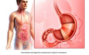
Anatomy of the gastrointestinal tract
The stomach and intestines are organs of the digestive system that provide the body with proteins, fats, carbohydrates and other nutrients. These are hollow organs with a thin wall - digestion occurs in the lumen of the gastrointestinal tract, during which food is broken down into easily digestible components. The movement of the food bolus is facilitated by constant wave-like movements of the middle wall of the stomach and intestines, consisting of smooth muscle fibers. This phenomenon is called peristalsis. Peristalsis is of great importance in magnetic resonance imaging of the stomach and intestines, as it affects the quality of the resulting images (see preparation for magnetic resonance imaging of the abdominal organs).
Advantages of the method
The advantages of MRI of the stomach over other techniques include the following scanning properties:
- no radiation exposure;
- non-invasive and painless;
- Possibility of carrying out at any age and during pregnancy with some restrictions;
- lack of long-term preparation and rehabilitation after the procedure;
- detail and clarity of images, the ability to view all tissues and organs within the scanning area;
- no side effects and a small number of contraindications to the procedure;
- speed of obtaining results in printed and electronic form.
Diseases of the gastrointestinal tract
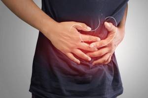
Diseases of the stomach and intestines are important for a person’s overall health and well-being. All of them lead to a decrease in quality of life. Constipation, diarrhea, abdominal pain, indigestion and other symptoms of stomach and intestinal diseases can be debilitating, leading to exhaustion and nutritional deficiencies.
Timely diagnosis of diseases of the digestive system is a prerequisite for their successful treatment. However, in reality, most patients seek medical help late, with severe violations of the function and anatomy of the stomach and intestines.
Reasons for late presentation include discomfort and pain associated with fibrogastroduodenoscopy or colonoscopy. MRI cannot completely replace FGDS, since this method is the “gold standard”, but even a single magnetic resonance imaging may be sufficient for timely diagnosis.
Indications for MRI of the abdominal cavity
MRI can be performed both for the primary diagnosis of diseases and to clarify the diagnosis identified using other, less advanced research methods. Magnetic resonance imaging is carried out if it is necessary to clarify or confirm a previously made diagnosis, if it is impossible to anticipate the pathology using ultrasound, as well as if the presence of a tumor is suspected.
Magnetic resonance imaging has also proven itself in diagnostics:
- possible hemorrhages in the abdominal cavity caused by injuries or diseases;
- stones in the gall bladder and ducts;
- accumulation of fluid in the abdominal cavity;
- damage and diseases of the liver, spleen and pancreas;
- congenital anomalies of organ development.
Magnetic resonance scanning is also used in preparation for surgery in the abdominal area.
Indications and contraindications for MRI of the stomach and intestines
MR imaging can be used in the diagnosis of the following gastrointestinal diseases:
- Complications of gastric and duodenal ulcers - perforation, penetration of ulcers, formation of fistulas and fistulas;
- Polyps of the stomach and intestines;
- Diverticula of the stomach and intestines;
- Intestinal abscesses;
- Inflammatory diseases - Crohn's disease and ulcerative colitis;
- Intestinal necrosis;
- Benign and malignant tumors of the stomach and intestines;
- Determining the stage of stomach and intestinal cancer;
- Preparation for surgical treatment, planning of surgical interventions;
- Assessing the effectiveness of the therapy;
- Anomalies in the development of the gastrointestinal tract.
Acute surgical diseases of the gastrointestinal tract, such as appendicitis or perforated gastric ulcers, are rarely diagnosed using MR imaging. Most often, computed tomography is used for this purpose, as a more accessible and faster method of diagnosing the causes of “acute abdomen”.
What symptoms may be an indication for MR imaging of the gastrointestinal tract:
- Abdominal pain of various localization and nature;
- Bloating, nausea, vomiting;
- Sudden weight loss, lack of appetite, aversion to meat foods;
- Constant constipation followed by diarrhea;
- Volumetric formations of the abdomen, which are determined visually or by touch.
MRI examination of the intestines and stomach is contraindicated:
- Persons whose bodies contain electronic devices - cochlear implants, neurostimulators, cardioverter-defibrillators, heart pacemakers, etc.;
- Persons with foreign bodies made of metals - these are fragments, particles of steel or iron stuck in the eye, medical devices (vascular stents, clips) made of stainless steel;
- Patients of early preschool age, infants;
- Obese people weighing more than 130 kg and having a body girth of more than 150 cm;
- Women in the early stages (up to 12 weeks) of pregnancy.
Features of the structure and functioning of the stomach and duodenum
Content:
- Features of the structure and functioning of the stomach and duodenum
- How does magnetic resonance imaging work and what is it used for?
- In what cases is the procedure necessary?
- When should a patient not be given an MRI?
- How to prepare for an MRI
- How does the process of magnetic resonance examination of the stomach and duodenum proceed?
- Analysis of research results
Both organs belong to the digestive tract. The stomach is anatomically defined by the fundus, body, cardia and pyloric part. Structurally, its walls consist of four layers - the serosa, the muscular layer, the submucosa and the inner mucosa. The organ performs several functions. It is here that the accumulation of absorbed food and its primary processing by gastric acid occurs. In addition, with the help of special enzymes, the stomach destroys some of the microbes and bacteria that enter the body with food. Among the secretions secreted by the stomach, there is a specific protein on which the absorption of vitamin B12 depends. The organ also produces hormones that directly affect the entire digestion process - gastrin and cholecystokinin.
The transition to the duodenum occurs after the pyloric part. The intestine looks like a hollow tube, and got its name due to the fact that its length is approximately equal to the thickness of a human finger in diameter, multiplied by twelve. The organ is represented by four parts - the upper part, descending, horizontal and ascending, and has a curved shape. It is primarily responsible for processing the bolus of food delivered from the stomach to bring it to a lower acidity level so that stomach acid does not irritate the small intestine. The intestine signals and controls the release of pancreatic enzymes and bile needed for the digestive process. In addition, the organ is also involved in the process of regulating the acidity and peptic activity of gastric secretions.
Together, these two organs represent interconnected cavities, the transition boundary between which is the sphincter in the pyloric part.
Preparing for MRI of the stomach and intestines
As stated earlier, gastric and intestinal motility can affect the quality of MRI images. Patient immobility is a prerequisite for obtaining clear and detailed images. Since the intestines and stomach are in constant motion, it can be difficult to obtain high-quality images - with intense peristalsis, they can be blurred, making diagnosis difficult.
Proper preparation for the examination helps reduce peristalsis and increase the quality of images. To do this you need:
- 2-3 days before the scheduled appointment, avoid certain types of food that increase the formation of gases in the intestines, which also contribute to increased peristalsis. These are products such as confectionery, white bread, alcoholic drinks, legumes, fresh vegetables and fruits, dairy, carbonated drinks;
- 8-12 hours before diagnosis (usually in the evening before the morning examination or in the morning if the examination is carried out in the evening), it is necessary to take an antispasmodic (no-shpu) and a carminative (espumisan, simethicone) in the dose recommended by the instructions for use of the drug. These medications relax the intestines and stomach, reduce the severity of peristalsis, and promote the elimination of gases;
- It is not recommended to smoke before the examination - nicotine increases peristalsis, do enemas, take laxatives;
- The examination itself is carried out on an empty stomach - you cannot eat 6-8 hours before the examination (you can drink water).
IMPORTANT! Magnetic fields generated during the examination can attract and heat metal objects and damage electronic devices. It is strictly prohibited to undergo examination with jewelry, piercings, watches, mobile phones, plastic cards, gadgets, etc. in your pockets or on your body. Failure to follow this rule may result in burns and injury.
Features of preparation for magnetic resonance diagnostics
The highest quality images are obtained from a properly prepared subject. Preparation begins a few days before the study.
You should adhere to a low-carbohydrate diet and consume a minimum amount of foods that promote gas formation - legumes, fruits, bread, cabbage, onions, juices, carbonated drinks. If a person is constipated, then care should be taken to cleanse the body by taking laxatives or an enema.
A light dinner is advisable the evening before the procedure, and on the day of the procedure it is better not to have breakfast if the procedure is performed in the morning. If later, a light breakfast is allowed.
The doctor may give an intravenous sedative if the patient is prone to seizures, agitated, or nervous, as this will make it difficult for the patient to remain still during an MRI of the bowel. What the monitor shows in case of improper preparation: unclear picture, distortion of images by the contents of the gastrointestinal tract. This makes assessment difficult and re-examination may be required.
Is it possible to do an MRI of the stomach and intestines at the same time?
In fact, MRI of the stomach and intestines is included in one standard examination of the abdominal organs, since the field of view of the equipment covers the entire abdomen. This is one of the advantages of magnetic resonance imaging, which takes images not of individual organs, but of the entire body as a whole. The equipment receives a series of layer-by-layer images of the human body in several planes with a step of 1-2 mm. In this way, a comprehensive examination of all organs of the abdominal cavity is performed, and not just the stomach and intestines, which cannot but affect the accuracy and efficiency of diagnosis.
How often can you undergo a magnetic resonance examination of the gastrointestinal tract?
It is not difficult to understand how often this study can be carried out, based on the distinctive feature of the phenomenon of nuclear magnetic resonance that underlies tomography. The magnetic field interacts with hydrogen atoms, which are part of the human body. The resulting energy is released into space, recorded by sensors, without causing any harmful effects on the body.
During scanning, a person is not exposed to ionizing radiation, which does not create a limit on the number of examinations. If it is necessary to monitor the dynamics of the development of the disease, track the stages of recovery after surgery or treatment, the doctor has no restrictions on the number of intestinal MRI procedures prescribed.
Do they do MRI of the stomach and intestines with contrast?
Magnetic resonance imaging can be performed with contrast. This technique allows you to increase the information content and sensitivity of the method to some gastrointestinal diseases. Contrast agents for intravenous administration can accumulate only in those tissues that are affected by an inflammatory or tumor process. The affected areas of the intestines or stomach will be highlighted in the images, making diagnosis easier.
In some cases, the use of contrast during MRI helps to clarify the preliminary diagnosis, confirming the results of the biopsy with high accuracy. The reason is that the dynamics of absorption and release of the contrast agent is directly dependent on the type of pathological process and is a constant indicator for a particular disease.
Tomography of the gastrointestinal tract with contrast
Despite the fact that tomography occupies a leading position among other diagnostic methods in its ability to clearly visualize the internal structures of the body, its capabilities can be expanded.
When performing an MRI of the intestine with contrast, you can examine small malignant tumors at the initial stage of development, assess the rate of metabolic processes in tissues, and examine blood vessels in detail.
The contrast agent enhances the signal received by the sensors from the body. For these purposes, gadolinium compounds are used for intravenous administration.
It should be remembered that MRI of the intestine with a contrast agent is not performed on pregnant women, people with allergic reactions to the components of the drug, or people with chronic renal failure.
Together with intravenous contrast, in some cases they resort to the infusion of 1-2 liters of liquid, which is poorly absorbed in the gastrointestinal tract. By filling the lumen and straightening the intestinal loops, the liquid provides a more detailed view of its walls. Additionally, medications are prescribed that slow down peristalsis (movement) in the gastrointestinal tract.
How is MRI of the gastrointestinal tract performed?
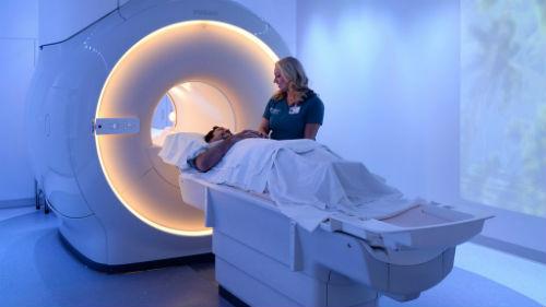
Magnetic resonance imaging of the gastrointestinal tract is an extremely simple, painless and safe procedure. All that is required of the patient is to lie still and follow the operator’s instructions exactly.
Before starting the procedure, you will be asked to put on headphones - they will protect you from the rather loud noise coming from the equipment. They also transmit operator instructions and music to help you relax and make waiting less tiring. The duration of the study is about 40 minutes (for studies with contrast - 55-60 minutes).
After completing the examination, you need to wait for the images to be decrypted. In our center it takes no more than 30 minutes. The images themselves on a CD, together with a specialist’s report, must be handed over to your attending physician. In addition, images can be obtained on film or a flash drive if you pay a little extra - CDs are not always convenient for storing research results.
Progress of the procedure
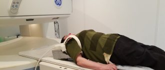
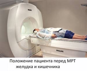
Before the procedure, the subject is given an iron-containing solution to drink (three times with an interval of half an hour). To conduct the study, open and closed type devices are used. The patient is placed on a special couch, which slowly moves into the tomograph tunnel, which has a diameter of 0.7-0.8 m.
The tunnel is provided with lighting, a supply of fresh air, and a means of communication with the doctor during the examination. To eliminate the loud noise that accompanies the operation of the device, the patient is asked to use earplugs.
The main requirement for the patient to obtain high-quality images is to remain still during the procedure, which lasts from 20 minutes. up to an hour (if contrast is necessary). A contrast agent is injected into a vein.
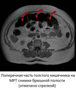
What does MRI of the stomach and intestines show?
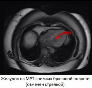
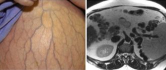
With proper preparation for the study, MRI images of the abdominal cavity show the stomach and intestines along its entire length. When decoding images, the doctor pays attention to the thickness of the wall of hollow organs, the presence of outgrowths, protrusions in the mucous membrane directed towards the lumen, nodes, pockets (diverticula) and neoplasms in the wall of the intestine and stomach. Normally, the mucous membrane should maintain a folded structure, and the large intestine retains haustration. The patency of the intestine, the presence of pathological narrowings and dilations in its lumen are examined.
When using contrast, pay attention to the local accumulation of the drug in the tissues of the mucous membrane (a sign of inflammation). In the presence of neoplasms, the dynamics of accumulation and washout of contrast are studied in more detail. Normally, contrast accumulates evenly in all tissues, which is why there are no areas of its accumulation in the images.
results
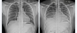
By analyzing radiofrequency pulses in a magnetic field, the tomograph makes it possible to study the stomach layer by layer and obtain high-quality images of the organ in 3 planes. The doctor examines the obtained images in detail and gives a conclusion within 1-2 hours.
When an ulcer is detected, its exact location, size, and depth (risk of perforation) are indicated. When a cancerous tumor is detected, its size, exact location, stage of development, and the presence of metastases are determined. MRI results make it possible to predict the possible development of the disease and decide on the advisability of surgical treatment in each specific case.
What is revealed during an abdominal MRI examination?
Thanks to its unique accuracy, magnetic resonance imaging allows you to see a whole range of diseases and identify abnormalities in the functioning of the internal organs of the abdominal cavity and retroperitoneal space.
Installed in a modern Philips Intera 1.5 Tesla tomograph, it is capable of diagnosing the following pathologies:
- Benign and malignant tumors of the abdominal organs.
Tomography can accurately determine the nature of the tumor. The contrast agents used for research stain benign and malignant tumors with different intensities. The malignant tumor grows into surrounding tissue and has jagged edges - these features appear on MRI scans.
- Metastases and enlarged lymph nodes.
With widespread cancer, metastases can occur in the abdominal cavity. It is quite difficult to identify them, since they can be located not only on the surface of the internal organs, but also grow into them or be localized in the soft tissue of the peritoneum (omentum and mesentery). It is extremely difficult to detect enlarged lymph nodes in this area. Magnetic resonance imaging allows you to see even the slightest malignant foci located in hard-to-reach places that cannot be visualized using other diagnostic methods.
- Liver lesions.
Diseases such as fatty hepatosis, cirrhosis, and hepatitis are primarily diagnosed using ultrasound. Using a magnetic resonance imaging scanner, you can get a more detailed picture of the damaged organ.
- Pathologies of the gallbladder and ducts.
The presence of stones is usually clearly visible using ultrasound. MRI, as a more advanced type of diagnostic study, is used when it is important to examine the lumen of the ducts, determine the size and location of the gallbladder, assess the condition of the mucous membrane lining the ducts and gallbladder, and the possible presence of cysts and polyps. For the most detailed examination of the ducts, MR cholangiography can be used.
- Condition of large vessels of the abdominal cavity.
MR images show the condition of the abdominal aorta, hepatic vein and other large vessels. The images may help identify an aneurysm or ischemia. For targeted examination of blood vessels, MR angiography using a contrast agent is usually prescribed.
- Enlarged spleen.
An increase in the size of the spleen can occur in a number of diseases. For a detailed diagnosis of the organ, magnetic resonance imaging may be prescribed.
- Kidney pathologies.
MRI displays the structure of the kidneys, changes in their structure caused by diseases such as polycystic disease and pyelonephritis.
What will a CT scan of the intestine show?
Computed tomography of the intestine is best suited to determine intestinal obstruction and identify masses and pathologies of all intestinal walls. In addition, this study helps to identify inflammatory processes and ulcerative lesions. The attending physician can give the patient a referral for a CT examination of the desired area of the intestine for differential diagnosis:
- causes of bleeding and damage to the walls of the small and large intestines;
- ulcerative processes and irritations of the mucous membrane;
- stenosis and diverticulitis;
- inflammation of tissue layers and colitis;
- volumetric formations of a benign or malignant nature;
- autoimmune diseases affecting the gastrointestinal tract, such as Crohn's disease.
Computed tomography is also prescribed to assess the condition of the digestive tract after injuries and when foreign bodies get inside as an emergency diagnostic method. MSCT data of the intestine may also be needed when planning surgical interventions, for example, intestinal resection, in order to accurately visualize the features of the anatomy and accurately determine the area of surgical intervention.
Contraindications for MRI of the abdominal cavity
Despite the modernity and safety of the MR scanning technique, the procedure is not without contraindications.
Research is strictly prohibited for people:
- having a pacemaker;
- insulin pump;
- metal structures in the body;
- weight more than 130 kg;
- in the first trimester of pregnancy.
You should approach this with caution in the following cases:
- late stages of pregnancy.
The procedure is possible, but you should first consult with your doctor;
- claustrophobia.
The patient may take sedatives shortly before the examination to avoid fear of the confined space of the tomograph;
- the presence of implants in the body.
Most modern medical structures are made of metals that are insensitive to magnetic radiation. Their presence in the body does not interfere with the procedure.
Before conducting the study, the radiologist will definitely ask you about possible contraindications.