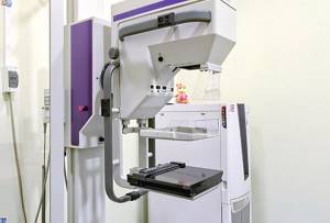
Almost every woman knows that breast health needs to be monitored especially carefully. The greatest attention should be paid to the breast in the postpartum period, at the onset of menopause, and also in case of an unfavorable prognosis for oncology. In addition to palpation and visual examination, as well as a number of tests, the doctor may prescribe you a type of examination such as mammography - this diagnosis is done using an X-ray, ultrasound or MR machine.
What is the mammary gland: anatomy, age characteristics
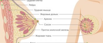
Studying the anatomy of the breast is mandatory in understanding the normal mammography image. In a general anatomical definition, it is a complex organ that consists of glandular tissue. If we look in more detail, the mammary gland is a series of cone-shaped lobes, about 15-20, which are separated from each other by connective tissue. Each lobe is internally divided into several more small lobes. That is, connective tissue is present both between the cone-shaped lobes and inside. The lobules, in turn, consist of alveoli and ducts. These are the two main components that ensure the functioning of the gland.
Alveoli are secretory units (they secrete secretions). Ducts are pathways that carry secretions.
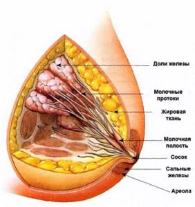
Rice. 2: Anatomical image of the mammary glands The mammary glands of young patients, before puberty, are normally high in density and contain little fat and connective tissue. Over the years, fatty tissue thickens, connective tissue stroma and glandular tissue grow, and fibrous tissue septa develop. During the childbearing period, the mammary glands undergo the greatest changes and differ in the amount of glandular tissue, which is characterized as follows:
- Well-defined or well-developed glandular tissue;
- Moderately pronounced glandular tissue - the ratio of “dense structures” that form the mammary gland and adipose tissue is approximately the same;
- Mildly defined glandular tissue – i.e. there is a predominance of adipose tissue over “dense structures”.
By the age of 35-40, the breasts undergo involutive changes, which are the norm for this age. The connective tissue becomes looser, the adipose tissue is at the stage of active growth. With the onset of menopause, fat accumulates between the lobules, adipose tissue significantly predominates over glandular tissue, and the ducts begin to experience so-called protein degeneration. With age, with the natural decline of physiological functions, involutive changes in the mammary gland develop in one of two types: fatty (more often) or fibrous. Against the background of fatty involution, diagnosing initial changes in the mammary gland is not difficult. It is more difficult to interpret changes against a dense background of fibrous involution, since the tissue has a heterogeneous structure due to alternating fibrous and fatty areas. During the premenstrual period, a woman's mammary gland swells and swells. In the postmenstrual period, the glandular tissue undergoes reverse development, it becomes denser, and the swelling regresses. During pregnancy, a woman's breasts also gain fat mass, which occurs on a hormonal background and due to large weight gain. Any physiological changes in the mammary gland associated with natural age-related and cyclical processes are necessarily reflected in X-ray images and are accepted as the norm.
Mammary cancer
What at the same time can make a woman run to the doctor or, conversely, delay this visit? Yes, you are right - this is fear of a diagnosis.
Of course, there are several types of patient behavior: some are panicky, so they run to the doctor for every little thing, others are disciplined, and this is very good, but doctors’ headaches are associated with those who are afraid and do not go. People call this “ostrich politics” - stick your head “in the sand”, and it seems like the problem is solved. But in life everything is not like that: if there is a disease, it must be treated, if not cured, it’s neglected, and you’ve wasted time.
Cancer is not a death sentence! And the number of surviving patients would have been much greater if women had come for examination on time. The absurdity of our Russian reality is that even when screenings are organized for women, they do not come for examinations, and as a result, cancer is in an advanced stage.
When talking with such patients, you hear that it hurt for a long time, or the density was palpable for a long time, but they hoped that everything would go away. It's sad because their treatment will be much more serious than if they came a year ago.
I'll show you a few photos, maybe this will make you come to the doctor!
Photo 1. Edema form of breast cancer
Photo 2. Skin ulceration in stage 3-4 breast cancer
Photo 3. Skin metastases after RME on the left
Of course, advanced stages and emerging problems are already visible during a clinical examination, and you don’t need to be a super diagnostician to understand the seriousness of the situation, but our goal is early diagnosis.
Cancer begins to grow very slowly, and sometimes it takes more than one year.
Each examination method has its own advantages, therefore, for greater reliability, it is advisable to conduct a comprehensive diagnosis, which includes a clinical examination, mammography in two projections and ultrasound.
Complex diagnostics is necessary! Let me give you a few examples.
Example No. 1.
Patient, 56 years old. Menopause 10 years. Complaints of discomfort in the right m/f. Palpation reveals a slight density of the left mammary gland. Mammography: against the background of fibro-adipose tissue, a node with uneven contours is clearly visualized.
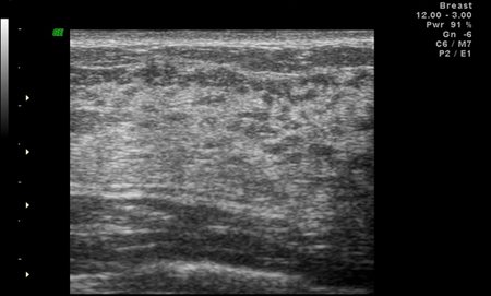
Ultrasound picture: against the background of connective tissue, an area of unpronounced restructuring with a single Doppler signal.
Diagnosis: Cancer of the left breast. Conclusion. Mammography is more informative in this case.
Example No. 2.
Patient, 35 years old. Births - 3 (last in 2007). There is no hereditary factor. Complaints of discomfort in the right m/f for 1 month. Mammography: areas of glandular tissue in the upper - outer quadrant and in the central zone against the background of fibro-adipose tissue.
Ultrasound examination: a hypoechoic area of vertical localization with a rupture of Cooper's ligaments. The contours are uneven.
Diagnosis: Cancer of the right breast. Conclusion. Ultrasound examination is more informative in this case.
Example No. 3.
Patient, 41 years old. Childbirth - 1, medical abortion - 2. There is no hereditary factor. Complaints of periodic sensitivity of the mammary glands before the cycle for 7 days. Clinical examination: the mammary gland is developed correctly, soft and painless on palpation. Mammography: dense glandular tissue; in the central zone there is an area with microcalcifications.
Ultrasound examination: glandular tissue. No data for the restructuring has been identified.
Diagnosis: Breast cancer. Conclusions. Microcalcifications are mainly visualized only on mammography. Mammography is more informative in this case. Get tested - save your life!
The mammology service at the Scientific Center for Children's Health invites you to undergo a complete examination of the mammary glands in one day. Waiting for you!
Description of mammography is normal
To begin with, it is worth understanding what anatomical elements the radiologist sees in the mammography image, which he describes in the conclusion. On radiographs, doctors distinguish:
- nipple;
- areola;
- skin;
- vessels;
- connective tissue structures;
- subcutaneous fat;
- glandular tissue.
Normally, the width of the shadow corresponding to the skin is uniform in all sectors of the gland and is 0.2 cm, increasing slightly in the area of the areola and nipple. The premammary space is represented by adipose tissue with the presence of connective tissue septa - Cooper's ligament. The width of the subcutaneous fat strip depends on the woman’s age and the condition of the glandular tissue itself: at a young age it does not exceed 2 cm, with increasing involution (age-related) changes it increases, with complete involution the fat “layer” merges with the rest of the gland mass. Shadows of veins are visible against the background of the fatty tissue of the gland. Arteries are usually visualized if their walls are calcified. Behind the subcutaneous fat layer, the so-called “body” of the mammary gland is differentiated, presented in the form of a triangle or semi-oval. This is a connective tissue glandular complex with a predominance of connective tissue elements, represented by vessels, milk ducts and lobules, which form a heterogeneous structure, expressed by shadows of various shapes, sizes and positions. At different age periods and for each stage of the menstrual cycle, their own radiological signs are also characteristic. In the premenstrual period, the connective tissue becomes more loose, therefore, the interlobular septa are clearly visualized in the photographs. The glandular part and the lumens of the glandular ducts increase. The structural pattern intensifies, the contours lose sharpness in mammography images. The mammary glands of a teenager are normally small and dense due to the poorly developed subcutaneous fat layer. The photographs show a small conical and hemispherical shadow with a small nipple. The connective tissue is poorly differentiated, glandular elements (alveoli, milk ducts) are insufficiently or almost not developed. The shadow is uniform, intense due to dense connective tissue and underdeveloped glandular tissue. The anterior contour of the shadow of the mammary gland at the border with the premammary space is smooth. Images of the mammary gland of adolescents aged 16-18 years are characterized by the same radiological signs, only the shadows of trabeculae appear additionally against the background of the shadow of the breast. After 35-40 years, fragmentation of the shadow of the glandular triangle is observed, which means tissue heterogeneity.
Adipose tissue gradually replaces functional glandular tissue, and under such conditions it is easier to detect “abnormality” than in dense breasts. Accordingly, in older women, lumps of various kinds are better visualized than in younger women, and there is less likelihood of mammography errors.
During menopause, the shadow of the glandular part is heterogeneous, the glandular lobules are unclear or not visible at all in the image. Connective tissue cords are clearly visualized. The shadows of the trabeculae are also clearer than before and are indicated in the images by sharp contours.
What diseases does mammography detect?
In addition to malignant neoplasms, mammography reveals a number of benign tumors, including:
- mastopathy;
- calcifications;
- fibroadenomas;
- cysts.
Mastopathy refers to a benign pathology of the mammary glands. The concept of “mastopathy” includes a number of fibrocystic diseases that are similar in many ways and have structural tissue disorders. Speaking in detail, experts identify more than 50 types of mastopathy, but more attention is paid to two:
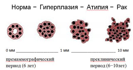
- mastopathy without atypical cells and proliferation
- mastopathy with atypical cells and proliferation.
Atypical cells (atypia) are understood as cells that differ in structure from healthy breast cells, and tissue cell division is called proliferation. The diagnosis of mastopathy is made in more than 42% of cases, but experts cannot indicate the exact reason why mastopathy occurs. But among the various reasons, the version about a violation of the hormonal status of a woman deserves the most attention. Cysts in the mammary gland are also detected using mammography. Unlike tumors, a cyst is a cavity filled with intracystic fluid, so it shows up quite well on the image. Pneumocystography may be prescribed as an additional study. Most often, this disease occurs in nulliparous women, and both group and single cysts can be detected. In addition, mammography can detect fatty cysts, which are mostly harmless, but when they become large, they can become inflamed and cause pain. Calcifications represent a concentration of calcium salts in the breast tissue. Calcifications cannot be felt by palpation, but they are easily detected by mammography. The size of calcifications may require additional examination methods. If calcifications reach large sizes, then this is not a sign of a malignant tumor, but at the same time, small calcifications indicate the activity of breast tissue cells. Fibroadenoma is a benign tumor-like formation and is formed from healthy cells. Moreover, the ability of the latter to increase in size often requires surgical intervention, and with the help of mammography it is possible to determine not only the shape and size of the formation, but also the need for surgical intervention.
You can make an appointment on the clinic website or by phone +7 495 982-10-10
, +7 495
982-10-60
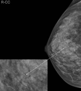
Mammography: the norm in examples (photo)
Rice. 3: Example of normal on a digital mammography image
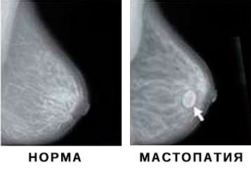
Rice. 4: Comparison of a normal image with a mastopathy image In Fig. Figure 4 clearly shows the difference between a normal mammographic picture and a pathological one. The photo on the left side shows a healthy mammary gland. The photo on the right shows a gland with signs of mastopathy. The image clearly visualizes a round formation of regular shape with smooth contours - a cyst.
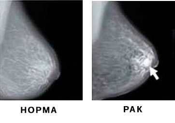
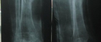
Rice.
5: Comparison of normal mammogram with cancer image In Fig. 5 you can see the differences between normal and cancer images. The image with the pathological formation is placed on the right in a direct projection. The image clearly shows an intense shadow of the formation and dilated ducts. Other articles from the section “Mammography: normal, cyst, cancer, mastopathy in pictures”