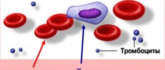
Myeloma is a malignant tumor that suppresses normal hematopoiesis, destroys bones and produces abnormal proteins that damage internal organs. When they talk about myeloma of the blood or bones, or the spine, or the bone marrow, they mean one disease with various manifestations.
Relating to hematological malignancies or oncohematological processes, that is, malignant diseases of the blood and lymphatic tissue, the disease has many names: multiple myeloma, multiple myeloma and generalized plasmacytoma, plasmacytic myeloma.
- Cause of myeloma
- What happens with myeloma?
- Diagnosis of multiple myeloma
- When the diagnosis of myeloma is beyond doubt
- Myeloma symptoms
- Treatment of multiple myeloma in young people
- Treatment of myeloma in the elderly
- Prognosis for multiple myeloma
Cause of myeloma
Myeloma consists of altered plasma cells. In normal bone marrow, plasma cells are born from B lymphocytes, but their number is limited to only 5%; a larger number is already pathological.
There is no certain clarity about the root cause of the development of a plasma tumor; poor heredity and a tendency to allergies against one’s own tissues, radiation and work with toxic substances are suspected of initiating the process; herpes virus type 8 has also come under suspicion.
True, reliable evidence of the participation of all of the above in malignant degeneration has not been presented. One thing is clear, something prevented the normal maturation of B lymphocytes or interfered with the multi-stage path from their “childhood” to lymphatic maturity, because of something the lymphocyte turned into a defective plasma cell, which gave rise to myeloma.
Multiple myeloma affects three out of 100 thousand Russians, usually elderly people - mainly in the seventh decade of life; in young people under 40 years of age, the disease is very rare.
Among those suffering from diseases of the blood and lymphatic tissue, 10-13% have plasmacytoma, but of all malignant processes existing in nature, patients with plasma cell tumors account for no more than one percent.
Causes
Science has not yet determined the exact reason for the development of the process. There are factors that increase the risk of multiple myeloma:
- Old age – 60 years and older.
- Having excess weight.
- Past exposure episode.
- Working with pesticides, insecticides and other toxic elements.
- Immunodeficiency (with HIV infection).
- History of autoimmune diseases.
- Burdened heredity.
The average life expectancy for multiple myeloma from the time of diagnosis is about 5 years.
The favorable prognosis of multiple myeloma depends on the type and stage of the process, the age of the patient, and his concomitant chronic pathology.
What happens with myeloma?
For some reason, abnormal cells appear in the bone marrow, multiplying and disrupting normal hematopoiesis, which is manifested by anemia. The lack of red blood cells affects the functioning of all organs, but especially strongly on the lung tissue and brain, which is manifested by a lack of their functions.
The function of normal plasma cells is to produce immunoglobulin antibodies to protect against pathogenic agents. Myeloma plasma cells also produce immunoglobulins, but defective paraproteins that are not capable of immune defense.
Paraproteins produced by malignant plasma cells are deposited in organ tissues; the favorite “storage site” is the kidneys, in which “light chain disease” develops, resulting in renal failure. In the affected liver, the production of blood-thinning substances decreases - blood viscosity increases, disrupting metabolic processes in tissues, and blood clots form. Deposits of immunoglobulins cause damage to other organs, but are not so fatal.
In bones, myeloma cells stimulate osteoclasts, causing osteolysis—the erosion of bone. From the destroyed bone, calcium enters the plasma, accumulating, leading to hypercalcemia - a serious condition that requires urgent measures.
Book a consultation 24 hours a day
+7+7+78
Infectious complications in multiple myeloma.
In multiple myeloma, the frequency of bacterial and viral infections increases by 7-10 times compared to population controls. Haemophilus influenzae, streptococcus pneumoniae, Escherichia coli, gram-negative bacteria and viruses (influenza and herpes zoster) are the most common culprits of infection in patients with multiple myeloma.
The increased sensitivity of patients to infectious diseases is the result of two main circumstances. Firstly, the influence of the disease itself, and secondly, old age and the side effects of the therapy. Lymphocytopenia, hypogammaglobulinemia, neutropenia due to infiltration of myeloma cells in the bone marrow and under the influence of chemotherapy cause increased sensitivity to infection. Disease-associated innate immune deficiency involves different parts of the immune system and includes B cell dysfunction as well as functional abnormalities of dendritic cells, T cells, and natural killer (NK) cells. Impaired kidney and lung function, gastrointestinal mucosa, and multiorgan disorders caused by the deposition of immunoglobulin light chains also increase the risk of infectious diseases. Finally, multiple myeloma predominantly affects older people with comorbid age-related diseases and a sedentary lifestyle, who are initially predisposed to infections.
Immunomodulators and glucocorticoids are part of the treatment for the most severe variants of the disease. In case of existing infectious contacts, the presence of neutropenia and hypogammaglobulinemia and suppressed cellular immunity, therapy with immunomodulators requires prophylactic antibiotics.
Diagnosis of multiple myeloma
The diagnosis is made by blood tests, where paraproteins are found and their total and type concentrations are determined. Paraproteins are designated as immunoglobulins - IgA, IgG and IgM. Plasmocytes produce immunoglobulins at their own discretion and in varying quantities; their changes in the production of pathological proteins subsequently assess the effectiveness of treatment and disease activity.
The degree of aggressiveness of plasma cells is determined by microscopy of the bone marrow; it is obtained from the sternum during a sternal puncture or during a biopsy of the pelvic bone. The study is especially relevant when the production of paraproteins is low or when the nature of the course of the disease changes.

A long-standing marker of the disease is Bence Jones protein in the urine, detected in 70% of patients. The protein is formed from chains of small molecular weight immunoglobulins A and G—“lungs”—that leak out of the kidney tubules. The Bence-Jones content also controls the course of the disease.
Often the disease is accidentally discovered during a routine chest x-ray based on lytic defects of the ribs. At the first stage, it is necessary to identify all destructive changes in the bones in order to further monitor the process and results of therapy, which is possible with highly sensitive low-dose CT of the entire skeleton.
MRI examines the condition of the flat bones - the skull and pelvis, which is necessary for smoldering and solitary tumors. MRI helps to evaluate not only bone defects, but also the presence of tumor infiltration of soft tissues and involvement of the spinal cord in the process.
A karyotype analysis is required to identify genetic abnormalities that affect the patient’s prognosis and the effectiveness of treatment.

When the diagnosis of myeloma is beyond doubt
The characteristic features of the cells determine the course of the process from a slow and almost benign gammopathy or smoldering myeloma to rapid plasma cell leukemia.
It is not always possible to initially classify the disease, which complicates the choice of optimal therapy. In 2014, an international consensus defined criteria that facilitate accurate diagnosis and separate one type of tumor process from others.
First of all, the percentage of plasma cells in the bone marrow is determined, so in case of symptomatic myeloma there should be more than 10%, and 60% indicates a high aggressiveness of the tumor.
For each variant of the disease, certain quantitative characteristics and combinations of criteria are provided, so to be completely sure that a patient has myeloma, it is necessary to detect specific “products”:
- M-protein in the blood, that is, IgA or IgG;
- immunoglobulin light chains;
- Bence Jones protein in urine;
- lesions in the bones of the skeleton.
If specific criteria are insufficient, diagnosis is helped by nonspecific, but often occurring effects of the activity of plasma cells and paraproteins on target organs:
- increased blood calcium levels as a result of massive bone destruction;
- decrease in hemoglobin due to tumor replacement of the bone marrow;
- increased blood creatinine, a marker of renal failure.
Anemia
Anemia is a manifestation of myeloma in approximately 75% of patients. The anemia is usually normochromic and normocytic, with signs of hypoproliferation (reticulocyte index <2.5%), with elevated ferritin levels (an indicator of inflammation). Hypochromic red blood cell counts >5% and low transferrin saturation are typical of iron deficiency. In these cases, the level of anemia is moderate. But 10% of patients with Hb<80 g/l experience a decrease in quality of life and an unfavorable prognosis. Anemia is rarely found in individuals with initial disease. The hemoglobin level determines the time of initiation of treatment for anemia in multiple myeloma. Several factors are responsible for the development of anemia. These are infiltration of the bone marrow by myeloma cells, leading to a decrease in the number of erythroid progenitor cells, erythropoietin deficiency in patients with renal failure, decreased response of proerythroblasts to kidney erythropoietin, impaired iron utilization due to high levels of hepcidin in chronic inflammation, increased plasma volume with increased levels of paraproteins, side effect of therapy. However, the main cause of anemia in myeloma disease is myeloma cell-induced apoptosis of erythroblasts.
In case of persistent symptomatic anemia and a hemoglobin level of less than 100 g/l, the possibility of other causes of anemia (Fe-deficiency, B12-deficiency, hemolytic, chronic infections, etc.) should be excluded. In the case of iron deficiency anemia, which is determined by the number of hypochromic red blood cells of 5% and a reduced level of transferrin saturation (TLS) of less than 20%, intravenous iron supplements are used.
The hemoglobin level determines the time of initiation of treatment for anemia in multiple myeloma. One of the methods for predicting the effect of erythropoiesis-stimulating agents, in particular erythropoietins, is to determine the integrity of bone marrow function. Since thrombomodulin, which stimulates thrombocytosis, is synthesized mainly by the liver, the bone marrow resource is preserved when the number of platelets in the blood is more than 150x10^9 cells/l. An initially reduced level of erythropoietin in the blood is important for predicting a positive response to recombinant erythropoietin therapy, which makes it possible to avoid red blood cell transfusions. Frequent side effects of the use of erythropoietins are thromboembolic complications and arterial hypertension.
Myeloma symptoms
It has been noted that each pool of plasma cells produces immunoglobulins with personal characteristics and according to its own schedule, which is why the clinical manifestations are very unique and deeply individual. No two patients are alike, and it is even more impossible to find two similar patients based on diagnostic criteria. However, there are several types of the disease. According to the number of lesions, the tumor can be generalized or multiple and solitary - with a single focus.
According to the course, a distinction is made between sluggish or smoldering, also known as indolet, and symptomatic plasmacytoma, which occurs with obvious clinical manifestations.

The main manifestation of symptomatic myeloma is pain in the bones due to their destruction, which does not appear immediately and often not even in the first year of the disease. Pain syndrome occurs when the periosteum, penetrated by nerve endings, is involved in the tumor process. With a slow process, several years may pass before the tumor is identified, since the patient experiences nothing but episodes of weakness.
In the advanced stage with multiple lesions, fractures in places of bone destruction and manifestations of renal failure, or amyloidosis of organs, come to the fore in different combinations and with individual intensity.
Treatment of multiple myeloma in young people
The indolent variant of myeloma does not always require treatment, since it is not life-threatening, and the therapy is not at all harmless. In this case, monitoring the process is more beneficial to the patient than toxic chemotherapy. Regular examinations allow timely diagnosis of the activation of the process.
Symptomatic myeloma is divided into stages from I to III according to the level of specific microglobulin and albumin in the blood; the strategy for stages I and II-III differs only in the drugs used and their combinations.
At any stage, the main thing that determines the tactics is the patient’s condition and his age. Thus, healthy patients up to 65 years of age and without severe chronic diseases are offered aggressive high-dose chemotherapy with transplantation of their own blood stem cells, scientifically called autologous transplantation.
Physically intact patients from 65 to 70 years of age can also qualify for high-dose chemotherapy, but not with a combination of drugs, but with a single drug - melphalan.
Before high-dose chemotherapy begins, several courses of polychemotherapy are carried out at normal doses, then a special drug is used to stimulate the bone marrow to produce its own stem cells, which are collected and preserved. Then the patient receives very high doses of cytostatics, as a result of which all blood cells - tumor and normal - die. Normal, pre-preserved blood elements are administered to the patient.
| More information about treatment at Euroonco: | |
| Oncologist-hematologist | 10500 rub. |
| Chemotherapy appointment | 6900 rub. |
| Emergency oncology care | from 11000 rub. |
| Palliative care in Moscow | from 40200 per day |
Treatment of myeloma in the elderly
Patients over 65 years of age and younger, but with concomitant diseases that affect their general condition and activity, also undergo cyclic chemotherapy at the first stage, including the use of targeted drugs. The result of treatment is assessed by blood and bone marrow tests, where the concentration of disease-specific proteins and the percentage of tumor cells are determined. The result of treatment is affected not only by age, but also by the presence of several chronic diseases, asthenia, which implies physical weakening with or without weight loss.
Our ancestors called an asthenized person “kvely”. Such patients run the risk of not being able to tolerate aggressive treatment, but respond quite well to milder options for antitumor chemotherapy.
In recent years, the range of chemotherapy drugs has expanded significantly to include targeted agents that have demonstrated good immediate results and increased life expectancy in study participants.
Skeletal lesions are subject to long-term therapy with bisphosphonates, which reduce pain, prevent fractures and hypercalcemia. Individual tumor foci are exposed to ionizing radiation; radiation therapy is required if there is a threat of compression of the spinal cord and damage to the cervical spine.
Thrombophilia
The risk of venous thrombosis is due to a number of reasons, and myeloma significantly increases it. Risk factors for thrombosis include advanced age, limited mobility due to pain, frequent infections, dehydration, renal failure, obesity, diabetes mellitus and other comorbid diseases.
Among the manifestations, the most dangerous is pulmonary embolism, which can be fatal.
The incidence of thromboembolism in myeloma is estimated at 5-8/100 patients.
This is due to the fact that myeloma is accompanied by increased blood viscosity, inhibition of the production of natural anticoagulants and hypercoagulation of the blood caused by infections, with increased levels of von Willebrand factor, fibrinogen and factor VIII, decreased levels of protein S, and so on. A course of drug therapy, including the prescription of erythropoietins, can also play a role as a trigger for venous thromboembolism. Therefore, in the first months of therapy, it is recommended to supplement traditional myeloma therapy with aspirin or anticoagulant therapy.
Screening for predisposition to thrombosis and venous thromboembolism in myeloma, along with a standard coagulation examination, should include a blood viscosity test.
Prognosis for multiple myeloma
In addition to the patient’s age and physical condition, the prognosis of myeloma and life expectancy is reflected in the sensitivity of the tumor to drug treatment and the biological characteristics of plasma cells, in particular genetic abnormalities with deletion of chromosomal sections and amplification - gene duplication.
The concentration of paraproteins and their fractions, the volume of the lesion at the time of detection of the disease and the degree of involvement of other organs in the pathological process play a role, so that already developed renal failure will “outweigh” all other favorable signs of the disease.
It is very important for the patient’s life to choose the right doctor and clinic, where they can conduct an accurate examination and the patient is treated by a whole team of doctors of different specialties who know the clinical problems of an elderly myeloma patient and know how to solve them.
Book a consultation 24 hours a day
+7+7+78
Bibliography:
- Davydov M.I., Aksel E.M./ Statistics of malignant neoplasms in Russia and the CIS countries 2007 // Bulletin of the Russian Cancer Research Center named after. N.N. Blokhina RAMS, 2009; 20 (3)
- Kyle RA, Rajkumar SV./ Criteria for diagnosis, staging, risk stratification and response assessment of multiple myeloma// Leukemia. 2009; 23(1)
- Durie BGM, Salmon SE. / A clinical staging system for multiple myeloma: Correlation of measure Myeloma cell mass with presenting clinical features, response to treatment, and survival// Cancer, 1975;36.
- Facon T, Mary JY, Hulin C et al./ Melphalan and prednisone plus thalidomide versus melphalan and prednisone or reduced intensity autologous stem cell transplantation in elderly patients with multiple myeloma (IFM 99-06): a randomized trial// Lancet 2007; 370
- Weber DM, Chen C, Nievisky R et al./ Lenalidomide plus dexamethasone for relapsed multiple myeloma in North America.// N Engl J Med 2007; 357.


