Rhinoscopy is a diagnostic instrumental technique that is used to study the condition of the nasal cavity. Can be anterior, middle or posterior. When performing anterior rhinoscopy, a nasal speculum is used, a middle one (for examining the middle nasal passage and the upper parts of the nasal cavity) is a special mirror with elongated branches, and a posterior rhinoscopy is a nasopharyngeal speculum. The procedure is performed in the process of diagnosing chronic rhinitis, polyposis and other diseases of the nasal cavity. No special preparation required. Middle and posterior rhinoscopy can be performed under local anesthesia.
Rhinoscopy is widely used in otolaryngology, it allows you to examine the nasal passages, turbinates, nasal septum, and nasopharynx. It is carried out using bicuspid nasal speculums, nasal dilators, nasopharyngeal speculums and a frontal reflector. Depending on the objectives of the study, the patient may be indicated for anterior, middle or posterior rhinoscopy (rhinopharyngoscopy). The data from classical rhinoscopy is supplemented by the results of fabrorinoscopy, microrhinoscopy, radiography of the paranasal sinuses, ultrasound, MRI, paranasal sinuses, as well as diagnostic puncture, examination of a smear from the nasopharynx or a biopsy of the nasal mucosa.
Anterior rhinoscopy
Anterior rhinoscopy is also called direct or external. This examination involves the use of a nasal dilator for examination. The patient sits opposite the doctor, who fixes the patient's head with his right hand and inserts a closed nasal dilator into the nostril with his left. In this case, the depth of insertion of the viewing dilator depends on the area of mucosa being examined and the age of the patient. In young children, an ear specula may be used instead. After insertion, the dilator is carefully opened.
For direct rhinoscopy, the patient's head must be in one of two positions. The first option is to examine the nasal cavity with the head in an upright position. In this position, the lower part of the nasal cavity, the lower nasal passage and the lower third of the septum are accessible for examination. The second option involves tilting the patient's head back. In this position, the middle nasal passage and the anterior cells of the ethmoidal labyrinth are accessible to inspection.

The middle nasal passage is examined most carefully, since the natural openings of the nose (maxillary, frontal) open into it.
During a rhinoscopic examination, the condition of the mucous membrane is assessed (wet, dry, atrophic, edematous, pale, hyperemic, cyanotic, with spots, hemorrhages), the size of the nasal turbinates, septum, properties and amount of discharge are characterized.
In some cases, with direct rhinoscopy it is possible to examine the posterior wall of the nasopharynx and the lymphoid tissue on it can be diagnosed with adenoiditis. In some cases, during the examination the patient is asked to utter some sounds (words) or tilt his head to the right or left, which improves the visual examination.
Normally, direct rhinoscopy should not cause pain. If the patient is in pain, for example after a nasal injury, the mucous membrane is irrigated with a local anesthetic before the examination.
The normal rhinoscopic picture should look like this:
- mucous pink;
- the partition is smooth;
- nasal passages are free;
- shells are not enlarged.
In addition to examining the nasal cavity, the mucous membrane is palpated with a button probe and its density, elasticity, as well as the shape, consistency, localization, and mobility of pathological formations are assessed. In this way, foreign bodies can be detected and, in most cases, removed.
Anemization facilitates improved examination of the nasal passages and differential diagnosis of hypertrophic and other forms of rhinitis. Anemization is the treatment of the nasal mucosa for several minutes with strong vasoconstrictors (ephedrine with adrenaline). After vasoconstriction, much more surface of the mucous membrane and structures of the nose are available for inspection. With hypertrophic rhinitis after anemization, the expansion of the nasal passages does not occur due to pathologically thickened mucosa, which distinguishes it from other forms of rhinitis.
In many cases, anterior rhinoscopy can be performed without additional nasal dilators. To examine, simply lift the tip of your nose and illuminate the nasal cavity with a reflector or other light source.
Average rhinoscopy

Using average rhinoscopy, the middle nasal passage, the upper two thirds of the nasal septum, the nasal openings of the maxillary (maxillary) and frontal sinuses, the semilunar cleft and, in some cases, the posterior wall of the nasopharynx are examined. For inspection, a nasal dilator with long jaws is used, which can be used to move the middle concha towards the septum, exposing the middle meatus for inspection.
After inserting the nasal dilator with closed jaws, they are carefully opened. During the examination, the following is assessed:
- color and condition of the mucous membrane;
- patency of the nasal passages;
- curvatures and defects of the septum;
- presence and characteristics of pathological formations;
- quality and quantity of discharge.
Since the procedure is unpleasant and can cause pain, the nasal mucosa is pre-treated with local anesthetics, and in case of severe swelling of the mucosa, with vasoconstrictors.
Posterior rhinoscopy
This procedure is carried out using a nasopharyngeal speculum, which is inserted deep into the oropharynx, behind the uvula. The tongue is pressed down with a spatula so that it does not interfere with the inspection. The patient should breathe through his nose if possible.
The light from the reflector is directed onto a mirror and the formations in the nasopharynx are examined. To prevent the patient from developing a gag reflex, the doctor must be careful during the examination and avoid touching the root of the tongue and the back wall of the pharynx with a spatula or mirror. If the gag reflex is pronounced, the patient should treat the back of the throat with a local anesthetic spray before the procedure.
Posterior (retrograde, indirect) rhinoscopy allows you to examine the choanae, pharyngeal openings of the auditory tubes, the posterior parts of the three nasal conchae, the nasal passages, the vomer (the posterior part of the nasal septum), the posterior wall of the nasopharynx and the soft palate.
Types of procedure
There are several types of rhinoscopy:
- anterior, in which the bottom of the nasal cavity is examined, the first two-thirds of the nasal septum, the anterior parts of the nasal turbinates are examined;
- middle - allowing you to get an idea of the state of the middle nasal concha, olfactory fissure and the central part of the nasal passage;
- posterior, during which the otolaryngologist clearly sees the posterior parts of all nasal passages, the nasopharynx and the entire nasal septum.
Also, such a study can be traditional (carried out using instruments) and endoscopic (carried out using an endoscope).
Indications for use

Knowing and taking into account which cavities and structures of the nose and nasopharynx can be examined by rhinoscopy, the indications for its implementation are:
- prolonged nasal congestion or dryness of unknown origin;
- purulent or profuse watery discharge from the nose or drainage into the throat;
- unpleasant smell in the nose;
- nosebleeds;
- suspicion of adenoid growths, polyps, neoplasms or foreign bodies;
- disturbances of smell;
- pain in the paranasal sinuses;
- deviated nasal septum;
- injuries to the nose and facial skull;
- anomalies of the development of the facial skull.
Rhinoscopic examination is carried out to make a diagnosis, dynamic monitoring of the effectiveness of the treatment, before surgical interventions on the ENT organs.
Anterior rhinoscopy has no contraindications. Middle and posterior rhinoscopic examination is not performed on newborns, children under one year old and children of primary preschool age. In case of severe pain in older children and adults, anesthesia is performed before the procedure or it is replaced by endoscopic examination or other diagnostic methods.
Why CELT?
Endoscopic rhinoscopy is a simple procedure, but requires the skill of a specialist. Our multidisciplinary clinic employs highly qualified otolaryngologists. They have extensive successful experience in conducting this diagnostic procedure in adults and children. They have modern endoscopic equipment and effective painkillers at their disposal.
How is rhinoscopy performed in children?
It is worth noting that in general the process of endoscopic rhinoscopy in children does not differ from the technique used for adult patients. What is important is how well the doctor knows how to establish contact with the baby and explain to him how to behave during the procedure. During the examination, an endoscope of a smaller diameter is used. For young children, examination with an endoscope in our clinic is performed under general anesthesia.
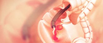
Endoscopic rhinoscopy
Rhinoendoscopy is a minimally invasive diagnostic and treatment study that allows you to examine the nasal cavity and carry out minor manipulations on intranasal structures that are difficult to reach with conventional rhinoscopy.
This study is carried out using a rhinoendoscope (flexible or rigid), and a magnified image of the area being examined is displayed on the monitor screen. Modern rhinoendoscopes allow photo and video recording of the examination, which is of particular value for assessing the dynamics of treatment.
Indications for rhinoendoscopy are:
- recurrent sinusitis (sinusitis, sinusitis, ethmoiditis, sphenoiditis);
- polyps, cysts in the sinuses;
- deviated nasal septum;
- disorders of nasal breathing and sense of smell;
- inflammatory diseases of the nose and nasopharynx;
- recurrent nosebleeds;
- nasal injuries;
- pain in the nose and paranasal sinuses;
- diagnosis of tumors.
An endoscopic examination is carried out after local anesthesia by irrigating the mucosa with local anesthetic sprays. It lasts no longer than half an hour and does not require any preliminary preparation.
Indications
Rhinoscopy is performed for sinusitis (sinusitis, frontal sinusitis, ethmoiditis, sphenoiditis), rhinitis of various origins, hay fever, suspected polyps, synechiae or adenoids, recurrent nosebleeds. The procedure is performed for injuries of the nose, deformations of the external nose or facial skull, curvature of the nasal septum, and foreign bodies in the nose. The technique can be recommended for headaches of unknown etiology, disorders of the olfactory function, naphthyzine addiction and other pathologies. In addition, a study is prescribed after rhinoplasty to evaluate the results of the operation.
Surgical rhinoscopy

If some areas of the nasal cavity are not accessible even for a rhinoendoscope tube, surgical rhinoscopy is performed. Surgical rhinoscopy is a special case of an endoscopic procedure. Examination of the nasal cavity using an endoscope is preceded by excision of a difficult-to-reach pathological area of the mucous membrane. After inserting the endoscope, minor operations in the nasal cavity are possible. Surgical rhinoscopy is used for:
- removal of polyps;
- restoration of patency of the outlet openings of the paranasal sinuses;
- removal of fungal masses in case of fungal infection of the sinuses;
- restoration of the correct anatomical structure of the nasal structures;
- removal of foreign bodies from the nasal passages and sinuses;
- treatment of cysts, paranasal sinus bullae;
- scraping the hyperplastic mucous membrane of the nose and sinuses.
In addition to therapeutic purposes, surgical rhinoendoscopy is used for diagnostic purposes - to diagnose neoplasms by biopsy.
Unlike a diagnostic endoscopic procedure, surgical rhinoscopy is performed under general anesthesia, since the operation requires complete immobilization of the patient.
Otolaryngologist services at the Federal Scientific and Clinical Center of the Federal Medical and Biological Agency
It is difficult to get an appointment with a specialized specialist in district clinics - it takes a lot of time and effort. At the Federal Scientific and Clinical Center of the Federal Medical and Biological Agency, paid ENT services can be obtained quickly, without queues, at a time convenient for the patient.
The advantage of the center is also its broad scientific base, which allows you to quickly introduce new medical technologies into practice, highly qualified doctors, modern equipment for diagnosis and treatment, versatility (the ability to quickly receive advice and assistance from a specialist in a related discipline).
At the initial appointment, the ENT doctor conducts:
- patient interview;
- palpation, visual, instrumental examination (rhinoscopy, otoscopy);
- issues a conclusion with a preliminary diagnosis;
- chooses examination and treatment tactics;
- gives therapeutic or preventive recommendations.
To clarify the diagnosis, the range of services is expanding and includes:
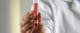
- laboratory tests (clinical, immunological, biochemical);
- X-ray;
- endoscopic examination of the nasopharynx, throat, trachea (insertion into the organs of a thin tube with a camera at the end that reproduces the image on the monitor);
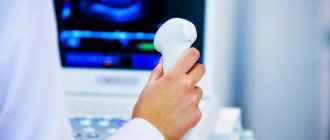
- Ultrasound;
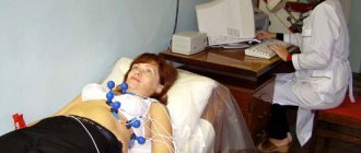
- functional tests to assess the state of hearing (audiometry), smell, coordination;
- MRI, CT.
Medical procedures and minor operations performed by an ENT doctor include:
- washing cavities with special solutions;
- sinus punctures;
- blowing out sulfur plugs;
- collection of biopsy material;
- removal of foreign bodies;
- opening of abscesses;
- tracheotomy;
- endoscopic examination.
Repeated consultations with a doctor are needed to monitor the dynamics of therapy.
To speed up the recovery process, treatment is often supplemented with a physiotherapeutic set of procedures. If a pathology is identified that requires extensive surgery, the patient is offered hospitalization in a hospital center equipped with high-precision equipment for interventions of any level of complexity, including microsurgical.
The final price of a specialist’s services depends on the additional procedures required for the diagnosis and treatment of diseases of the ENT organs.
Features of examination of the nasal cavity in children
Rhinoscopy in children under one year of age and children of primary preschool age has its own characteristics. Children at this age categorically do not accept such manipulations, so the procedure must be carried out as quickly and painlessly as possible. Most often, when examining the nasal cavity in young children, nasal dilators are not used, and if necessary, ear specula are used, since they have a small diameter. When using dilators, it is advisable to pre-treat the nasal mucosa with a local anesthetic spray.
If there is no need for a nasal dilator, the doctor lifts the tip of the child’s nose with his finger and examines the accessible areas of the nasal cavity: the lower nasal passage, the inferior concha. To prevent the child from resisting, the parents or the doctor’s assistant sit him on their laps and fix his arms and head.
Posterior rhinoscopy in young children is recommended to be performed by palpation of the nasopharynx, however, if the child is not properly fixed, there is a risk of injury to the doctor himself (a bite). In difficult cases, rhinoscopy is performed for children under anesthesia, combining examination of the nasal cavity with the collection of biomaterial or surgical manipulations.
Process
The nose examination follows a standard procedure. There are two types of rhinoscopy:
- anterior (straight);
- posterior (indirect).
The difference between these methods is access to the area under study - through the nasal cavity or pharynx. There is also a middle rhinoscopy, but most doctors classify it as anterior. The set of tools depends on the age of the person and the type of research. Other medical devices are used for pediatric rhinoscopy.
The price of the examination will depend on its complexity and the devices used. Procedures using an endoscope have a higher cost.
Anterior rhinoscopy
This is the easiest and fastest diagnostic method, for which the main instrument is used - a rhinoscope. It looks like a tube with handles, capable of expansion.
Children under 2 years of age are examined using ear specula. Two health workers are involved in the examination process: the nurse holds the child, and the doctor examines.
Technique:
- The patient sits on a chair, the doctor places his hand on the crown of the patient to move the head in specified directions.
- If swelling is present, the nasal mucosa is treated with vasoconstrictor drugs.
- The branches of the nasal dilator are inserted into the nostrils 2 cm deep and carefully pulled apart.
- The doctor evaluates the mucous membrane, the anterior and partly the middle compartment of the nasal turbinates.
- For a deeper examination, the doctor may use anesthetics and a device with longer jaws. The instrument is advanced under the middle turbinate or between it and the septum. This type of examination is called intermediate rhinoscopy.
If boils are found in the nose, the procedure is stopped.
Posterior rhinoscopy
This method allows you to examine the nasopharyngeal space, the posterior edges of the nasal concha and septum, and the mouths of the auditory tubes. Tools used:
- otolaryngological spatula;
- nasopharyngeal speculum.
The last device is a small mirror on a long handle.
Technique:
- The patient sits on a chair.
- The doctor presses the tongue with a spatula without touching its root.
- The nasopharynx is treated with an anesthetic. It will relieve pain and reduce the gag reflex.
- The mirror is placed against the wall of the pharynx behind the soft palate. To prevent the device from fogging up, it is preheated.
- During manipulation, you need to breathe through your nose, opening your mouth wide.
Posterior rhinoscopy in children has its own characteristics. Children do not tolerate such manipulations well and may bite doctors. In difficult cases, when examination of the child is necessary, general anesthesia must be given.
Endoscopy
Modern medicine uses an alternative option for examining the nasal cavity - endoscopy. The doctor has the opportunity to see the condition of the area being examined on a color monitor. Also important is the device’s ability to record what the camera sees on the screen.
The procedure involves the use of an endoscopic device - a flexible tube (probe) equipped with optics. It is not associated with problems with the patient's gag reflex.
Endoscopes are equipped with a camera, as well as surgical devices for performing operations, electrocoagulation and taking biopsy material.
Progress of the procedure:
- The patient takes a semi-sitting position in a special chair.
- Local anesthesia is applied to the area being examined.
- The endoscope is inserted into the vestibule of the nose, the common nasal passage.
- The doctor then slowly moves the device towards the nasopharynx. The condition of the inferior nasal concha and the mouths of the auditory tube are examined. An endoscope allows you to determine the proliferation of adenoid tissue.
- The device is moved towards the middle turbinate. The doctor has the opportunity to see the outlet of the sphenoid sinus.
- Next, the upper nasal passage and the olfactory fissure are examined. Sometimes it is possible to recognize the efferent openings of the cells of the posterior compartment of the ethmoid bone.