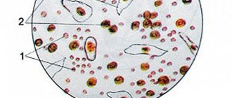
Down Syndrome is a genetic congenital disorder considered a form of mental retardation. Children with Down Syndrome develop slowly, have poor memory and are visually different from their peers. Some couples decide to give birth to such a baby and raise him with pleasure. Others are shocked by the diagnosis and decide to have an abortion. To understand what to do if an ultrasound reveals Down Syndrome in the fetus, let's look at the causes and consequences of the pathology.
What is Down Syndrome
The content of the article
Pathology implies a congenital, genetically determined, and therefore incurable, delay in physical and mental development. In such children, bone growth is disrupted, which leads to changes in body structure.
Children with diabetes have a characteristic appearance:
- small round head;
- slanted, narrowed, swollen eyes, ears and nose are smaller than normal, lips are thickened, tongue is elongated;
- short stature, short fingers;
- the skin is swollen, the hair is thinning;
- genitals are underdeveloped.
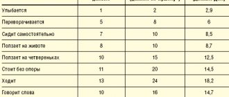
The mental development of a person with diabetes, even in adulthood, remains at the level of a preschooler - this is the main reason why many couples choose abortion.
It should be noted that people with this diagnosis are very kind, affectionate, love animals and diligently do various simple things. For their openness and good-naturedness they are called “sunny”. Many of the patients can take care of themselves - get dressed, clean up after themselves. Some people can count and read at a first-grader level. But they will not be able to survive without a guardian.
Important facts about Down syndrome
- The English doctor John Down was the first to describe this disease, so there is nothing offensive in the name of the pathology - the disease is named after the great scientist.
- The frequency of the genetic abnormality does not depend on gender and is detected with equal frequency in both girls and boys.
- Most scientists believe that diabetes is not a hereditary pathology; one family can have both healthy and sick children. But doctors advise to pay attention to cases if sick children have already been born in the family, and to be more thoroughly examined in the early stages of pregnancy.
- Down syndrome is detected before 12 weeks of pregnancy by screening ultrasound. Therefore, by undergoing a timely examination with a good ultrasound machine, it is possible to detect pathology even during the period when abortion is permitted.
- According to statistics, up to 10% of newborn babies suffer from significant disorders of brain activity, including mental retardation, in various forms.
- Many parents, having given birth to “downies,” love them very much and are glad that they once refused to have an abortion. Some families, on the contrary, get tired of difficulties and break up. The men leave, tired of the endless care of the baby, with whom they have to deal a lot and patiently.
Causes of Down syndrome development
The true cause of the development of Down syndrome was determined quite recently - in sick people, not 46, but 47 chromosomes are detected. An extra chromosome in the karyotype is the result of disturbances during the maturation and division of germ cells.
The sex cell divides, giving each of the new cells 23 chromosomes. If at this moment one of the chromosome pairs could not separate (the phenomenon of trisomy), an extra chromosome remains. Most often, this problem occurs in marriages of older age - in women over 35 years of age, the risk of having a baby with Down syndrome is increased.
Risk groups requiring mandatory screening for Down Syndrome:
- The presence in the family of a child with genetic diseases or developmental defects;
- The presence of hereditary genetic and chromosomal diseases in one of the spouses or any close relatives;
- The woman’s age is more than 35 years and/or the man’s age is more than 40 years;
- Unfavorable living or professional conditions;
- A history of spontaneous abortions (miscarriages, recurrent miscarriages) or missed abortions;
- Consanguineous marriage;
- Taking medications in the early stages of pregnancy, incl. medications for medical abortion, antibiotics,
Second trimester
The second screening is carried out in the second trimester, at 16 - 18 weeks, it also consists of several procedures - determination of the level of hCG, a-fetoprotein, free estriol, inhibin A, ultrasound, as well as genetic studies - transabdominal aspiration and cordocentnesis. All these procedures are harmful for both the baby and the pregnant woman.
HCG level
They donate blood again to check the level of hCG - chronic gonadotropin. The results, as in the first trimester, depend not only on chromosomal defects, but also on many other reasons. Doctors use some deviations in the level of a substance in the blood to refer a pregnant woman for further research.
α-fetoprotein level
The level of alpha-fetoprotein (AFP) is determined in the blood. This is a protein compound that is formed in the child’s stomach and liver. This substance affects a woman’s immunity and helps protect the child from rejection by the mother’s body. The level of this substance increases in the blood throughout pregnancy, reaching a maximum before childbirth. If a-fetoprotein is below normal, in isolated cases this may indicate a risk of a genetic failure, but other reasons are also possible:
- pathology of embryo development, the so-called hydatidiform mole;
- possible fetal death;
- possible miscarriage;
- false pregnancy or incorrect gestational age;
- other reasons - you should not worry too much about the tests, this is only one of the diagnostic methods, even bad tests on paper can be situational and not indicate any problem, and the possibility of an error cannot be ruled out.
In many cases, a low level of this protein may indicate an incorrect timing of pregnancy.

An increased AFP may indicate various reasons:
- An analysis error cannot be ruled out; blood sampling is done according to special rules that are easy to break;
- a viral infection in a child that can damage his liver;
- with a rather rare disease - spina bifida, deviations from the norm in tests can also indicate problems in the development of the child’s neural tube, which is also rare;
- if two or more children develop in the mother’s tummy;
- if a purulent-septic complication develops, which happens if the anterior wall of the abdominal cavity does not heal;
- problems with the child's urinary tract.
The peculiarity of this analysis is that the level of a-protein can be higher or lower than normal even in healthy babies and in the absence of problems in the mother.
Free estriol
Free estriol testing is also done to determine the likelihood of Down syndrome. But, as is the case with other tests, the lack of this female sex hormone may indicate various processes occurring in the mother’s body, and may also simply indicate the peculiarities of the course of pregnancy.
Possible problems with low levels of free estriol in the blood serum:
- the threat of premature birth, such tests may need to be kept in a hospital setting;
- post-maturity, in our case in the second trimester this reason is irrelevant, but if the timing is late and post-maturity actually occurs, you need to think about inducing labor if the woman is tormented by constant contractions;
- an infectious process that can take place in the mother's womb;
- aencephaly in a child (complete or partial absence of the cerebral hemispheres);
- fetoplacental insufficiency (a set of symptoms that may be a consequence of disorders of the placenta, and may also be a consequence of various obstetric characteristics);
- adrenal hypoplasia in a child.
There is no need to worry about the tests again - estriol can decrease when taking antibiotics, glucocorticoids, or with liver problems, even when the baby in the tummy is large, or there are twins or triplets. The analysis must be taken under certain nutritional conditions, and even if all requirements are met, it is not a fact that the analysis will be accurate.

Inhibin A
In the second trimester, screening involves measuring the level of inhibin A. This is a protein hormone that is practically absent in the male body; in women, its synthesis occurs in the follicles during pregnancy, as well as in the tissues of the child and in the placenta. The concentration of the hormone reaches its maximum by week 10, then its level decreases, reaching a minimum by week 17, then its amount increases slightly.
The analysis is taken from a vein under special conditions - you cannot eat heavy food, or overexert yourself physically. A reduced level of the hormone may indicate a risk of miscarriage if it is done in the first trimester, when the concentration of the hormone is quite high, as well as a possible hydatidiform mole (a disease of the embryo when the chorionic villi degenerate into vesicles with liquid, while the epithelium of the villi grows; this disease occurs approximately once case per 1000 births). The analysis can also show a predisposition to preeclampsia - this is late toxicosis, which occurs at the end of the second trimester - at the beginning of the third trimester, this toxicosis is accompanied by high blood pressure and edema.

In Down syndrome, Edwards syndrome, and other genetic diseases, the level of inhibin A in the blood is higher than normal. But still, if the tests are bad, there is no need to worry, since it is influenced by various factors that are not related to genetic diseases. Doctors may recommend various invasive studies that involve interfering with the body of the mother or child; it is better to ignore these recommendations, since they can cause irreparable harm to the health of the mother and child. And the test results, even if they are negative, should be the basis for improving the mother’s life - improved nutrition, greater comfort, more frequent walks, perhaps needing to go to the hospital if there is a threat of miscarriage. Bad tests are a signal to loved ones to show more concern for the pregnant mother and not think about terminating the pregnancy.
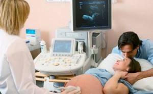
Ultrasound
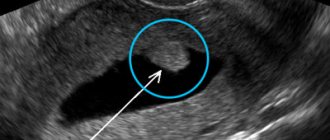
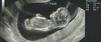
In the second trimester, ultrasound is performed as planned. The child is quite small, and ultrasound diagnostic technology, even new, does not give a complete picture of what the child looks like in detail. And it’s not just the “noise” that reduces the image clarity by half. The fact is that each person has many individual characteristics, so diagnosing Down syndrome by the appearance of a child, and even one in the womb, is fundamentally wrong. This method of determining a genetic defect or confirming with ultrasound the results of other examinations undertaken as part of screening for Down syndrome has a low degree of reliability, so you should not get too carried away with ultrasound diagnostics.

An ultrasound should be viewed primarily as an opportunity to get to know your baby better while eagerly awaiting his birth. Some people wonder whether it is possible to detect Down syndrome using an ultrasound method and at what time it can be done. What arguments do some doctors give to determine trisomy using ultrasound:
- The child is smaller than normal. There may be an error in determining the term, as well as weight and body length - a very individual feature that depends on the child’s nutrition and heredity. Determining the health of a child by its intrauterine size is at least not serious. The baby can be smaller than the norm, larger than the norm, and also correspond to the norm. It’s not about the size, but about the established norm; it shows average figures, without adjustments for individual characteristics. It is very difficult to recognize any disease, not just trisomy, in this way.
- The nasal bone is shortened or absent - this is an inconspicuous detail on ultrasound. The child's bones are weak, especially on the head. This is the body's preparation for childbirth. Ossification actively takes place after birth. This structural feature of the head bones certainly cannot be considered significant in the prenatal diagnosis of Down syndrome.
- The upper jaw is smaller than normal - this feature can be genetically determined in a completely ordinary child, not to mention the fact that it is incorrect to determine body size relative to the norm in the second trimester, because at 16 weeks, the beginning of the second trimester, the child measures about 15 centimeters and weighs - about 130 grams.
- Shortened bones of the shoulders and hips - the baby is in a fetal position, in which it is difficult to measure the size of the bones of the upper and lower extremities, and these features are individual for all people.
- When diagnosing Down syndrome, they can measure the size of the bladder, which is very small, and count the arteries near the umbilical cord; these arteries are difficult to see.
- Little or no water in the mother's tummy. Oligohydramnios occurs in 3 percent of pregnancies and has many causes, with Down syndrome occurring in about 0.1 percent. Not drinking enough water can pose a threat to the child’s health, and is not a sign of a genetic disease.
- Rapid heartbeat in a child can have various causes and most often is not associated with trisomy, as claimed by proponents of prenatal screening.
Chorionic villus biopsy
Geneticists, to whom doctors refer in all doubtful cases, may suggest doing a chorionic villus biopsy (transcervical chorionic biopsy, chorionic analysis), or transabdominal aspiration of placental villous tissue. This is an extremely harmful procedure that has the following complications:
- Bloody discharge occurs in a third of women who have undergone this painful procedure, which is useless and also humiliating. Bleeding in every twentieth woman can develop into a hematoma, which can soon resolve. Excessive bleeding, which is hazardous to health, is also possible.
- It is possible to get an infection that causes chorioamnionitis, inflammation of the amniotic membranes and amniotic fluid. The body of women and the baby in the tummy are quite well protected from bacteria. But senseless intervention in the abdomen can lead to rupture of the membranes and the loss of the child, even if a caesarean section is performed (the period is very short, it is difficult for such a small child to survive).
- The development of chorioamnionitis in early pregnancy often leads to rupture of the membranes and premature birth. After childbirth (both by cesarean section and natural), chorioamnionitis can develop into endometritis, that is, inflammation of the inner mucous membrane of the uterus. Even if the child was healthy and everything was in order, problems can begin just with such tests, and this harm could have been avoided by refusing to perform a biopsy.
- Violation of the integrity of the membranes.
- Increased levels of afetprotein in the blood.
- The development of alloimmune cytopenia in a child (destruction of blood cells) requires drug intervention after chorionic villus biopsy.
- Every thirtieth woman has a miscarriage after chorionic villus biopsy - this is a lot, they forget to talk about it (often they don’t want to talk about the dangerous statistics of deaths of children after this dangerous procedure), proposing to perform a dangerous invasive intervention in the body of a pregnant mother.

Even if in each case the probability of complications occurring is insignificant (and often the complications are very dangerous), the total risk is very strong, often fatal. Also, a woman can expect a large number of side effects. Danger also awaits a woman who agrees to undergo another procedure as part of the screening - transabdominal cordocentesis with puncture of the umbilical cord vessels; this procedure can also cause serious harm. Those who take such studies lightly, calling them “Down tests during pregnancy,” “Down tests during pregnancy,” may not take into account all the risks and make erroneous actions that can lead to serious problems. You just need to keep in mind that Down syndrome can be determined with 100% certainty only after the birth of the child.
A woman's happiness lies in motherhood. There are no special or non-special children, all children are different, different from others and are the most precious to their mother. The child needs to be accepted as he is - very talented or with genetic differences (people with Down syndrome can also be very talented). A baby is a piece of mom and dad; to destroy a life is to destroy both yourself and society. After all, a child is the weakest creature that cannot protect itself. How can you cruelly punish criminals who commit atrocities against people who can stand up for themselves, and at the same time remain silent about the murder of children in the wombs of mothers who are so defenseless that they cannot even cry. Give life and love to your child and do not doubt his eternal love for you. There is no need to do any tests, screenings, analyzes whose purpose is abortion; such studies are dangerous in themselves and can have negative consequences for many years. Moreover, the risk of having a child with Down syndrome is very small.
How is diabetes detected - effective diagnosis during pregnancy
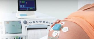
Currently, all pregnant women, regardless of whether they have hereditary diseases, must undergo fetal screening for ultrasound and biochemical diagnostics. The World Health Organization (WHO) recommends at least one screening procedure during the second trimester of pregnancy.
Down syndrome during pregnancy can be determined by several methods:
- Fetal screening ultrasound. The doctor measures the size of the fetal collar zone, determining the presence of subcutaneous fluid, fetal growth and other parameters characteristic of various gene-chromosomal pathologies.
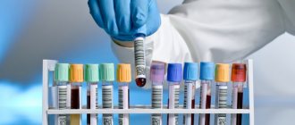
- Non-invasive antenatal diagnosis - blood test using biochemical markers. Blood is taken from a vein and treated with special substances.
- Amniocentesis is the study of amniotic fluid. The technique requires intervention in the body, therefore it is classified as an invasive procedure. The liquid is taken with a thin needle through a microscopic puncture in the abdomen and sent for examination.
If you suspect diabetes, you need to undergo at least two types of diagnostics in order to be 100% sure of the presence of pathology and only after that make a decision about abortion.
Ultrasound results are analyzed together with biochemical screening data, allowing a more accurate assessment of the likelihood of genetic abnormalities in the fetus. If, according to biochemical and ultrasound screening, the risks of having a particular genetic disorder are sufficiently high, an invasive genetic analysis procedure is prescribed, which gives results with an accuracy close to 100%. Based on the results of the invasive analysis, a decision is made to terminate the pregnancy.
First trimester
In the first trimester, mothers who were offered a combined screening for signs of Down syndrome in the fetus undergo blood tests - they measure the level of chronic gonatropin, protein A, and use ultrasound to measure the thickness of a special parameter - the child’s nuchal translucency. In case of doubt (there are often errors in tests), the geneticist suggests doing various tests that involve gross mechanical intervention in the woman’s body, and this is dangerous for the health of the mother and baby.
HCG level measurement
Blood is taken from a vein, in which the level of the beta subunit of the pregnancy hormone human chronic gonadotropin (hCG) is determined. It is assumed that chromosomal abnormalities in a child are possible with elevated levels of chronic gonadotropin. But Down syndrome is very strongly associated with this indicator, although other reasons for the increased content of this hormone are possible:
- the gestational age is set incorrectly, and accordingly the normal concentration of the hormone is calculated incorrectly;
- complications of pregnancy in the form of gestosis, that is, late toxicosis (every woman can have such a complication and it is not a fact that it will harm);
- the mother has diabetes mellitus;
- various anomalies in intrauterine development, among which may be Down syndrome (or may not be, mind you);
- pathology in the child (this is one of the possible signs, although the child may actually be healthy);
- taking various drugs, including gestagens of synthetic origin, as well as hCG drugs, they are used to maintain pregnancy when there is a threat of miscarriage;
- post-maturity (in the first trimester this is hardly relevant).
This analysis is taken several times, since analysis errors are possible, including during blood sampling. Despite the results, there is no need to directly link an elevated level of hCG with Down syndrome; this method does not accurately diagnose the disease, but speaks about possible features of the child’s development and the course of pregnancy.

Protein A level
The level of PAPP-A (plasma protein A) is determined in the blood; if the content of this protein is low, doctors talk about possible Down syndrome. But the level of this substance can be low for many reasons, including possible analysis error. But mothers who have been diagnosed with low plasma protein A begin to visit doctors during the day, and in the evening they may cry, worrying about the child, this can lead to severe stress and negative consequences for two people connected by the umbilical cord.
The mother is sent for an ultrasound, to a geneticist and other specialists who may suggest that the mother undergo amniocentesis - the collection of amniotic fluid for genetic analysis using a long needle inserted through the abdomen. This procedure is very dangerous and can harm the baby and cause miscarriage. The main problem is that low levels of PAPP-A occur in many mothers whose baby is born completely healthy. But however, such an analysis may not be independent, but only prepare the mother for the second screening, which is carried out in the second trimester - the mother might not have done the second screening if there were no negative results of the first screening. So a negative test result may not necessarily be reliable, but doctors consider it a basis for further research - and this means additional worries, financial costs and danger to the health of the mother and child.
This is the prenatal medicine industry - one doctor gives a negative test, then many other specialists get hired, although all this could have been avoided if no screening was done at all. Moreover, even after drawing blood, they often suggest piercing the stomach with a needle for further research, and this already jeopardizes the health and life of the mother and child. It is possible that plasma protein A levels are indeed low with trisomy 21, but this genetic disease is generally rare, and poor tests can occur under various circumstances not related to hereditary diseases. But such analyzes lead to a woman’s hysterical state, in which she is easy to manipulate; parents are ready at this stage to part with a significant amount to continue research, although these studies are generally useless.
Collar thickness
In the first trimester, the thickness of the child’s collar space is determined. The measurements refer to the distance between the inner surface of the child's skin and the soft tissues that cover the spine in the cervical region. Deviations of this parameter from the norm occur when a child has Down syndrome, however, based on the measurements, they talk about a possible “risk group”, and not about an exact diagnosis. Other possible reasons for deviations in the size of the collar space:
- Turner, Edwards, Pattau syndromes
- heart defects, problems with lymph circulation, connective tissues, bone structures with a normal chromosome set
- complications that the mother's body experiences during pregnancy - anemia, also called taxoplasmosis, cytomegalovirus, parvovirus, although the latter viruses are unlikely to affect anything, their presence in tests, in principle, does not mean anything
- a completely normal child may also have deviations from the norm, which also needs to be taken into account when measuring the thickness of the collar space.

A measurement error is quite possible, since the norm ranges from 0.7 to 3 millimeters, and ultrasound has very loud noise, even modern and expensive devices, so it is very difficult to accurately determine this parameter, since during ultrasound everything is difficult to see, including the child may take a position in which the collar space cannot be accurately determined - the head may be pressed to the chest, or turned to the side.
A woman whose child has some wrong parameter according to ultrasound comes under the radar of doctors - she needs to repeat the diagnosis every month. The woman is also referred to a geneticist, who begins to persuade the mother to undergo completely harmful procedures that can harm the baby or cause premature birth - among such harmful procedures are amniocentosis, in which water is taken from the fetus, chorionic biopsy and placental biopsy, in which placental cells are taken for analysis, cordocentesis - analysis of blood from the umbilical cord and villi of a human embryo. If such studies, according to statistics, lead to miscarriage in one case out of three hundred, then damage to the child with subsequent pregnancy complications occurs even more often.
Particularly dangerous is chorionic villus sampling, which is taken through the cervix. With such a “research” the child may die. With transcervical amnioscopy, which geneticists also suggest doing, complications are possible for both the child and the mother - rupture of the membranes with premature rupture of amniotic fluid, bleeding due to mechanical damage to the cervical canal.
Even if we take into account three parameters - hCG level, protein A, nuchal translucency, the likelihood of identifying Down syndrome is very low, even according to official data. But in fact, the likelihood of determining trisomy 21 using these methods is much lower, because no one examines aborted children for the presence of Down syndrome. And a woman who finds herself in a difficult life situation can easily be pushed to rash actions, even such serious ones as killing a child. But screening in the first trimester is not everything; doctors suggest doing many tests in the second trimester. Many expectant mothers must go through all the negative medical research, and this is to determine an incurable disease (there is no point in knowing about an intrauterine incurable disease), the probability of which is 1 in 1000, but this can be avoided with adequate information. You need to take care of the health, well-being and future upbringing of the child, and not succumb to the persistent proposals of financially interested people.
Determination of markers in the blood of a pregnant woman
Biochemical screening involves a mother's blood test, which identifies certain substances that appear in the presence of genetic abnormalities in the fetus. Their proportion constantly changes depending on the duration of pregnancy.
Biochemical screening is carried out in two stages:
- In the first trimester of pregnancy - for the content of the free β-subunit of human chorionic gonadotropin (hCG) and plasma protein A associated with pregnancy (PPP-A);
- In the second trimester - for the content of hCG or the free β-subunit of hCG, free estriol, inhibin A and alpha-fetoprotein (AFP).
For each of the substances listed above, there are certain blood levels depending on the stage of pregnancy. Deviations in the number of markers from certain norms do not 100% confirm a gene-chromosomal disease, but only indicate a high risk of genetic defects and anomalies. Therefore, the study is supplemented by the results of fetal ultrasound. And if doubts remain, additional examination using invasive methods will be required.
In addition to blood test data, biochemical screening also takes into account a number of general data about the pregnant woman (age plays a particularly important role) and ultrasound readings done at the same period. One way or another, the result is only the probability of the existence of a genetic disease in the unborn child. Reliable information can only be obtained as a result of invasive procedures, that is, amniocentesis, cordocentesis or chorionic villus biopsy.
Composition of the analysis
As mentioned above, to establish a diagnosis, a triple screening test is performed, which includes:
- Ultrasound examination of the fetus.
- Blood tests, namely hormones.
If the results are questionable, amniocentesis, cordocentesis or villus biopsy is performed.
The first two indicators are mandatory types of research. For this reason, gynecologists strongly recommend registering for pregnancy before the 12th week of gestation.
The main screening is carried out no later than 13 weeks. If non-invasive manipulation is necessary, no later than 20 weeks, ideally from 16 to 18 weeks.
Ultrasound
Ultrasound diagnostics is one of the proven and “old” ways to detect fetal anomalies. Ultrasound is performed in combination with biochemical parameters.
Subsequently, the data is entered into a special program, where a blood test is performed and the risk or percentage of the pathological phenomenon is established.
The method is informative, but may be unreliable (approximately 20% of false positive results).
During an ultrasound examination, the following indicators are determined:
- Fruit size. With a chromosomal abnormality, the size of the fetus is significantly below the permissible norm.
- Rapid heartbeat, sometimes the cardiac system has serious developmental defects.
- Deterioration of the bony base of the nose and enlarged collar zone. These two indicators are fundamental, since all “sunny children” have an expanded collar area with signs of swelling, and an absent or too small nose.
Ultrasound examination reveals fetal hypotonia and possible pathology of the renal system. There is often echogenicity in the intestinal buds.
In severe situations, the fetus may not be viable and die in the first trimester.
Blood biochemistry
The blood collects for 3 types of hormone: hCG, AFP and free estriol:
- free b-hCG is a hormone produced by the chorion. It grows rapidly in the presence of chromosomal pathology, however, the concentration of the hormone is not taken into account separately, but is compared with other indicators.
- AFP is a specific protein that decreases in Down syndrome. Alpha-fetoprotein is synthesized in the liver and is a fundamental test for the presence of fetal abnormalities.
- Free estriol is the third screening test that is reduced to extremely low results. Estriol can drop sharply, in which case we are talking not so much about a possible genetic pathology, but rather about fetal abnormalities that are incompatible with life.
- As an additional test, PAPP-P, a protein produced by the placenta, is monitored.
Blood must be donated in the first half of the day on an empty stomach, and an ultrasound scan is performed either on the day of blood donation or the next morning.
To obtain reliable results, you should not neglect the simple rules for collecting venous blood.
Amniocentesis, cordocentesis and chorionic villus sampling are invasive genetic screening methods
If diabetes is not detected before 15 weeks (the woman missed an ultrasound or neglected tests), but there is a suspicion of pathology, the gynecologist refers the pregnant woman to unpleasant invasive procedures.
- Amniocentesis is a method of genetic screening of the fetus, performed starting from the 15th week of pregnancy. The doctor takes amniotic fluid from the uterus, monitoring the process with an ultrasound. The amniotic fluid contains fetal cells, so after amniocentesis it is possible to directly study the chromosome set of the unborn child. The procedure is practically painless, but its use slightly increases the risk of premature birth and the development of intrauterine infection. In addition, the process itself is very unpleasant for the woman, as well as waiting for the test results.
- Chorionic villus biopsy. Doctors obtain cells from the fetal amniotic sac (chorion), which are genetically identical to the cells of the embryo. The risks of infection and premature birth remain.
- Cordocentesis. The doctor takes umbilical cord blood, which also contains fetal cells.
The resulting cellular material is cultivated in the laboratory under special conditions for approximately 2 weeks and then subjected to full genetic analysis. Tests may vary depending on the geneticist's orders.
There is a more urgent diagnosis of fetal cells - fluorescence hybridization (FISH), the result of such an analysis is usually ready within 2-3 days. However, the capabilities of the method are limited, and the number of deviations determined by this method differs from the complete list of hereditary diseases. The FISH method usually determines abnormalities in the number of chromosomes from 13, 15, 17, 18, 21, 22 pairs of chromosomes, as well as among the sex chromosomes X and Y.
The accuracy of invasive genetic screening methods is 97-99%.
HOW TO DO NIPT
| Call us for a free consultation. We will help you correctly calculate your gestational age. | Do an ultrasound and sign up for Analysis in your city More about the analysis | Donate regular venous blood. (Without the use of dangerous invasive procedures!) | Make a payment and wait for the result | Your result will be ready in 14 days and notified to you in any convenient way. |
Is it necessary to terminate a pregnancy?
Let's start with the fact that a woman decides on her own whether or not to have an abortion with Down Syndrome. Those. No one will force her to terminate her pregnancy due to fetal pathology. But the gynecologist and geneticist will definitely warn about the consequences - the disease is incurable and will progress.
Some women do not want to have an abortion, planning to place their sick child in an orphanage. If previously it was enough to sign a waiver for the baby, now the law obliges unscrupulous mothers to pay child support. Therefore, having given birth to a child with diabetes and abandoning him, you will not be able to live happily and carefree.
BENEFITS OF NIPT
| EARLY Analysis can be performed from the 9th week of pregnancy* | SAFELY The mother's normal blood is used. The risk of fetal infection is excluded. | EXACTLY High Accuracy 99%. More reliable than the first screening. | AVAILABLE The analysis can be taken in any city in Russia and Kazakhstan | NIPT No. 1 IN THE WORLD Officially introduced into the social system. medicine in many countries of the world. | RELIABLE Recommended for all pregnant women |
When is an abortion performed if the fetus has diabetes?
If a woman decides to have an abortion, it is prescribed urgently in order to carry it out in a gentle manner. The longer the period, the more possible negative consequences for the psyche and health can be expected.
Normal timing for abortion:
- Up to 6 weeks - medical abortion. This is done by taking pills that cause miscarriage;
- Up to 7 weeks - vacuum abortion. It is done using special equipment that does not affect the walls of the uterus;
- Up to 12 weeks - surgical abortion. The fetus is removed using a surgical instrument. The walls of the uterus are scraped out.
If the timing is missed, a late abortion is performed - the permissible period is up to 22 weeks of gestation.
If a woman decides to continue the pregnancy, all subsequent examinations are carried out according to a standard plan. Childbirth is also no different from normal.
If you find an error, please select a piece of text and press Ctrl+Enter