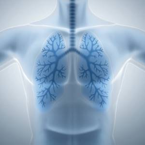
Smoking has a negative impact on human health. The lungs initially suffer from harmful addictions.
The smoke that smokers inhale settles on the walls of the organ as a toxic nicotine film and is difficult to remove from the body.
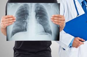
Each cigarette contains many carcinogens, which cause various mutations and cancer. It is advisable to check the lungs of any person annually using fluorography.
Find out what angiography is, to whom, when and why it is prescribed -
Features of the procedure
In order to answer your questions, you must first understand what fluorography is and what can be diagnosed with it.
A fluorogram is a photograph of organs, which is obtained through the use of X-rays. Fabrics differ in their permeability to radiation. This makes it possible to see in the image organs of different densities and any changes that have occurred in their structure. A fluorogram helps show:
- foreign bodies and neoplasms;
- tissue sclerosis and fibrosis;
- abscesses;
- various pathologies;
- tuberculous cavities and others.
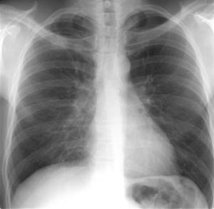
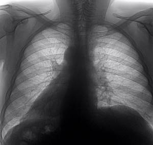
The lungs of a healthy person appear light gray in the photo. The organs have clear outlines, and the silhouette of the ribs is reflected against their background. Therefore, if smokers take a fluorographic photograph of the lungs, then in the absence of pathologies, the fluorography will not be able to show anything. But in an experienced smoker, you will notice changes that can lead to a conclusion about the presence of an addiction.
conclusions
You have received the answer to the question: “is it possible to smoke before fluorography?” I guess, yes. But after the picture shows the presence of serious pathologies, maybe we really need to reconsider our attitude towards cigarettes?
DO YOU WANT TO QUIT SMOKING?
Then you need a strategy for quitting cigarettes. With its help it will be much easier to quit.
Modern medicine has many diagnostic methods. Some procedures are preceded by careful preparation, and some are performed on the day of treatment. Fluorography is the simplest examination with a minimum set of rules that should be followed. Many people are interested in whether it is possible to smoke before a planned fluorography? Let's look at the risks of smoking on the day of a chest X-ray scan, how this will affect the results, and what experts think about this.
What are the differences?
A lung photo of a person who has been smoking for a long time looks denser than a photo of a healthy organ. On the pulmonary background, you can notice the presence of lighter small areas formed by bronchiectasis. In a smoker, this organ looks like a mesh. This occurs due to the effect of smoke on the walls of the alveoli, which in turn are transformed. You can see them in the photo because they are outlined in black. The walls become less elastic, and disturbances in gas exchange occur. Smoking causes problems not only with the lungs, but also with the heart tissues. In the photo you can see that the volume of the heart is much larger than average, which is a pathology.
The image of the vessels takes shape in the peripheral areas of the lungs. Due to the inhalation of toxic substances and nicotine, the lung tissues die out. Impaired gas exchange and oxygen transfer can lead to pulmonary failure.
Changes also occur in the roots of the lungs. They become less clear, various shadows appear, the tissue loses its structure, the density becomes greater and the shape of the roots changes.
Problems with clarity indicate the prevalence of fibrous tissue, as well as the fact that the clearance between the vessels has increased.
If the roots of the lungs have undergone changes, this indicates the appearance of inflammatory processes. They become deformed, tortuous, and have a swollen appearance.
When tissue density begins to increase, then we are talking about problems with the self-cleaning function, enlarged lymph nodes and stagnation of lymphatic fluid. Due to the negative effects of nicotine and toxic substances found in tobacco smoke, the cilia of the epithelium are not able to cleanse the lungs and bronchi of soot, dust and mucus. The accumulation of these substances leads to inflammation and congestion. This is how bronchitis begins to develop.
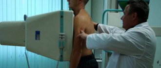
Description of the technique
This is a diagnostic method that began to be used in Russia back in the 30s. The essence of the technique is to obtain photographs of areas or organs. The method is based on the ability of X-rays to pass through tissues and be retained in them depending on their density.
The procedure is completed in a few minutes. The man undresses and stands at the machine. The doctor indicates what position to take. Then he goes into another room and directs the process from there. After turning on the device at the doctor’s signal, the person must hold his breath for a few seconds. Once the pictures are taken, the results are ready within 24 hours.
The appearance of shadows on a fluorogram
If additional shadows are found on a fluorographic image, this may not always indicate a person’s addiction to cigarettes. Some professions have specific working conditions that can lead to:
- diaphragm hernia;
- bronchitis;
- pulmonary tuberculosis;
- bronchial asthma and others.
Round light spots on the image indicate the presence of inflammation. If the inflammatory process occurs constantly, the body becomes weaker, its protective functions deteriorate, which can cause the development of severe tuberculosis infection.
What is shown on fluorographic images?
Fluorography is prescribed to detect gas, infiltration and many pathologies in the lungs:
- abscesses;
- tuberculosis;
- sclerosis;
- inflammation;
- fibrosis;
- pneumonia;
- hearts;
- emphysema;
- cystic formations;
- pulmonary obstruction;
- tumors.
The photographs are taken in black and white in different shades. This is achieved due to the ability of tissues to absorb radiation in different quantities.
Uniform color indicates the absence of disease. Any color changes or the appearance of gray shades of varying intensity indicate pathology.
What is special about the method?
A fluorogram is an image of organs obtained using x-rays. Since different tissues have different permeability rates, the structure of the image is heterogeneous and the presence of shadows is a natural feature.
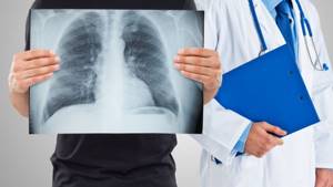
Attention! A specialist must decipher the image; it is impossible to read the information on your own.
With the help of such an examination, the following is revealed:
- the presence of neoplasms;
- the presence of an inflammatory process;
- fibrosis and sclerosis of organ tissues;
- cysts and other cavities;
- areas of necrosis;
- presence of foreign bodies;
- cavities and tuberculomas;
- occupational pathologies.
Patients should be aware that healthy lungs in the image have clear outlines and a light gray color. The silhouette of the ribs is visible against the background of the organs.
If the patient is a heavy smoker, his image may show pathological changes that appear due to prolonged exposure to tobacco smoke. Fluorography of a smoker and a healthy person has differences.
Will FLG show whether a person smokes or not?
It is important to note the fact that cigarettes have the most detrimental effect on the health of the entire human respiratory system. If a person smokes for many years, active changes occur in his body, not for the better in terms of health. they can manifest themselves after 1 year or after 10 years. This difference in time frame is associated with the individual characteristics of each organism.
Attention! Teen smoking is a real problem nowadays. Changes in the structure of the respiratory tract at a young age occur quite actively. The harmful effects of nicotine change the order of all stages of the formation of the respiratory system.
Indeed, heavy smokers “can be identified” by fluorography. This fact is confirmed by displaying the condition of the lungs, but does not constitute confirmation. Smokers should undergo FLG annually. If there are any problems with the functioning of the organs, the specialist will determine the need for further manipulations.
It should be noted that FLG cannot unambiguously determine whether a patient smokes. This is due to the fact that changes can occur in the human body under the influence of various factors, and nicotine addiction can only aggravate the patient’s condition and contribute to the progression of the disease.
Fact! The lungs of a smoker on fluorography differ significantly from the lungs of a non-smoking patient.
Based on the information described, it should be concluded that fluorography cannot definitively confirm the fact of smoking, but pictures of a smoker and an absolutely healthy person will be fundamentally different.
Irregularity of the image, as evidenced by the appearance of shadows?
The appearance of a shadow on fluorography is not only the result of the effect of nicotine on a person’s lungs.
The following factors can influence the manifestation of such changes:
- dust bronchitis associated with harmful working conditions;
- tuberculosis;
- bronchial asthma;
- diaphragmatic hernia;
- expansion, deformation of the bronchi.
It should be noted that patients who are heavy smokers face not only chronic bronchitis. They face a more serious danger - pathological dilation and deformation of the bronchial tubes.
The video in this article will tell the reader about the dangers of smoking:
Attention! Bronchial deformation is a protective reaction of the human body, manifested as a response to the influence of an external stimulus. By expanding the bronchioles, the respiratory system tries to compensate for the lack of functional lung volume. The danger is that the expanded cavities act as some “containers” for the accumulation of mucous secretions and resins. In such an environment, pathogenic microorganisms multiply fruitfully.
Against the background of such a process, some prerequisites are created for the development of a serious inflammatory process. Against the background of such changes, the protective function of the body decreases, and there is a possibility of tuberculosis infection. Such cavities appear on fluorography as round, light-colored spots.
Can fluorography show that a person smokes? The study will not reveal a novice smoker; structural changes appear more pronounced in patients who have been addicted to the bad habit for many years.
Does fluorography show that a person smokes?
Young smokers tend to worry about their smoking status being detected by X-rays. But, since their smoking history is low and the degree of lung damage has not reached a pathological level, fluorography will not be able to detect smoking.
Changes in the size of the heart muscle can indirectly indicate the use of tobacco or electronic cigarettes. Narrowed bronchi, which are under the constant destructive influence of smoke, can also indicate the fact of smoking.
An electronic cigarette or hookah makes the same changes to the structure of the lung walls as regular tobacco smoked through a cigarette or pipe. The difference can be considered the absence of acute or chronic forms of bronchitis caused solely by tobacco smoking. Detection of this form is possible if a person smokes for a large amount of time without breaks.
Fluorography will show the structure of the lungs, and not the fact of smoking. Determining whether a person is a smoker or a non-smoker is not the responsibility of the radiologist.
A fluorogram can show the acute or chronic stages of such diseases. One or more of these symptoms may indicate whether a person smokes or not.
In the video from the author of the “Health-Saving Channel” you can learn about the diagnosis of smoking through the fluorography procedure
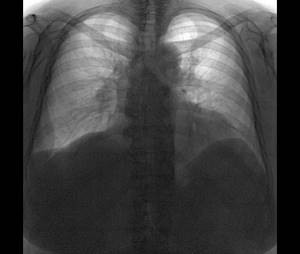
The difference between healthy lungs and a smoker's
In a healthy person who is not addicted to tobacco, the lungs look light gray and without obvious pathologies. They do not have clearly defined vessels, there is no darkening against the background of the lungs. The roots of the lungs are straight and show no signs of inflammation or swelling.
The lungs of a tobacco or hookah smoker, exposed to prolonged toxic effects of smoke and their decay products, will have a denser, more pronounced pattern. Small light areas will be especially noticeable against the background of a dense structure. The radiologist recognizes this pathology as bronchiectasis in the lung tissue and the pulmonary bags themselves. Long-term destructive effects on the lungs can reveal a reticular pattern of the respiratory organs.
The fact of smoking tobacco, an electronic cigarette or a hookah is indicated by:
- Black areas on x-ray. In the affected parts of the lungs, the elasticity of the walls significantly degrades, which, in turn, can provoke gas exchange. The heart muscle is also subject to destructive effects. An increase in the volume of the heart in the image indicates the presence of pathologies and changes in the tissue structure.
- A more distinct manifestation of the structure of blood vessels. Cells, oppressed by the action of nicotine and combustion products, die and are not removed from the lungs, forming growths on the blood vessels. With prolonged smoking, the peripheral parts of the respiratory system can be impaired and subject to changes.
- Non-functional tissue formed due to clogging of the pores of the lungs under the influence of resins. On an x-ray, it will be marked by alternating light and dark areas, making the lung pattern appear mottled. Such changes can provoke disruption of the oxygen transport process and, as a result, oxygen starvation of the brain.
- Loss of clarity. Indicates the emergence and spread of fibrous tissue, an increase in the lumen of blood vessels. In conditions of constant oxygen deficiency and starvation, the human respiratory organs begin to adapt and change their structure.
Mechanisms of adaptation and compensation for oxygen deficiency negatively affect the density of the tissue of the pulmonary bag. The process of change begins in the lower part of the lungs and moves upward, gradually filling and replacing the lung tissue with a fibrous, non-functional protective film.
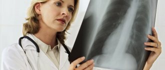
Photo gallery
In the photo you can see the differences between healthy lungs and a smoker's.
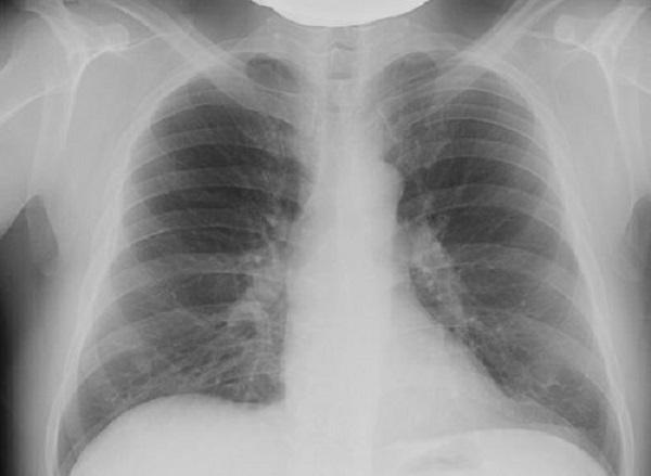
X-ray of healthy lungs
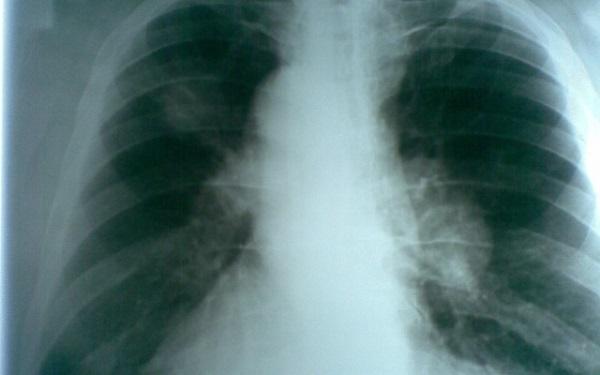
X-ray of a smoker's lungs
Indications for the examination and interpretation of the results obtained
The following are required to undergo fluorography:
- patients over 16 years of age once every 2 years;
- patients undergoing treatment in a medical institution in the absence of the necessary data on FLG;
- persons living with pregnant women and newborns;
- conscripts;
- persons applying for work or applicants;
- HIV infected.
Unscheduled FLG is carried out:
- if you suspect the development of pulmonary tuberculosis;
- in the presence of tumors in the lungs and heart;
- inflammatory processes in the lungs;
- pathologies of the heart and blood vessels.
Differences between the lungs in the picture
If a person has recently become acquainted with a cigarette, then there will be no pronounced changes. However, a specialist with extensive experience will immediately identify a heavy smoker from the image.
The image of healthy lungs appears light gray, and the organs have clear boundaries. Fluorography of a smoking person is characterized by the appearance of a “mesh” of alveoli. Excessive expression of blood vessels may indicate the presence of an inflammatory process, bronchitis, or pneumonia. Inflammation in the respiratory system is typical for people with nicotine addiction.
What pathologies can be detected
The fluorogram reflects the condition of the chest organs. Most often, fluorography is used to check for the presence of lung tissue cancer and tuberculosis. Normally, healthy lungs are gray in color, evenly colored, and have clear boundaries. Against this background, you can see the pattern of veins, arteries and vessels, the heart shadow, the root of the lung, and the position of the diaphragm.
The following parameters are assessed:
- Clarity of the vascular network and contours of the lungs. Too clearly defined vessels and organ boundaries indicate a problem - perhaps bronchitis, pneumonia, the first stage of neoplasm development. Insufficient functioning of the cardiovascular system also gives a picture of increased clarity.
- The presence or absence of fibrous tissue. If fibrosis is present - the growth of lung tissue into connective tissue - this may be a consequence of operations performed, diseases of infectious etiology, or injury. The same process occurs in liver cirrhosis.
- The roots of the lungs, which have cords, often give away experienced smokers. This kind of pathology indicates the occurrence of inflammation in the lungs. People who smoke usually have a chronic inflammatory process that affects all respiratory organs.
- Changes in the position of the diaphragm, indicating obesity, diseases of the gastrointestinal tract, and hereditary predisposition.
Based on the state of the shadows, one can assume the development of such ailments as:
- bronchitis of unspecified etiology;
- diaphragm hernia;
- tuberculosis;
- bronchial asthma;
- bronchiectasis.
What other changes are typical?
Smoking affects not only blood vessels, but also organ tissues. This manifests itself in the following:
- Increased work of the heart muscle caused by respiratory failure leads to hypertrophy of the heart muscle. Therefore, fluorography shows an increase in the volume of the heart.
- Damaged areas of the lungs that cannot perform their main function - gas exchange - will be darkened.
- Lymphatic drainage is disrupted, so the boundaries of the roots of the lungs become blurred. The shape of the roots may also change, causing shadows to appear.
- Loss of image clarity indicates fibrosis - the replacement of lung tissue with connective tissue. This is often observed after pulmonary infections.
Changes are visible even in novice smokers, the difference is only in the degree of their severity.
Why are changes visible?
Lung tissue can be compared to a sponge. Tobacco smoke, tar, particles of burnt paper and glue - all the rich contents of the cigarette settle on them, destroying their health. These substances penetrate deeply and cannot be removed. Normally, special cilia are responsible for cleansing the lung tissue, which, by moving, expel incoming contaminants. However, a smoker's lungs, subject to multiple inflammatory processes, are unable to cope with this task. The cilia gradually become thinner and lose their ability to cleanse the organ of dirt.
How smoking affects the lungs
It is well known that smoking has a detrimental effect on the entire body, and especially on the lungs. Nicotine and tar, which smokers inhale with each cigarette, settle on the mucous membrane of the respiratory system and then leave the body for quite a long time, leaving behind thousands of harmful substances. The lungs of a smoker can be compared to those of a person who lives next to an industrial area. Smoking addiction can lead to diseases such as lung cancer. In addition, every cigarette contains carcinogenic substances that provoke cellular mutations and stimulate cancer processes in various systems of the human body.
Smoking before the procedure
Sometimes people are interested in how to deceive fluorography. Unfortunately or fortunately, this cannot be done. It is also questionable whether fluorography can show that a person smokes. Changes in the bronchopulmonary system are not a direct indication that a person is addicted to a cigarette. Yes, characteristic changes will be observed, but the fluorogram only reflects the condition of the patient’s body, and does not indicate the cause of their occurrence.
The listed changes can also be observed in non-smoking people. They may be associated with harmful production conditions or developing pathologies.

An x-ray of a smoker's lungs can reveal the following changes:
- compaction;
- tumor-like formations;
- accumulation of fluid;
- thickening of the walls.
Detection of additional shadows may indicate the development of:
- bronchitis (not only the chronic form, characteristic of smokers, but also dust);
- diaphragm hernia;
- bronchial asthma;
- tuberculosis;
- bronchiectasis.
A cigarette smoked immediately before an x-ray will not cause any distortion in the image - all pathological processes in the smoker’s body are persistent and it will not be possible to achieve any sharp improvement or deterioration in the overall picture.
This will not affect the reliability of the conclusion - if you really want to smoke, then you can do it.
There will be changes in the body of a passive smoker. After all, he inhales all the same harmful substances. If you are constantly in a smoky room and systemic penetration of tobacco smoke into the lungs, they will react with the same symptoms.
What the lungs of a smoker and a non-smoker look like on FLG
A fluorogram is a picture of the lungs, heart and surrounding tissues, which has a heterogeneous appearance. This directly comes from the principle of conducting any x-ray examination, which consists in the uneven absorption of x-ray radiation by organs. If a person’s lung tissue is in normal condition, then on the film (or matrix) it will look smooth. If an inflammatory process occurs in it, the picture will contain darkening of varying intensity, and areas of increased airiness, on the contrary, will appear lightened.
Thus, an increase in the pulmonary pattern indicates the development of pathologies such as bronchitis, pneumonia, tumor, and cardiovascular failure. The presence of fibrous tissue indicates a history of severe infectious disease or surgery in the recent past. But stringy roots are a sign not only of an acute inflammatory process or a chronic disease, but also evidence of an addiction to smoking.
Usually, fluorography of a smoker looks like a regular photo without pathologies. In avid smokers, changes in the lungs may be noticeable, which is determined by the color on the photographic film.
A doctor cannot say with certainty that a person smokes based on an image. The darkening that is visible on a fluorogram has many other reasons for its appearance. Therefore, just by looking at the picture, it is impossible to unambiguously determine the presence of an addiction.
Smoking can make the situation worse. Air pollution and many other factors already put strain on your lungs every day. If we add to this the influence of cigarettes, an extremely unfavorable picture can emerge.
Is it clear from fluorography that a person smokes?
Such a conclusion can be drawn, but only indirectly. There can be no absolute certainty about this. Whether fluorography will show smoking or not largely depends on the length of service.
Often, with prolonged abuse, the harm from cigarettes reaches its peak and leads to serious illness. Then such changes will definitely be visible on the fluorogram. But no doctor can prove that the device shows a negative result precisely because of addiction to cigarettes.
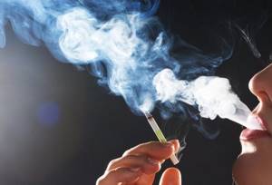
Is it possible to smoke before fluorography?
There is a category of patients for whom the burning question is whether smoking is allowed before fluorography. Sometimes these are teenagers who are afraid that “their parents will find out.” Dependent smokers think about this in order to know whether it is possible to smoke a few minutes before screening. So, what is the effect of smoking immediately before the procedure?
In fact, the results of the image do not change depending on the cigarette you smoke. The device does not detect smoke that remains in the lungs; it detects serious pathologies or changes in the body.
If the patient’s health is not in danger, not a single cigarette can affect the quality or reliability of the image. Therefore, it is impossible to designate a time during which you should not smoke before FLG.
Related question: is it possible to smoke hookah the day before or on the day of such a study? The effects of smoking using such a device on the body, and in particular on the lungs, have not been precisely proven. And perhaps this method is more harmful than regular cigarettes. But in this case the same rule applies. Hookah smoke is not visualized in the image. A doctor will be able to guess about hookah smoking only if they find specific pathologies.
How to improve the result of fluorography?
It is impossible to get rid of contaminants that have entered your lungs over the years. You can try to improve the survey results a little. It is highly advisable to give up the habit after using these methods. Of course, it will not be possible to obtain any significant effect in a few days, but gradually the condition of the organ will improve, provided there is no negative impact.
- Inhalations using pharmacological drugs. Direct impact on the pulmonary epithelium with means designed to eliminate the symptoms of shortness of breath and cough will stimulate the removal of some of the contaminants.
- Diving, yoga, aerobics classes. Any of these directions will help increase lung capacity.
- Cardiovascular training and breathing exercises. They improve the condition of lung tissue, stimulate blood supply and remove accumulated tars from the respiratory system. You should start with low-intensity loads, but gradually increase them.
- Regular visits to the sauna. Many toxins and wastes are eliminated through the skin - this is the largest organ in area. Removing toxins through the pores and sweat glands will promote rapid cleansing.
- Walks in the open air. It is especially recommended to walk in parks and forests with coniferous trees. Inhaling phytoncides will increase the concentration of oxygen in the blood and improve lung tone. In addition, the depressed ciliated epithelium will gradually recover.
You can use herbal medicine methods. Teas and decoctions with juniper, oak bark, mint, and lavender will have a beneficial effect on the condition of the lungs.
Where and how to check your lungs
You can check your lungs using one of the x-ray methods: x-ray, fluorography, computed tomography. Fluorography is the most accessible and safe method for assessing the condition of the bronchopulmonary system. This procedure is mandatory for every adult. It is carried out in order to detect serious pulmonary diseases: tuberculosis, cancer, bronchitis (chronic and obstructive), cysts, abscesses, emphysema, fibrous formations, as well as indirect signs of problems in the heart (reduction of the lumen between the lungs).
An X-ray of the lungs of a moderate smoker is not much different from an X-ray of the lungs of an ordinary person, provided there are no pathologies.
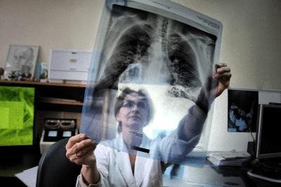
FLG (fluorography) is an x-ray examination. Its results are presented in the form of a small picture of the internal organs. The method is based on the fact that different tissues absorb Ro-rays differently.
Nutrition
When quitting smoking, you should pay attention to your diet. There are products that will remove toxins faster and improve the condition of the respiratory system.
It is recommended to include in the diet:
- Pineapples. The bromelain they contain helps cleanse the lungs and also has a beneficial effect on the nervous system.
- Garlic. Contains allicin, which dissolves accumulated mucus and removes it out.
- Apples. Thanks to its saturation with a large number of vitamins, it strengthens the immune system and consolidates the effects obtained from other cleansing methods.
- Water. Drinking plenty of fluid helps eliminate toxins from the body.
- Green tea. The catechin it contains reduces the likelihood of developing cancer and also cleanses the body of toxins.
Medicines with mucolytic and expectorant effects can be used. These are Ambroxol, Mucaltin, Gedelix.
How to clean a smoker's lungs
The lungs of a smoker, namely a heavy smoker, are difficult to restore. First of all, you need to immediately quit smoking, start leading a healthy lifestyle and try to use lung cleansing methods. This may take a long time. It is usually possible to fully rehabilitate within 10 months.
What can you do at home?
Breathing exercises will be very useful for former smokers. It promotes ventilation of the lungs and activates their work. There are entire systems of breathing exercises that were created by experienced doctors. The simplest exercise is “deep breathing”. It will help clear the tar deposits that remain in the lungs after smoking.
Physical exercises are great for cleaning the lungs. Sports activities keep the entire body in good shape, and it, in turn, is a complete system. Thus, the functioning of the lungs will be restored along with other parts of the body. Finally, physical activity provokes accelerated gas exchange, which also improves respiratory ventilation. For example, running in the fresh air can “restart” the lungs and, in combination with other actions, give a good result.

Experts recommend that people who have quit smoking regularly visit baths. This will help you forget about the bad habit and its consequences. The bath, due to its elevated temperature, provokes increased sweating and helps the body get rid of harmful substances. The brooms that are used when visiting bath complexes contain beneficial herbs. They promote even faster detoxification.
Nutrition
Recommended products for quitting smoking:
- pineapples. They contain bromelain, which perfectly cleanses the lungs, actively rejuvenates the body and forces it to get rid of harmful substances and toxins. Pineapples have a positive effect on the nervous system;
- garlic. It will help stop pathological processes caused by smoking. The active ingredient is allicin. It dissolves toxic mucus in the lungs and removes it from the body;
- apples. They contain a large amount of vitamins. It is they, in combination with other products, that will be able to consolidate the result of cleansing the body from the influence of tobacco smoke;
- water. For any unstable conditions, doctors advise drinking more fluids. This is what helps the body get rid of toxins;
- green tea. It includes the antioxidant catechin, which helps prevent cancer, and is also good at removing toxins introduced by tobacco smoke into the human body.

Medicines
There are a number of medications designed to cleanse the lungs of accumulated harmful agents. Usually these are mucolytics and expectorants. Among them: Ambroxol (Lazolvan), Acetylcysteine, Gedelix, Mucaltin and the like. They are available in a variety of forms - tablets, capsules, syrup or inhaler.
ethnoscience
Traditional medicine recommends making self-inhalations from herbal infusions.
The composition of such collections includes leaves of black currant, oak, birch and eucalyptus, chamomile, sage, pine, fir, juniper and cedar needles, mint, wormwood. The easiest option is to brew the mixture in any container and breathe over it under a towel. This procedure should be performed for 10-15 minutes daily for two weeks.
Prevention
Smokers should have their health checked with a fluorogram every year. It reflects the degree of existing deviations from the norm and can indicate a developing serious disease.
The attending physician will definitely ask, looking at the picture, whether you smoke. In such cases, you cannot hide the truth - this will help in making the correct diagnosis.
Smoking grass, hookah, cigars - all this leads to structural changes in the lungs and their clogging. In any case, pathological processes and inflammation will be reflected in the image. The specialist will determine the reasons for their occurrence based on a dialogue with the patient. Therefore, the doctor will not find out what exactly you smoked when deciphering the fluorography, and the task before him is different. It is important for a specialist to find out what danger the existing disorders pose to the patient’s life and, if possible, eliminate them. You will make this task easier if you give up a bad habit.
Is it possible to smoke before the procedure?
If you smoke even a few cigarettes before the examination, this will not be noticeable in fluorography images, unless the pathology has already begun to develop earlier. In this case, its outbreaks will be displayed. In good health, a cigarette smoked does not affect the examination results.
Fluorography is necessary for the treatment of pulmonary diseases or some other pathologies.
Diagnosis is also carried out as a preventive measure. In any case, you can smoke before the examination; without pathology, this will not be shown on the images. Next entry Ultrasound of the bladder in a pregnant woman: types, preparation, indications