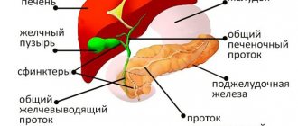
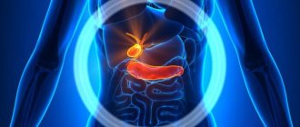
The gallbladder is classified as an auxiliary organ of digestion. Its main task is the accumulation of bile, which is produced by the liver. Our body needs bile to digest animal fats. Therefore, disturbances in the functioning of the bladder are accompanied by digestive disorders and pain in the right hypochondrium. In this case, the patient undergoes an ultrasound of all abdominal organs, during which the gallbladder is examined.
- What it is?
- Who is prescribed an ultrasound of the gallbladder?
- How should you prepare for an ultrasound?
- How is the procedure done?
- What are normal indicators for the gallbladder?
- What pathologies of the gallbladder can be detected on ultrasound?
What is an ultrasound examination of the gallbladder?
The gallbladder is located just below the liver, approximately at the level of the last rib, on the right side of the abdominal cavity. In normal condition, this organ cannot be palpated. Instrumental and laboratory methods (ultrasound, radiography, computed tomography) are used to diagnose it. The simplest and most accessible way to examine an organ is ultrasound.
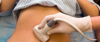
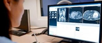
Ultrasound, or echography, is a fast, informative and safe diagnostic method for the patient’s health. It is based on the ability of ultrasonic waves to penetrate the structures of our body and be reflected from them. The denser the tissue or organ is in the path of ultrasound, the more the characteristics of the reflected wave change. The ultrasound machine detects these changes, processes them and displays an image of the organ on the monitor.
Echography of the gallbladder allows you to determine the shape and location of the organ, its size, examine the ducts and identify stones and tumors. For ultrasound examination of the gallbladder, the transabdominal method is used, that is, scanning through the anterior wall of the abdomen. The procedure is carried out using ultrasound sensors with a frequency of 2.5-3.5 MHz. With this scanning mode, ultrasound waves can penetrate only to a depth of 23-25 cm. Therefore, for very obese people, ultrasound is a little informative method.
In addition to the standard procedure for ultrasound scanning of the gallbladder, echography is also prescribed to determine the function of the gallbladder (ultrasound with a choleretic breakfast). The essence of such an ultrasound is to monitor how much the organ contracts when eating food. During the study, the patient is asked to eat choleretic food (eggs or sour cream), then the release of bile is observed. Diagnosis of bladder function takes place in several stages. This ultrasound takes about an hour.
Preparing for the examination
For an accurate diagnosis, careful preparation will be required before the examination. First of all, three days before the procedure, you must give up unhealthy, salty and heavy foods. The patient also excludes from the menu all foods that can cause gas formation. Avoid alcohol completely.
On the eve of the procedure, you must have your last meal no later than 7 pm. This is so that bile can accumulate. In general, at least 8 hours should pass between the meal and the examination. Therefore, if the procedure takes place in the evening or after lunch, a light breakfast is allowed.
Stop smoking 3 hours before and stop taking liquids during the same time. Any violation will lead to the production of bile and a decrease in the size of the organ being examined.
Who is prescribed an ultrasound of the gallbladder?
The gallbladder is examined in cases of liver dysfunction, as well as during ultrasound diagnostics of the abdominal organs. In addition, the reasons for prescribing an ultrasound are:
- pain on the right side of the body,
- discomfort or heaviness in the liver area,
- icteric syndrome,
- injuries and damages,
- monitoring of the treatment performed,
- suspicion of stones or tumors formed.
The only contraindication for ultrasound is an open wound in the scanning area. Using an ultrasound sensor in such a situation can lead to infection in the wound.
What conclusions can be drawn based on the results of the ultrasound?
Based on the results of ultrasound of the bladder, the most dangerous dysfunctions of the organ are often identified: cholelithiasis, cancer, cholecystitis, pathologies of the excretory tract, cholecystocholangitis, inflammatory processes. Immune and hormonal disruptions, which are accompanied by gallbladder dysfunction, are also recognized in ultrasound images.
The huge advantage of this manipulation (taking into account the fact that the load test does not cancel the x-ray, but complements it) is the possibility of verifying a specific degree (one out of 5) of dyskinesia of the biliary system (disturbed rhythm of contractions), which can lead to serious violations of its systems, especially in children.
How should you prepare for an ultrasound?
In order for the echography of the gallbladder to be accurate and informative, patients need to go on a diet. The main goal of the diet is to reduce the process of gas formation in the intestines. Gas bubbles interfere with the passage of ultrasound waves, which affects the quality of the ultrasound image.
A few days before the ultrasound scan you should avoid:
- carbonated and alcoholic drinks,
- dairy products,
- fatty and spicy foods,
- fresh vegetables,
- sweet fruits,
- legumes,
- black bread and yeast baked goods.
If patients are prone to flatulence, they are recommended to take adsorbents (activated carbon) and enzyme preparations (pancreatin). If you need to take any medications, you should tell your doctor about them. Taking medications may affect the accuracy of the results. The procedure is carried out on an empty stomach. The last meal is allowed 8-10 hours before the ultrasound. During this period of time, the gallbladder has time to fill with bile again, so it will be easier to examine it.
Dietary restrictions also apply to children. Remove sweets and fruits from your child's diet a few days before the ultrasound. Children, like adults, undergo the procedure on an empty stomach. Children under one year old should not be fed 3 hours before an ultrasound scan, children under 4 years old should not be fed 4 hours before, and children under 8 years old should not be fed 6 hours before. Preparation for an ultrasound examination for children over 8 years of age is the same as for adults.
If you have previously undergone an ultrasound examination of the gallbladder, then take with you copies of the protocols; by comparison, you can evaluate the dynamics of recovery or worsening of the organ’s condition. Bring a small amount of food to the clinic before your ultrasound function test. This could be boiled chicken egg yolks, cottage cheese or sour cream. You will be told exactly what to take and in what quantities when you schedule an ultrasound.
Our advantages
In the Alfa Health Center network of clinics, you can undergo an ultrasound of the gallbladder in the most comfortable conditions. We provide:
- Prompt service without queues or long waits. We value our patients’ time and offer convenient hours for gallbladder examinations and specialist consultations;
- quick receipt of ultrasound results. Within 15–20 minutes after diagnosis you will receive a transcript of the results of the ultrasound examination. With them, you can immediately get an appointment with a specialized doctor to receive further recommendations on treatment, diet and lifestyle changes;
- modern technical equipment. The diagnosticians of our clinics have at their disposal the equipment of the world leader - General Electric. Ultrasound devices from this manufacturer are distinguished by advanced functionality, convenience, high information content of the data obtained and safety. Innovative technology allows you to obtain the most accurate information about the structure, functional capacity of the gallbladder, the number and nature of neoplasms;
- information support from clinic specialists. Experienced doctors and consultants answer questions, talk about how to properly prepare for the procedure and avoid inaccuracies during the examination of the gallbladder.
How is the procedure done?
As a rule, ultrasound is performed in the first half of the day, always on an empty stomach. Under no circumstances should you drink or eat before the procedure. Even a small amount of water or food provokes the release of bile from the bladder. The organ decreases in size, which makes it difficult to diagnose. You should also refrain from chewing gum before visiting the clinic; it also provokes the secretion of gastric juice and bile. If you are scheduled for an ultrasound examination in the afternoon, then a light breakfast is allowed 6-8 hours before the ultrasound.
The duration of the procedure is 10-15 minutes. The patient is asked to remove his outer clothing and lie on his back on the couch. Before the ultrasound, the sonologist applies a small amount of gel to the patient's skin in the area being examined. The gel ensures continuous contact between the ultrasound transducer and the skin and improves the transmission of ultrasound waves.
During the scan, the doctor moves the sensor over the patient's skin at the location of the organ. To view the gallbladder from a different angle, the patient is asked to change position (sit down or roll over on his left side). The monitor displays the organ and surrounding tissues. The data obtained is included in the study protocol, which is given to the patient. Your attending physician will decipher it and make a preliminary diagnosis.
Carrying out an ultrasound with a choleretic breakfast differs from the standard procedure. During the study, the patient needs to eat several chicken eggs, a glass of full-fat sour cream or cream. Instead of food, a sorbitol solution can be used. First, the organ is scanned at rest. Then the patient must eat a choleretic breakfast, after which 4 ultrasounds are performed at intervals of 15 minutes. On each of them, the ultrasound specialist notes how much the gallbladder has shrunk. Ultrasound with determination of function allows you to determine which type of biliary dyskinesia the patient has (hypomotor or hypermotor disorder).
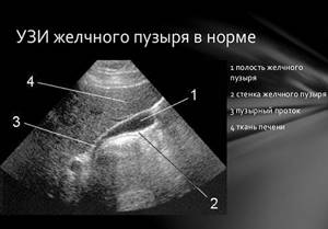
What are normal indicators for the gallbladder?
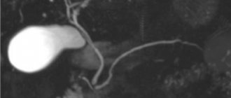
During an ultrasound, first of all, the shape, contours and size of the organ are determined. The gallbladder has the shape of a hollow pear, with smooth and clear edges. Its length ranges from 7-14 cm, and its width from 3 to 5 cm. The thickness of the bubble walls is 2-3 mm. The volume of the bile is 40-70 ml. These parameters are considered normal ultrasound for an adult. Normal sizes for the gallbladder of children depend on the height and weight of the child; they are determined using specially compiled tables.
In addition to determining the size of the organ, during the scanning process its ductal system (common bile and lobar bile ducts) is studied. Their diameter, permeability and the presence of concretions (stones) in them are determined. The diameter of the lobar ducts is 2-3 mm, and the common bile duct is 6-8 mm. The common bile duct unites with the Wirsung (pancreatic) duct and flows into the duodenum. Bile enters the gastrointestinal tract along this route.
In normal condition, the organ and its ducts do not have stones or other formations. If any deviations from normal values are detected, the patient is prescribed additional diagnostics.
The norm for ultrasound to determine function is considered to be a reduction of 70% from the initial level, which suggests the absence of dyskinesia.
What is ultrasound with function determination?
This is a method of diagnosing gallbladder diseases using ultrasound waves. The examination helps to measure the organ by taking longitudinal and transverse sectional photographs, recording the length, as well as the thickness and width.
Based on the images, the doctor draws conclusions about the condition of the bile ducts. During an ultrasound to determine function, the doctor performs not one, but 4 stages with breaks. As a result, in one hour, the specialist receives 4 scan results, which allows him to analyze the functional characteristics of the organ in a given patient.
What pathologies of the gallbladder can be detected on ultrasound?
Ultrasound can detect the following pathologies:
- cholecystitis (acute or chronic),
- organ dyskinesia,
- cholelithiasis (presence of stones),
- tumors
- congenital pathologies (absence or atypical position of the gallbladder, protrusion of its walls).
Cholecystitis is inflammation of an organ. Inflammation of the bladder can occur in chronic or acute form. The acute form is accompanied by intense pain in the right side, the chronic form is accompanied by nausea, heaviness, discomfort and dull pain in the area where the organ is located. Acute cholecystitis on an ultrasound image is determined by thickening of the walls and an increase in the size of the organ, while blood flow increases in the arteries of the bladder. In the chronic form, thickening and deformation of the walls, as well as a decrease in the size of the organ, are visualized.
Dyskinesia is a disorder of motor function in which there is insufficient contraction of the muscles of the organ. The ultrasound image shows an inflection of the neck of the organ and an increase in the tone of its muscles.
Cholelithiasis, or cholelithiasis, is the formation of calculi (stones) in the bile duct or its ducts. On echography, stones are displayed as areas with a dense echo structure, which can shift depending on the position of the body. Since stones reflect the signal well, an acoustic shadow from them can be seen on the echogram. In case of tumors, ultrasound shows formations on the walls of the organ that are larger than 2 cm in size. There is also a thickening of its walls and deformation of the contours.
The ultrasound procedure is sometimes not enough to make an accurate diagnosis, so patients are prescribed additional tests. Ultrasound allows you to quickly identify any abnormalities that have arisen, which makes it an indispensable method for diagnosing gallbladder diseases.
Sphincter of Oddi examination
Despite the fact that ultrasound allows a fairly accurate assessment of the functional state of the gallbladder, diagnosing sphincter of Oddi dysfunction using ultrasound is difficult.
Dysfunction of the sphincter of Oddi is a violation of its motility, manifested by a clinical symptom complex, evasion of liver and pancreatic enzymes, dilatation of the common bile duct or episodes of pancreatitis. Dysfunction of the sphincter of Oddi is confirmed (according to the Rome III criteria, 2006) by ultrasound examination (choledochus > 8 mm, increase in its lumen after stimulation), magnetic resonance cholangiopancreatography, cholescintigraphy, ERCP (choledochus > 12 mm, basal pressure > 40 mmHg.).
With biliary sludge and hypokinesia of the gallbladder, hypertonicity of the sphincter of Oddi is observed.
In case of flatulence, severe excess body weight, ultrasound examination is difficult; in these situations, it is necessary to supplement ultrasound with endoscopic ultrasonography, the information content of which in diagnosing biliary sludge is significantly higher (sensitivity 92-96%, specificity - 86-100%). Endoscopic ultrasonography is also indicated in cases where there are difficulties in making a differential diagnosis between a clot of putty-like bile fixed to the wall of the gallbladder and a parietal formation, primarily of tumor origin.