Fluorography is a mandatory procedure prescribed for every adult. It helps to identify any tumors in the lungs, as well as tuberculosis. Fluorography is a necessary procedure that is included in the medical examination. It is mandatory to prescribe it to people who are suspected of having heart and vascular diseases.
Since most people smoke, before undergoing fluorography, they have a question: “is it possible to smoke a few minutes before such a procedure?” and “Is it clear from fluorography that the person smokes?” The second question especially often worries teenagers because of the fear that their parents will find out about their bad habit. At the same time, many adult smokers are interested in whether smoking affects the indications for the procedure.
Briefly about fluorography
To maintain human health and timely detection of any pathologies, specialists prescribe fluorography once a year. No matter how many people believe that this procedure is harmful to the body and should be performed as rarely as possible, only x-rays can detect changes in the lungs at the initial stage. The results of the procedure are the basis for further serious medical examination.
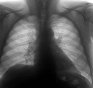
The X-ray image has a non-uniform color. Do not be alarmed, this means that all organs located in the chest area do not absorb x-rays evenly. When a person’s health is in perfect order, the image shows a uniform coloration. If any changes are observed in this area, this means that the person has pathologies or an inflammatory process is occurring.
In cases of increased pulmonary airiness, light spots will predominate in the image. Significant differences in the image also show the lungs of a smoker and a healthy person.
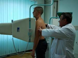
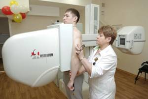
Brief description of the procedure
The examination refers to diagnostic procedures carried out for the purpose of prevention. It is based on low-power X-rays, which pass through tissues of varying densities and produce shadows, which doctors use to determine normality or pathology.
The reason why fluorography is so strongly recommended by doctors and controlled by health workers and employers is the high level of tuberculosis. If previously the disease was of a social nature and affected mainly the poor, now tuberculosis occurs even among wealthy people. The prevalence of pathology in children is also high. Therefore, fluorography is performed to diagnose tuberculosis.
The lungs have a uniform, homogeneous structure. The bronchial tree in the fluorographic image is clearly limited, smooth outlines are visible. The inside is light and “transparent”. The color of the lung tissue will be light gray, and the ribs will be clearly visible against the background of the lungs. With this picture, doctors confirm the norm.
40 million people out of 147 million in our country smoke. Skipping the point of danger, let's look at whether it is possible to see a smoker's lungs on fluorography?
Does fluorography show smoking?
You should know that even the very first cigarette affects a person’s health, especially his respiratory system. When a person smokes for a long period, changes in his body occur very actively, but are not always evident. Only over time, internal organs will begin to fail and the consequences of smoking will manifest themselves very actively. To prevent exacerbation of the disease, smokers should first undergo regular x-rays.
Fluorography shows that the person is an active smoker, as this is revealed by the lungs. If any problems are detected in the respiratory system, the doctor prescribes additional x-rays.
Only in such cases can images show that a person has a disease called “active smoker’s bronchitis.” At the same time, it is worth knowing that not a single change in the lung area can prove that a person smokes. This is explained by the fact that diseases can have a large number of causes, and this is not always the active influence of nicotine. Regular smoking habit only aggravates the situation and helps the disease progress. All this confirms that even the most active addiction to smoking cannot be detected by a fluorographic image.
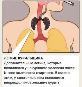
Why does this happen, or where do such lungs come from?
What causes such lung pollution in smokers?
In general, the problem of the purity of inhaled air is a global problem of anthropogenic origin.
Due to human activity, suspended particles and aerosols (that is, the same particles, but deposited on moisture droplets) are constantly present in the air.
It is estimated that the annual composition of these particles weighs approximately 100 million tons - approximately 14 grams per person, based on the total population of 7 billion people living on Earth. However, if we take into account that many of the Earth’s inhabitants are far from civilization or live in areas where industrial emissions are minimal, then on average for each potential “inhaler” of dust and other particles there is a much larger number of them. via
WHO experts have calculated that 70% of the urban population in developing countries breathes polluted air. Of the total number of air polluting particles, approximately half appeared in the air as a result of incomplete combustion of fuel. Fuel is hydrocarbons, the combustion of which produces soot.
The size of soot particles ranges from 0.01 to 10 microns, with an average of about 1 micron. At the same time, the human lungs are able to filter and remove particles 5 microns in size and larger. That is, most of the soot particles remain in the lung tissues.
Tobacco smoke is a typical aerosol, which contains products of incomplete combustion of all components of a cigarette - from tobacco to paper and glue. The formation of soot and soot during combustion is inevitable, and given that these combustion products are not sprayed into the air, but are concentrated directly near the smoker’s respiratory tract, it is obvious that they enter in a large and, most importantly, constant volume directly into the lumen of the bronchial tree.
Normally, even in a metropolis, the lungs cope with their filtering functions due to the active work of cilia - a special ciliated epithelium in the form of the finest hairs on the surface of goblet cells.
The cilia are part of a protective barrier that prevents potentially harmful and irritating particles from entering the alveoli. They are in constant motion, and it is directed from the inside to the outside, that is, in the direction from the lungs to the external environment.
Pathogenic agents (dirt, dust, microorganisms, etc.) fall on the mucus surrounding the cilia, stick to it and are carried out as the cilia move. The same mucus (sputum) coughed up during illness is mostly a product of the work of goblet cells and cilia.
But eyelashes are very sensitive to damaging factors - both mechanical and chemical, of which there are many in cigarette smoke. Their chronic damage by smoke leads to the fact that the cells of the ciliated epithelium are gradually replaced by basal cells, more and more mucus is produced, and it is more difficult to cough it up, chronic bronchitis is formed.
How can you easily recognize a smoker?

You can determine whether a person smokes or not in different ways; no special procedures are needed for this.
- A smoker's skin has a yellowish or grayish tint.
- Even at a very young age, people who smoke regularly show fine facial wrinkles.
- An unpleasant odor of tobacco from your breath during a conversation also indicates that you smoke.
- The peculiar smell of clothes also gives away a smoker. Even the skin has a specific smell when smoked regularly.
- Yellow plaque and frequent inflammation of the gums, sometimes accompanied by bleeding.
- Prolonged coughing attacks, which sometimes lead to vomiting.
- The tips of the thumb and index finger of the right hand are yellowish from regular smoking.
Pictures on topic
Pictures are not proof for “non-believing Thomases” - they always have a lot of counterarguments. But photos from the websites and blogs of practicing pathologists and forensic experts are not the kind of tales, but, alas, sad evidence of human nicotine stupidity.
Proper nutrition
A citizen’s complete diet depends on his age, gender, body weight, and physiological characteristics. It differs in what it contains:
- nutrients (fats, carbohydrates, proteins);
- minerals;
- vitamins
Garlic should be a must-have product on the menu. It acts as a powerful stimulator of cleansing processes. Apples and citrus fruits should also be included in the diet daily. They contain ascorbic acid, which is responsible for the regeneration of connective tissue.
Another essential nutrient is water. It should be ingested in its pure form up to 2 liters per day. Tea, coffee, cocoa, juices are not included in the calculation. Plain water washes away toxins and decay residues of dead cells. Its action is irreplaceable.
Traditional medicine
The main folk methods of detoxification and cleansing:
- Bathhouse. A universal, common detoxifier.
- Herbal decoctions, infusions. Plants used: chamomile flowers, black currant leaves, sage, eucalyptus, birch leaves, pine needles and cones, sweet clover.
- Inhalations with leaves and roots. Herbal decoctions that have a characteristic, persistent, strong aroma help best. These are peppermint, wormwood, pine needles, cones.
The use of alternative treatment methods is recommended to be coordinated with the doctor and the prescribed course of treatment. They may not be beneficial in all cases.
The essence of the method
Does fluorography show cigarette smoking? To answer this question, it is necessary to understand the essence of this diagnostic method.
Fluorography is a type of x-ray examination. Nowadays, diagnostics are carried out using special digital devices, which allows the use of lower radiation doses. However, the essence of the method remains the same. X-rays are passed through the patient's body and are unevenly absorbed by the lung tissue. The image of the bronchi and lungs is displayed on a fluorescent screen and an image is taken.
Classic X-ray of the lungs and fluorography differ in the doses of X-rays required for the study. Conventional x-rays expose the patient to more intense radiation. For this reason, the image resolution is higher than with conventional fluorography.
Fluorography shows only gross and pronounced changes in the respiratory organs. This is a safer, but less reliable method. It is used during preventive examinations. As for classical x-rays, this examination is more often used to diagnose diseases. It shows a clear and accurate picture of pathological changes.