MRI is a non-invasive, informative diagnostic procedure. With its help, you can examine the condition of the abdominal organs, pelvis and head. MRI is performed by applying magnetic radiation to the human body. It provokes a displacement of hydrogen atoms in the desired direction. They will concentrate in the cavity of the body tissues.
The signals received from the protons will be transmitted from the tomograph to a computer program, where they are recorded in the form of images converted into a three-dimensional image. Due to the fact that each type of study has certain advantages, disadvantages, and limitations, before undergoing magnetic resonance therapy it is worth studying all the nuances so as not to encounter unpleasant consequences.
MRI and the menstrual cycle

Is it possible to do an MRI during menstruation? This is a question that girls often ask gynecologists and diagnostic specialists. In general, if we consider the point of view of the impact on women’s health, then this procedure during “critical days” turns out to be completely safe. MRI of the abdominal organs, in which this part of the body is exposed to magnetic radiation, does not contribute to hormonal imbalance and will not lead to destabilization of the functioning of the reproductive system. The only thing you might encounter when doing research during menstruation is a distorted result. Such phenomena occur in 5 cases out of 10.
Diseases of the pelvic organs in women
Main diseases of the female genitourinary system:
- Inflammatory diseases (salpingitis, oophoritis, endometritis, cervicitis, cystitis);
- Ovarian apoplexy;
- Polycystic disease;
- Torsion of the uterine appendages;
- Polyps, synechiae of the uterine cavity;
- Endometriosis, adenomyosis;
- Ectopic pregnancy;
- Endometrial hyperplasia;
- Bleeding;
- Infertility;
- Bladder polyposis;
- Benign and malignant neoplasms of the female genitourinary tract;
- Malformations of the female genitourinary system.
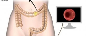
In addition, MRI is used to diagnose diseases of the rectum - Crohn's disease, polyps, perirectal abscesses, fistulas, and neoplasms.
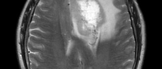
Is it permissible to examine the brain on MRI during menstruation?
It is quite possible to detect pathological changes in the brain during menstruation, and there is no need to immerse the entire woman in the tomograph capsule. Only the area of the body that is being examined will be affected by the magnetic field. The small pelvis is not involved. In the question of whether critical days and MRI of the brain are compatible, the answer will contain some clarifications.
- The diagnostic session will affect the glands that are responsible for controlling the cyclicity of the menstruation process.
- The pituitary gland is a lower cerebral appendage, which is considered one of the important control organs of the endocrine system.
- If there is a malfunction in the functioning of the gland, this will directly affect the regulation of menstruation.
- Magnetic radiation can interfere with the life processes of the body.
The restoration of this process occurs immediately after magnetization activity ceases.
You can minimize the risk if you properly prepare for how the MRI will be performed. It is also important to remove all metal objects from the body, no matter how far away they are from the scanning area. In addition, pay attention to the fact that the pituitary gland functions constantly, promoting the formation of hormones. If the study could interfere with the normal course of menstruation, then the disturbances would be noticeable throughout the entire cycle, and not just at its end.
It is almost impossible to determine the moment of diagnosis that will be ideally safe. Doctors are convinced that it is acceptable to do an MRI of the brain during menstruation. Negative effects on the functioning of systems such as reproductive, urinary, endocrine are excluded.
Brief description of the method
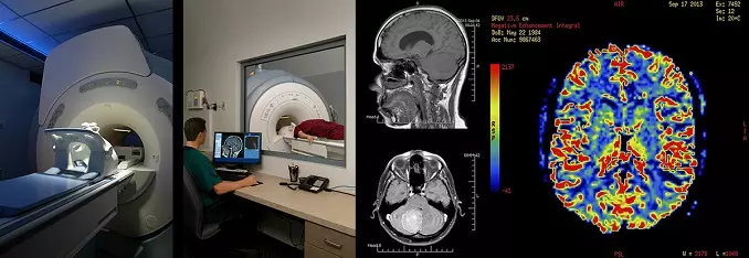
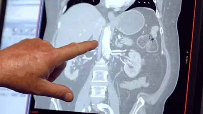
As a diagnostic method, magnetic resonance imaging can detect various pathological changes in the human body. This study is based on computer measurements of the activity of cells exposed to an electromagnetic field. Under the influence of magnetic pulses, healthy and diseased cells emit different waves. The device records the measurement results and converts them into a picture. If affected cells are present, the area of pathological changes is highlighted in the image. The visual representation of the organ under study can be three-dimensional or presented in the form of sections in various projections. Next, a radiologist works with the received images, describing them and providing a transcript.
An MRI scan can check all organs and systems of the human body. This method allows you to study the following parameters:
- physical and functional state of the brain and spinal cord;
- state of the circulatory system and blood flow speed;
- flow of cerebrospinal and lymphatic fluids;
- the condition of the internal organs of the thoracic region, abdominal cavity and small pelvis;
- assessment of bone and muscle structures.
The advantage of this diagnostic method is that it allows you to study any part of the body without resorting to traumatic invasive methods.
The MRI procedure is a safe diagnostic method that helps to quickly and painlessly localize pathological changes and determine their nature. Unlike X-rays, electromagnetic radiation does not have a destructive effect on cells. It can be performed on all categories of patients, with minor restrictions.
MRI of the abdominal organs during menstruation
Examination of the abdominal cavity or hepatobiliary system using MRI during menstruation is highly undesirable. The limitations are directly related to the factors below.
- During the activity of the menstrual cycle, the reproductive organs are renewed, and this negatively affects how reliable the research will be.
- Many women experience increased gas formation during menstruation. This is especially undesirable, since, again, it will distort the results obtained.
- When the area under study is heated, the pain experienced by a woman during her “critical days” increases. This causes additional discomfort for the patient.
- During the MRI, the subject's body must remain motionless. Considering the presence of psycho-emotional stress and pain in the lower abdomen, it is especially difficult to comply with this requirement.
Based on these data, we can conclude that if there is no emergency need, it is better to postpone the diagnosis to the 7-10th day of the menstrual cycle.
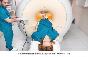
How is an MRI of the pelvis performed in women?
The duration of the study is 40 minutes (55 when using contrast). Diagnostics begins with placing the patient in a tomograph - a cylindrical device with a tunnel in the center 60 centimeters long and wide. To accommodate the patient, the tunnel is equipped with a movable table. After laying the patient down, the medical professional will ask you to put on headphones - they are needed so that the noise from the tomograph does not interfere with the examination (as well as to communicate with the operator and listen to music, which helps to relax and reduce the wait). The patient is given a button on a wire - by squeezing it, you can contact the operator via intercom.
Please remain still throughout the entire examination. Follow all operator instructions - you may be asked to exhale or hold your breath. Try to relax and remain calm - the procedure is absolutely safe and will not cause harm.
After completing the procedure, you must wait 15-30 minutes until the results of your examination are deciphered. After this, you will receive a written report from the radiologist and images on a CD. For convenience, images can be ordered on film or on a flash drive - not all doctors have the opportunity to view CDs (you need to pay a little extra for this).
MRI examination of the pelvis and menstruation
If it is necessary to study the condition of the pelvic organs, MRI during the monthly cycle is undesirable for a number of reasons.
- With intense blood flow to the reproductive organs, vasodilation occurs, which leads to local edema, and this can interfere with the diagnostic procedure.
- There will be difficulties with an objective assessment of the state of the endometrium. The old layer begins to be rejected, but the new one has not yet formed.
- There are difficulties in studying the condition of the vagina and cervix, as there will be an excessive amount of discharge.
- Hydrogen atoms that react to magnetic radiation during MRI will move in a chaotic manner due to congestion in the pelvic area. This also provokes distortion of the results obtained. This is an additional reason to refuse magnetic resonance imaging of organs related to the reproductive system during menstruation.
There are many cases when doctors specifically give a referral for such a session in order to determine or, conversely, to exclude hormone-dependent diseases that appear during this cycle. It can be:
- cysts;
- a number of other neoplasms in the ovarian cavity;
- endometriosis;
- myomatosis.
The decision about whether an MRI should be done or not will be made by the doctor.
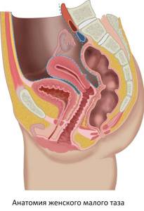
Anatomy of the female pelvis
The small pelvis is the space bounded by the symphysis pubis in front and the promontory of the sacrum in the back. On the sides, the boundaries of the small pelvis run along arcuate lines separating the body of the ilium from the wings. The pelvic cavity in women serves as a container for the genital organs - the uterus and its appendages (ovaries, fallopian tubes). There are a large number of blood vessels, nerves and fiber. The pelvic cavity also contains the rectum and bladder.
Additional contraindications
MRI is magnetic resonance imaging, which is a non-invasive procedure. The interaction of body structures and the magnetic field occurs without surgical intervention, but due to the formation of the magnetic field of the tomograph. Patients who have no idea how the MRI device works consider it to be especially dangerous during menstruation. However, there are two main limitations.
- Many girls experience emotional instability during menstruation. Such tension provokes difficulties in the study, as the patient will be restless.
- A number of women may panic due to being in a confined space.
In addition, as already mentioned, due to the duration of the study, discomfort arises from prolonged lying down.
Rate this article: (1 rated 5 out of 5)
MRI of the pelvis in women with contrast
Contrast is a special solution for intravenous administration containing nanoparticles of gadolinium, an element of the periodic system that can accumulate in pathologically altered tissues. Contrast enhancement is an MRI technique that involves the administration of a contrast agent during the examination. Contrast helps detect some diseases of the pelvic organs, such as acute and chronic inflammatory processes, cysts, benign and malignant neoplasms. This technique is indispensable in controversial and complex cases, as it can increase the sensitivity and specificity of MR imaging.

Contrast is injected midway through the pelvic MRI into the cubital vein using a routine intravenous injection. After administration of the drug, the study is repeated with contrast enhancement, which increases the duration of the procedure by 15 minutes. Contrast agents are safe, well tolerated by patients, and rarely cause allergic reactions.
Is contrast needed?
Standard magnetic tomography itself is highly accurate and sensitive to tissue, but in some cases it is necessary to apply amplification. This concerns the course of latent inflammation, which does not affect the density of soft tissues (difficult to see on regular screens) or the initial stages of oncology. Contrast helps to recognize the formation of a tumor at the very first stage of its inception with an accuracy of 98%. For this purpose, a coloring agent is introduced into the human bloodstream, which spreads through the bloodstream and tints inflamed areas and objects well supplied with small and large vessels (neoplasms). To undergo a contrast scan, you need to get a direct referral from a doctor, as the technology has special contraindications.
What can be seen in the photographs
Tomography scans are obtained in high resolution, so the following objects can be seen in layer-by-layer sections in transverse projection:
- protrusions and intervertebral hernias;
- degeneration of cartilage and bone tissue;
- complications or residual effects after a recent, old injury;
- sclerotic foci in the spinal fibers;
- primary tumors of various types;
- metastatic spread from adjacent or distant foci;
- purulent processes in bones, soft tissues, osteomyelitis;
- inflammation of spinal structures;
- degeneration of vertebral elements;
- narrowing of the spinal canal.
The most common cause of lumbar pain is considered to be osteochondrosis, when the vertebrae become deformed and the cartilaginous discs between them become flattened and worn out. Modern methods of containing the disease help slow down the degenerative process, but for this it is necessary to know the exact location of the lesion, the degree of its spread and the severity of the course. Magnetic tomography helps with this.
Another common disease of the lumbosacral region is protrusion and hernia formation. Often this occurs due to the progression of the same osteochondrosis, as well as with a sharp lifting of weight, turning the torso with a load. The pathology is accompanied by pain in the middle part of the back, numbness in the legs and groin areas. Visualizing the problem on an MRI helps detect intervertebral disc damage at a very early stage. Conservative treatment in the form of taking certain medications, manual therapy, massages, and neurological blockades helps to cope with this condition. If the annulus fibrosus of a disc ruptures, the contents of the disc bulge into the outer or inner side of the spine. In the second case, the spinal cord is compressed, which requires immediate surgical intervention.
When the disease affects the spinal region, severe radiating pain occurs. The patient is not always able to understand from which part of the spinal column the primary pain sensation comes. Your legs, groin, tailbone, buttock, collarbone and even shoulder may hurt. To obtain comprehensive data on the location of the abnormal zone, it is recommended to undergo an MRI of all parts of the spine.
Is it necessary to use a contrast agent when performing MRI of the lumbosacral region?
Magnetic resonance imaging of the lumbar spine usually does not require additional use of contrast; they are well visualized on images without the use of contrast agents.
If the indication for an MR examination is the suspicion of the possible presence of a tumor or metastatic screenings, the use of a contrast agent is justified. The staining substance helps to assess the size of the formation and its possible growth into the spinal cord tissue