Diagnosis of diseases affecting the brain and the vessels that supply it is difficult due to the inaccessibility of the study area. Various pathologies may have similar symptoms or occur latently, without characteristic manifestations. An informative diagnostic method is magnetic resonance imaging, which reveals changes in gray, white matter, vascular wall and allows you to differentiate diseases in the early stages.
MRI procedure of the brain and blood vessels
MRI of the brain: preparation for the study
The essence of MRI is the use of a magnetic field that affects water dipoles in the body's cells. The intensity of the response directly depends on the moisture saturation of the tissues. The examination takes about 30 minutes; when using a contrast agent, the duration of the procedure increases to 45 minutes.
The patient lies down on a mobile table, his head is fixed with restraints. The magnetic field generator and sensors that read information are located in the wide part of the tomograph, which has the shape of a pipe. The table with the patient moves to the annular section of the device, where the brain is scanned in sagittal, frontal and axial projections.
To improve the quality of the examination, a gadolinium-based contrast solution is used. The drug is administered intravenously, it has no toxic effect and is eliminated from the body within 1-2 days. The contrast illuminates the vascular system of the brain and visualizes the slightest changes in the area of soft tissue, veins and arteries.
During the procedure, the tomograph creates a lot of noise, so the patient is recommended to use special headphones provided by the clinic. Communication with medical personnel is maintained using an intercom. During magnetic resonance imaging, the doctor and technicians are located in an adjacent room, separated by a partition. The data obtained as a result of the study is received on a computer monitor in the form of layer-by-layer images.
Layered MRI photographs of the brain printed on film, axial projection
A tomogram visualizes the condition of internal organs and helps diagnose the following diseases:
- neoplasms in the head area;
- vascular development abnormalities;
- pituitary diseases;
- consequences of head injuries;
- pathologies associated with impaired blood supply to the brain;
- inflammatory changes in cerebral structures and membranes, cranial nerves;
- congenital pathologies.
MRI of the brain does not require complex preparation. The patient should consult a doctor and exclude the presence of factors that are absolute contraindications to magnetic resonance imaging. These include:
- the presence in the body of the subject of metal products used for medical purposes (pins, dental crowns, implants, dentures, knitting needles, etc.);
- the presence of implanted electromagnetic devices (pacemaker, insulin pump, etc.);
- the presence on the patient’s skin of tattoos made with paints with a high metal content.
If contrast is used, the patient must warn the doctor about the tendency to allergic reactions, the presence of renal and liver failure, diseases of the cardiovascular system and other previously diagnosed pathologies. Any medications taken immediately prior to the MRI should be reported. Patients suffering from claustrophobia should also warn their doctor. A specialist will help you avoid panic attacks during the procedure.
Women are not recommended to undergo an MRI during the first trimester of pregnancy. This is due to the insufficiently studied effect of a magnet on the embryo during the formation of vital organs. Nursing mothers should express milk for the next 2 feedings, which will avoid getting gadolinium salts during contrast MRI into the baby's food.
The subject prepares for the scan in advance: removes uncomfortable clothes, jewelry, accessories, and piercings. The patient then takes a horizontal position on the table and follows the doctor’s recommendations during the procedure.
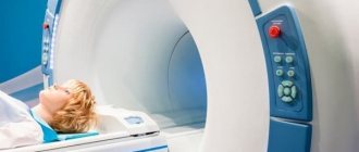
MRI of the brain in a child, images in a sagittal projection
How to prepare children for MRI
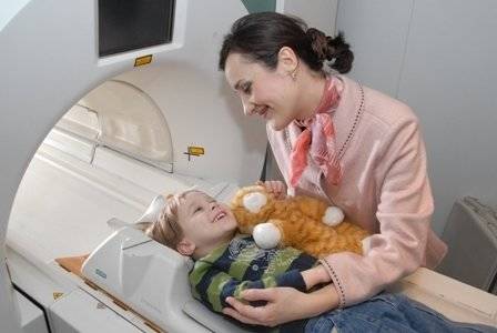
Before going to the procedure with your child, relieve him of internal fears about an unfamiliar situation. The day before, you need to talk to the baby, tell him what awaits him. You need to convince him that the process is safe and painless.
MRI in children has some peculiarities. Since it is difficult for a child to remain motionless for a long time, the session is carried out with the help of mild sedatives or under general anesthesia. During the entire tomography process, the child’s condition is monitored by an experienced specialist – an anesthesiologist.
Before conducting diagnostics with a contrast agent, the presence or absence of allergic rejection to the drug is determined. A severe form of bronchial asthma in a child’s diagnosis may also be a contraindication.
What should you not do before an MRI of the brain?
On the eve of the study, it is prohibited to drink alcohol; this measure is especially relevant when preparing for contrast MRI of the brain and blood vessels. Ethyl alcohol affects the tone of the walls of veins and arteries, which distorts the picture and complicates the diagnosis of pathological processes.
48 hours before the test, you should not take energy drinks, you should reduce the amount of coffee and other tonic drinks. It is not recommended to overeat on the eve of an MRI, change your usual diet, or eat new foods that can cause an allergic reaction.
Self-administration of medications is prohibited without prior approval from the attending physician. Patients are advised to maintain a sleep schedule 2-3 days before the examination and avoid overwork and stress.
Indications for MRI of head and neck vessels
As a rule, such an examination is not prescribed by specialists for no apparent reason. Reasons for prescribing a scan may include:
- ischemic disease;
- very frequent headaches;
- the appearance of dizziness;
- sensation of ringing and noise in the ears;
- vegetative-vascular dystonia.
After the scan, the doctor evaluates the resulting images, where it is possible to clearly visualize the blood supply to various areas of the brain and neck.
How often can I have a head MRI?
Magnetic resonance imaging is safe for the health of patients; the number of procedures is determined by the doctor. The frequency of head MRI depends on the clinical picture and the disease being diagnosed:
- hydrocephalus requires scanning every 5 years, if necessary, dynamic monitoring is carried out, the frequency of which is determined by the attending physician;
- neoplasms are scanned up to 4 times during the first year, then 1-2 times a year if there is no tumor growth;
- for multiple sclerosis, examinations are prescribed 1-2 times a year;
- stroke - an MRI is performed to establish a diagnosis, then sent for preventive examinations every 4 years;
- Alzheimer's disease - a one-time scan is recommended to confirm the nature of the pathology;
- To monitor recovery processes after surgery, 3-4 procedures are carried out during the first year; in the future, the frequency of MRI depends on the clinical picture.
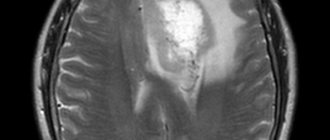
Alzheimer's disease on MRI
Scanning the brain with a magnet does not carry radiation exposure and does not have a negative effect on organs and tissues. The noise and the need to lie still during the entire examination create some inconvenience, but in general the method is painless and comfortable for the patient.
Preparing for an MRI with contrast
Contrast is a special dye solution administered intravenously. Before performing an MRI using a contrast agent, specialists need to make sure that the patient does not have problems with the kidneys or liver. A preliminary opinion from a urologist may be required.
It is important to exclude the presence of allergic reactions to the contrast agent. To do this, tests are carried out in advance for rejection of the composition.
Modern drugs are produced based on gadolinium salts. They do not cause allergies, but are excreted through the liver and kidneys. If you have chronic diseases of these organs, problems may arise. Therefore, preliminary consultation with a specialist is necessary.
What can you eat and drink before an MRI of the brain and blood vessels?
2 days before MRI of the brain, the patient should limit the intake of fatty, fried, salty, and spicy foods. It is not advisable to overload the digestive system. Experts recommend stewed vegetables, lean meat and fish, cereals, light soups, and fresh fruits.
It is advisable to maintain a drinking regime and avoid dehydration. Drinks allowed include still water, compotes, and juices. The volume of fluid consumed on the eve of an MRI should not exceed 1.5-2 liters, which will help avoid swelling. It is recommended to eat 3 hours before the procedure; you can drink water 1 hour before the scan.
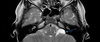
Tumors (probably craniopharyngioma) of the pituitary gland on MRI of the brain with contrast
Compliance with medical recommendations affects the quality of the study and increases the information content of the resulting images.
Is it possible to drink alcohol before an MRI?
Magnetic resonance imaging involves layer-by-layer scanning of a certain area of the body to obtain detailed images with a slice step of 1-2 mm. The study lasts 15-20 minutes. During this time you need to lie still. Otherwise, the pictures are unclear and it is impossible to objectively assess the condition of the internal organs and tissues.
Alcohol intoxication reduces the ability to concentrate. Withdrawal syndrome often manifests itself as trembling throughout the body. It is difficult for a person to remain calm, control movements, and follow doctor’s recommendations, which can negatively affect MRI results.
During the examination, the radiologist and the patient communicate via speakerphone. If there is severe discomfort, the procedure can be stopped, but this is not advisable, as time will be wasted and the scanning will have to start over. A hangover may cause nausea, vomiting, and panic, which increases the risk of interrupting the study.

Drinking alcohol on the eve of an MRI is not recommended.
In the process of deciphering scans, ambiguous situations may arise. If a tumor or inflammatory changes are suspected, it is necessary to increase the contrast of the images using intravenous administration of the drug gadolinium. Based on the specific distribution of a substance in the body, a pathological process is preliminarily judged. Under the influence of alcohol, the kidneys are subject to excessive stress. Since the enhancer is excreted from the body through urine, the drug will take longer to be eliminated, as will alcohol metabolites.
The negative answer to the question of whether it is possible to drink alcohol before an MRI is due to the factors listed above, and not to the ability of alcohol to cause fluid imbalance in the body (which is often mentioned on the Internet). You should refrain from drinking strong drinks 36-48 hours before the test. In case of chronic alcoholism, the process of preparing for an MRI should be discussed with your doctor. Detoxification therapy and the prescription of sedatives (under the supervision of a specialist) may be required. If the patient drank alcohol the day before or immediately before the test, it is better to reschedule the procedure.
Contraindications
There are relative and absolute contraindications. In the first case, research is possible under certain conditions, in the second it is unacceptable.
Absolute contraindications:
- pacemaker;
- middle ear implants;
- metal body prostheses made of ferromagnetic alloys;
- presence of insulin pumps;
- pregnancy in the early trimester;
- Ilizarov apparatus.
Relative contraindications:
- claustrophobia;
- mental disorders in the patient;
- prosthetic heart valves;
- Bracket system;
- patients under 5 years of age.
The examination is performed as prescribed by a doctor to clarify the diagnosis, as well as to monitor the dynamics of treatment. In most cases, MRI of the brain makes it possible to correctly diagnose and identify diseases at an early stage.
Can I drink alcohol after an MRI?
During scanning, the body is exposed to a powerful magnetic field. Under the influence of the latter, the state of hydrogen atoms in water molecules changes. The process is reversible; immediately after completion of the procedure, the effect disappears. The properties of the elements do not change, as do the biochemical reactions in the cells, so the method is considered absolutely safe. Upon completion of the scan, you are allowed to lead your normal lifestyle. The possibility of drinking alcohol after the procedure depends on the reason for which the study was ordered and what the MRI showed. If any serious illness is detected, drinking alcoholic beverages is undesirable. At the very least, this issue should be discussed with your doctor.
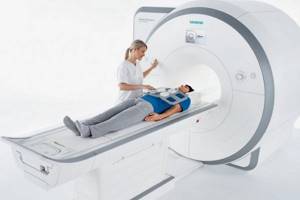
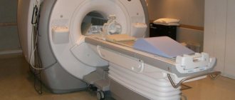
Positioning the patient for MR scanning
Can I drink alcohol after an MRI with contrast?
Preparations of the rare earth metal gadolinium are used as enhancers. The substance does not enter into chemical reactions in the body and is excreted unchanged by the kidneys. The elimination process takes 6-12 hours.
After MRI with contrast, alcohol can be consumed after gadolinium elimination is completed, taking into account the results of the study. If a person does not have serious chronic diseases and plans to attend a celebration, an increased drinking regime will help speed up the elimination of the drug. Alcohol should be excluded after an MRI with contrast that shows liver cirrhosis, tumor, or kidney damage.
You can undergo examination of any anatomical zone in the shortest possible time at the Magnit DC. You can make an appointment for a diagnostic procedure during a personal visit to the clinic or by calling +7 (812) 407-32-31.