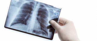
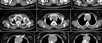
Chest X-ray
(radiography, fluorography) is a highly informative diagnostic method that uses X-rays - electromagnetic radiation with a wavelength of about 10-8 cm.
The method is based on the ability of radioactive X-rays, passing through human tissues and organs, to be partially or completely retained, leaving an image on the X-ray film. X-ray diagnostics
is widely used in clinical medicine and is also an important component of the minimum required testing in adults and children.
Indications for chest x-ray
- Cough, shortness of breath, chest pain.
- Pulmonary edema.
- Suspicion of inflammatory and non-inflammatory lung diseases (pneumonia, tuberculosis, tumors).
- Suspicion of the presence of fluid in the pleural cavity.
- Suspicion of diseases of the hematopoietic organs.
- Pulmonary embolism.
- Diseases of the spine and joints.
- Injuries to soft tissues and skeletal bones, fractures, dislocations.
- Contraindications to chest x-ray.
- There are no absolute contraindications.
- Relative contraindications – pregnancy.
Why do you need a lung x-ray?
COVID-19✓ Safe
Doctors advise, for preventive purposes, to undergo fluorography or x-ray examination once a year, mainly used to identify diseases of the chest organs.
With modern X-ray examination, the dose of radiation is minimal and does not harm the human body. X-rays help detect malignant tumors in the lungs and promptly identify dangerous diseases such as tuberculosis and pneumonia.
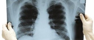
The tissues of the human body vary in density and have different abilities to transmit x-rays. The X-ray film shows an image of the organs being examined. At the same time, dense tissues and bones look lighter, and tissues containing air, that is, various natural or pathological cavities, look darker.
X-ray is considered an informative method for studying and diagnosing various diseases; as is known, it is early diagnosis that allows the doctor to prescribe the necessary treatment and quickly relieve the patient of the emerging disease. An experienced specialist can identify from the image, in addition to the main one, other so-called concomitant diseases, on which the effectiveness of the prescribed treatment also depends. For example, the image shows foreign bodies trapped in the lungs. An X-ray of the chest can show rib fractures and identify spinal diseases - scoliosis and osteochondrosis.
The doctor may also order a survey or targeted x-ray. An overview image shows the condition of an individual organ or organ system as a whole, for example, the general condition of the chest organs. A plain radiograph is usually ordered if pneumonia is suspected. The image shows pathological accumulations of fluid in the lungs; changes in the size of this organ can be determined.
With a targeted X-ray, you can see a certain area of the lungs much better, focusing on a suspicious tumor. This is especially important for prescribing a course of treatment, since during the treatment process a series of photographs can be taken and the effectiveness of the therapy used can be observed.
With an X-ray examination performed using modern equipment, the image can show not only pathological foci in the lungs, but also the condition of the blood vessels. This is very important - in some cases, an increase in the size of the vascular branch is the only symptom of the disease.
Today, many enterprises provide for an annual preventive examination, which all employees must undergo. During fluorography, which is done in a matter of minutes, a huge number of different diseases are revealed not only of the bronchopulmonary system, but also of cardiac pathologies.
Many people are afraid to undergo fluorography, considering it harmful. These fears are unfounded. When using modern equipment, the radiation dose is very small, and modern digital devices are equipped with a dosimeter. The permissible dose of X-ray radiation is 0.1 roentgen/year. With fluorography done on modern equipment, the radiation dose is only 0.005 roentgens/year.
Telephone
Where to get fluorography, chest x-ray in Moscow?
Of course, at GUTA CLINIC!
If you are looking for a clinic to do fluorography, chest x-ray in Moscow, choose GUTA CLINIC!
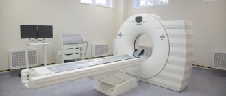
Only here we perform chest x-rays using the most modern x-ray machine
TELE DIAGNOST, which allows you to significantly reduce the radiation dose by eliminating the X-ray film exposure stage from the process - the image is read from the screen of the electron-optical converter and stored in the database.
TELE DIAGNOST is a digital X-ray device that allows you to obtain high-quality images in various modes, equipped with a system of objective and 100% accurate dosimetric control of each examination. For more accurate diagnostics, a special high-resolution film printer DRY VIEW 8100 from KODAK is used, which allows you to enlarge the image many times and subject it to computer processing.
X-ray examination of the chest
Diagnostic cost
RG-graphy of the chest (1 projection)
RUB 1,815
RG-graphy of the chest (2 projections)
2420 rub.
Description of the x-ray image by the doctor (1 image)
770 RUR
This X-ray examination is one of the most popular and in demand. With its help, you can obtain data about the organs of the chest and mediastinum.
What does a chest x-ray show?
The examination reveals pathological changes in the organs of the chest cavity (bronchi, lungs, pleura), shows the condition of the blood vessels and lymphatic ducts of this area. The image shows inflammatory processes, accumulations of fluid or gas, tumors, purulent foci, congestion and pulmonary heart failure. A chest x-ray is recommended if pulmonary tuberculosis is suspected.
X-rays are used to diagnose pathologies of the mediastinum - the space in the middle of the chest where the heart, esophagus and the largest artery - the aorta - are located. With its help, occupational diseases are identified in people working in conditions unfavorable for breathing.
The method is used to diagnose injuries to the ribs and sternum. In children, x-rays show the condition of the thymus gland, located in the mediastinum.
How is a chest x-ray performed?
The procedure does not require special preparation and can be done at any time of the day. Chest radiography is often performed in 2 projections, allowing you to examine the chest from all sides:
- straight (front or back) – the patient stands with his face or back to the X-ray machine;
- side (right or left) – the person stands sideways to the device.
In some cases, the examination is carried out in other positions:
- if the presence of fluid in the pleural cavity (hydrothorax) is suspected, photographs are taken on the side in a supine position;
- To clarify the condition of the apexes of the lungs, the patient is asked to bend back.
The doctor conducting the examination warns the patient about all the intricacies of the procedure.
The examination in our clinic in Moscow is carried out using modern digital equipment that provides minimal radiation exposure. The results are recorded on magnetic media, so the patient can copy the image onto his own media to show it to the attending physician.
Immediately after the thoracic x-ray, the patient receives ready-made images. Modern radiography is an informative and inexpensive procedure. You can find out about its price and sign up for an examination on the website or by calling the numbers provided.
Share on social media networks:
Advantages of digital fluorography
- High information content of the image.
- Minimum dose during examination.
- High throughput of the device.
- Affordable cost of examination.
Radiation exposure reduced by almost 10 times
, which allows you to perform it without restrictions for short periods of time. Compared to a film X-ray scanner, a digital device has a high throughput, which means benefits for both the doctor and the patient.
Chest X-ray
at GUTA CLINIC it is performed by highly qualified radiologists who have undergone special training and regularly improve their qualifications, candidates of medical sciences, and doctors of the highest category. The equipment used and the doctor’s skills will make the fluorography procedure easy and safe for you.
The dose of x-ray radiation used is fully consistent with the permissible values and is reduced through the use of an expert-level digital x-ray machine, as well as through individual selection of doses depending on the body density of each patient. We do everything to ensure that you return to your normal routine as quickly as possible and are sure that everything is in order with your health!
At GUTA CLINIC it is carried out as a survey radiography
, for example,
chest x-ray
, and targeted x-rays of specific organs and systems (heart, urinary system, gastrointestinal tract, musculoskeletal system).
X-ray of the chest organs (lungs, trachea, bronchi, mediastinum)
X-ray examination of the chest organs
This group of studies includes:
- fluoroscopy and radiography of the chest organs,
- plain chest x-ray
— digital fluorography.
Indications for chest X-ray examination:
- Preventive examination (including for patients with hazardous work conditions)
- Preparing for planned surgery
- Prolonged cough, shortness of breath of unknown origin, chest pain
- Dynamic monitoring of the condition of the lungs and pleural cavities during treatment
- Chest trauma
Methodology for examining the chest organs:
Fluoroscopy, radiography and fluorography, if necessary, can be performed in 3 projections - direct, right and left lateral, followed by taking radiographs in the same projections or targeted (increasing the area of interest).
Preparation and time of the study:
Fluoroscopy and radiography of the chest organs, fluorography and plain radiography of the chest do not require special preparation, the examination time is 10-15 minutes.
Dose load:
0t 0.01 to 0.03 mSv.
Advantages of X-ray examination:
- the ability to detect in real time (with fluoroscopy) the presence of pathological changes, their localization, assess the presence and localization of free fluid in the pleural cavity (pleurisy).
- with radiography - less radiation exposure due to the short duration of the exposure (image).
Disadvantages of X-ray examination:
— With fluoroscopy, there is no objective documentation of the detected changes, except for the doctor’s direct recording of what he saw on the screen, and the dose load on the patient also increases (but with a digital X-ray machine, the difference is almost minimal compared to radiography).
— X-ray examination is less informative in assessing the trachea and bronchi, the shadow of the mediastinum (lymph nodes), identifying small formations (less than 3-4 mm in size) of the lungs and pleura, assessing the amount of fluid in the pleural cavity. More informative research methods in this situation are MSCT of the chest and mediastinum, ultrasound of the pleural cavities.
— X-ray examination for heart defects or diseases is always performed in conjunction with other methods of radiation examination, such as ultrasound diagnostics (ultrasound).
Preparation for fluorography. How is a chest x-ray performed?
No special preparation is required if you are prescribed a chest x-ray or fluorography. If you wear jewelry or metal objects, they must be removed before the examination.
The following factors may influence the results of the study:
- Inability of the patient to hold his breath for a long time during inhalation or remain still during exposure;
- Incorrect centering of the patient's chest, which causes difficulty visualizing the costophrenic angle;
- Incorrect exposure (underexposure or overexposure).
Radiologists at GUTA CLINIC
It is recommended to undergo an annual
chest x-ray
for preventive purposes starting from the age of 15. Moreover, you need to pay attention to your health if you are at risk of developing diseases of the chest organs or have already had a history of these diseases, if you are an employee of a medical institution or a military personnel, and also if you are in contact with pregnant women or small children