Modern medicine has in its arsenal quite a lot of techniques with the help of which high-quality diagnostics of various diseases is carried out. The spine is the main axis of the body as a whole. The main load falls on it, so this structure is susceptible to various disorders that manifest themselves as a result of certain diseases or after injury.
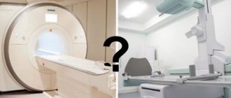
Examination of this area is possible in various ways, but the most effective methods are computer scanning and nuclear resonance screening. Many people mistakenly believe that these methods are absolutely the same, so there is no significant difference which method to give preference to. In fact, this is not true; CT and MRI have striking differences. Despite the external similarity in the equipment with which the described types of diagnostics are carried out, as well as similar results in the form of black and white images, these methods are used for different pathologies and to solve different goals and problems.
The question of which method is better and more informative is posed incorrectly, since different types of tomography study different areas and structures. The choice of the best diagnostic method in each individual case always remains with professional doctors, since both methods have both their limitations and advantageous indications.
If we compare the two types of tomography in terms of cost, there are no significant differences. Both diagnostics are classified as expensive. The price in each clinic specializing in hardware scanning will be slightly different, since each institution has its own pricing policy. Much depends on the modernity and power of the equipment, on the technical characteristics, on the number and qualifications of personnel and medical staff, on the availability of related procedures and the quality of consumables.
In what cases is CT or MRI of the spine prescribed?
There are a number of indications that require immediate diagnostic clarity. Traditional radiography is often prescribed as an initial step to examine the condition of the spinal column and adjacent structures and tissues. It will help identify obvious problems. However, to clarify the picture of the disease or in case of ineffectiveness of classical medical methods, the patient is additionally sent to a CT or MRI of the spine. This is necessary to find out the root cause of the disease even in the early stages of its onset.
On a regular x-ray you can see only noticeable fractures and cracks, the general condition of the bones and vertebrae. Microscopic lesions and soft tissues, as well as nerve fibers, are not visible on x-rays. If the cause of the disease is not identified and symptoms persist even after treatment, the doctor will necessarily refer the patient to a CT or MRI.
Indications for undergoing a CT scan of the spine are the following pathologies:
- osteomyelitis;
- constant pain syndrome;
- stiffness in movements;
- curvature of the spinal column of varying severity;
- dislocations and fractures;
- osteochondrosis;
- bone tissue dystrophy;
- implantation of ferromagnetic systems to maintain the spinal column, monitoring their correct location;
- determination of bone density;
- monitoring the migration of metastases, the degree of their spread in oncology;
- identifying the source of deformation and compression of the bone marrow.
An MRI of the spine is required if the following disorders occur:
- hernias between the vertebrae;
- tumor processes of varying nature and degree of spread;
- vascular pathologies;
- formation of liquid-containing capsules and cysts in the area of the spinal column and coccyx;
- change in the diameter of the spinal canal towards narrowing;
- injuries of adjacent soft tissues;
- genetic or acquired disorders of the development of the skeletal system;
- monitoring the condition of internal structures after surgery;
- detection of cancerous processes in soft fibers;
- spinal brain degeneration.
It should not be surprising that if you suspect the same diagnosis, different specialists may refer you to different types of tomography. The root cause of the disease can be pathologies localized in tissues of different density and nature. The doctor, based on the collected medical history of the patient, as well as based on his individual health characteristics, refers for diagnosis in one way or another. It should be remembered that tomography is not prescribed simply for the sake of idle curiosity. If the doctor strongly recommends undergoing a hardware examination, then there are indeed good reasons for this. Under no circumstances should you ignore a full examination; your subsequent condition and health, and sometimes life, depend on it.
In some complex cases, it may be necessary to jointly study the area under study using both methods; this will give the most complete picture of the developing pathology and the most accurate result.
How is an MRI of the lumbosacral region performed?
If it is planned to conduct a magnetic resonance scan of the lumbar region using contrast, it is recommended to follow a rational diet on the day of the procedure.
In other cases, no preliminary preparation for the study is required.
Before the scan, the patient will be asked to remove all metal items. This could be: accessories, jewelry, clothing containing metal fittings, glasses, removable hearing aids.
You must bring with you to your appointment your previously taken photographs, if available. The radiologist questions the patient about the presence of contraindications to the study and concerning symptoms.
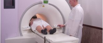
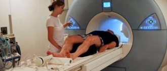
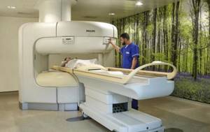
The patient lies down on a couch, which is placed in the tomograph tube.
You cannot move during the procedure, so the area being examined can be additionally secured with straps, although this is usually not required. The device produces quite loud sounds, but at the DiMagnit medical center they take care of the comfort of patients and provide special headphones to minimize the unpleasant sensations from the sounds of the operating device.
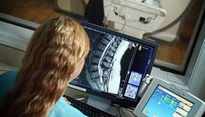
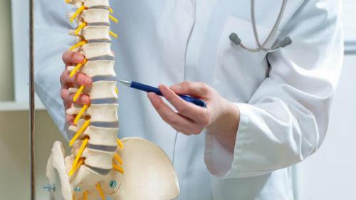
The radiologist carefully studies the images before making a conclusion
Once the scan is completed, results can be obtained in as little as 30 minutes. The radiologist draws up a detailed report in which he describes all the anatomical structures of the area under study, including those in which no abnormalities were detected. Possible pathologies are also described in detail, indicating their size and location in relation to other organs. The images are recorded on disk and sent to the patient along with a paper report.
Differences between CT and MRI of the spine
The main difference between diagnostic methods lies in the principle of their operation, hence the difference in results. In CT and MRI images, specialists see completely different things, although the same area is depicted. Let's take a closer look at how hardware research methods work:
MRI of the spine. The foundation of the operation of a tomographic MR installation is the generation of a high-frequency natural magnetic field. Once in the zone of influence of such a field, biological tissues begin to emit a resonating signature at the molecular level. The smallest particles of cells resonate with the magnetic field, which is detected by the equipment. These signals are processed by a computer program and transmitted to the monitor in the form of high quality and resolution images. MRI of the spine resonates best with particles that make up soft and dense tissues, as well as formations with a liquid substance in their composition (for example, the brain). The images clearly show muscles, tendons, cartilage, nerve fibers and the vascular network. The magnetic field does not have a damaging effect on the living fibers of the body, therefore, if necessary, the tomography procedure using nuclear resonance can be prescribed as many times as desired to a wide range of patients, including pregnant women and children from the first days of life.
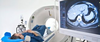
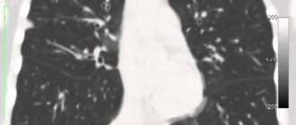
CT scan of the spine. This type of scanning is the modern successor to classical radiography, since the operating principle of computer equipment is based on the use of ionizing radiation. Small doses of X-rays have the ability to penetrate any human tissue. The denser the tissue, the more clearly it is visible in the photographs, since adjacent soft and liquid fibers and substances transmit x-rays almost unhindered. Therefore, a CT scan of the spine shows bone structures much better and more clearly in photographic images. Also, the images show contrasting internal cavities filled with air or liquid. Since the human body is capable of accumulating radiation, the number of possible examinations using computer screening is possible no more than twice a year.
CT or MRI of the spine: advantages and disadvantages
Each technique has both its pros and cons. Thus, MRI is more effectively used to identify pathological processes developing in soft tissues, for example:
- degenerative processes in the bone and spinal cord;
- infectious lesions of a bacteriological or viral nature;
- protrusion and compression of cartilage tissue between the vertebrae;
- early stages of oncology, degree of tumor ingrowth and spread of metastases into soft fibers;
- changes in configuration and destruction of the walls of blood channels;
- disorders in soft internal organs.
Important advantages of MRI of the spine are:
- complete safety in use by a wide range of patients, the ability to repeat sessions in multiple quantities;
- high quality of acquired images at any depth and from different angles, the ability to compose images into multidimensional projections;
- qualitative assessment of the state of organs and structures of the studied area.
The disadvantages of this diagnostic method include:
- high price for the provision of services;
- long duration of the procedure, the need to remain in a stationary position for a long time to obtain the correct result.
CT scan of the spine allows you to quickly recognize such anomalies as:
- old and new fractures and cracks in bones;
- prediction of the possibility of subsequent damage as a result of compression effects;
- determining the degree of operability of the patient in the preparatory period before surgery;
- postoperative monitoring of the condition of internal structures;
- the ability to quickly see internal bleeding, hematomas, and complex injuries.
The advantages of CT scan of the spine include:
- maximum degree of visual assessment of bone fibers;
- high resolution and quality of photographic images;
- painlessness and high diagnostic speed;
- possibility of diagnosing for patients with implanted metal implants.
The disadvantages of computer examination are:
- low financial accessibility, high price;
- high degree of ionizing radiation, the impossibility of frequent use of CT.
Indications for MRI of the lumbosacral region
The presence of pathologies of the spine in the lumbar and sacral regions and damage to the intervertebral discs can be manifested by the following symptoms:
- the occurrence of sharp pain in the lumbar region;
- chronic or periodically occurring lower back pain of varying intensity and nature: nagging, sharp, dull, aching, shooting, etc.;
- shooting or aching pain radiating to the thigh, buttock, lower leg, groin, ankle or even collarbone;
- pinching, loss of mobility, inability to straighten after bending or sudden movement;
- urinary and fecal incontinence;
- erection problems;
- weakness in the lower extremities or sensory disturbances.
If the symptoms described above occur, patients in most cases turn to a neurologist, who can prescribe an MRI diagnosis, based on the results of which a further treatment plan will be drawn up. If serious spinal cord lesions or neoplasms are detected, the patient may be referred to a neurosurgeon or oncologist for further consultation.
Contrast in CT or MRI of the spine
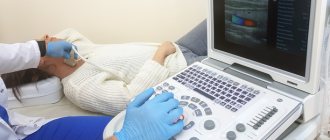
In case of complex pathologies, inflammatory processes, as well as at the first suspicion of oncology, specialists may prescribe a contrast scan. This mode involves the introduction of special coloring substances into the bloodstream, which make internal structures more visible in the images. For different types of tomography, different types of “dyes” are used. Thus, during MRI it is necessary that the intensifying solution be ferromagnetic. Therefore, a substance such as gadolinium salts is most often used. This solution is considered as safe as possible for use, as it gives an allergic reaction in exceptional cases. Before the procedure, the patient must be tested for an immune reaction to the contrast agent.
During CT scanning, the most common enhancer is an iodine-based solution. This element is more allergenic than its ferromagnetic counterpart, however, when used, the internal structures of the spine appear most clearly in the photo. If you have any allergies to medications, you should inform doctors in advance, both the treating doctor and the specialist on duty at the diagnostic center.
Contrast examinations are not suitable for everyone. In addition to allergy sufferers, coloring preparations are prohibited for administration to such categories of patients as:
- Pregnant women at any stage of pregnancy, as well as during breastfeeding.
- Young patients under 14 years of age.
- People suffering from chronic diseases of the urinary system and kidneys. If the function of timely removal of fluid from the body is impaired, the contrast remains in the tissues longer, which leads to complete intoxication.
- Persons suffering from asthma, heart and liver failure.
What to choose for qualitative research
The choice of the type of tomography remains with the treating specialist, who determines the diagnostic program for the patient personally. CT is better suited for examining the respiratory, urinary system, bone structures, tumors, blood vessels, heart and organs with cystic formations. It is best to check soft tissue systems, the brain of the head and spine, various types of inflammation, and oncology throughout the body using MRI.
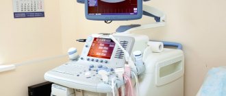
The quality of diagnostics is also influenced by the choice of equipment. Both computer and magnetic resonance technology use high-performance and low-performance devices. The clarity of MRI images is affected by the power of the equipment.
Low-floor machines produce a reduced inductive field. This is sufficient for visualization of obvious disease, confirmation of known disease, bone examination and preventive screening. However, to search for small, deep-seated foci of pathology, inflammation and degeneration in the brain, high-frequency equipment with a power of 1.5 Tesla and higher is required.
Regarding computed tomography, there is also a division of devices into step-by-layer, spiral and multispiral. The first type is outdated, since it takes one picture per full revolution of the tomograph ring around the studied area. Spiral and multispiral devices work faster, producing multiple scans in one pass of sensors along the body, which speeds up the procedure and reduces overall radiation exposure.
When should CT and MRI of the spine not be performed?
Any medical procedure has general and individual contraindications. In this case, both types of tomography are no exception. Screenings have both different prohibitions and general ones relating to the course of the procedure.
A CT scan of the spine is prohibited in the following cases:
- pregnancy, regardless of duration;
- children up to 14 years of age.
MRI of the spine is contraindicated:
- if there are any metal parts or electronic devices in the patient’s body;
- women in early pregnancy, up to 12 weeks.
General prohibitions for both types of examination are:
- Excess body weight from 120-200 kg, obese patients do not have the opportunity to fit in the inside of standard-sized tomographs. In this case, it is recommended to resort to open-configuration machines that do not have weight restrictions.
- Mental and neurological disorders accompanied by involuntary motor activity. For the results of the study to be correct, it is necessary to remain completely still during the session. If this is not possible for physiological reasons, it is better to resort to immersion in medicated sleep.
- Intense fear of confined spaces. To keep you calm and still, you can take sedatives or use an open-wall CT scanner.
- Emotional instability, state of shock;
- Diabetic syndrome.
Recommendations when choosing CT or MRI of the spine
The biggest mistake some patients make is choosing one type of examination or another on their own. In many cases, it is enough for a specialist to listen to the patient’s complaints, analyze the symptoms and see the results of x-rays in order to make the correct diagnosis. In such cases, spending significant amounts of money on an expensive procedure is simply not practical. But if a general examination is still required, then only a doctor can reliably determine which hardware method will be safer and more informative in each specific medical case.
The general condition of the body and all internal organs depends on the health of the spine; you should not take risks and self-medicate. A CT or MRI scan of the spine is designed to dispel or confirm the initial doubts of medical specialists, identify the cause of the pathology, help make a correct diagnosis and promptly begin treatment and rehabilitation measures. The earlier an anomaly is detected, the easier it is to completely get rid of it.