General information
The content of uric acid in urine is an indicator of uric acid excreted from the body in the urine, by which the nature and extent of purine metabolism can be assessed. This test is widely used in the diagnosis of gout, other metabolic pathologies, endocrine disorders, blood diseases, severe intoxications, etc.
Uric acid is a product of the breakdown of purine bases, which are actively involved in the production of nucleic acids (RNA, DNA), energy compounds (ATP), etc.
Purines enter the body from the following sources:
- food products (offal; meat; fish; legumes; cereals)
- body cells (as a result of disease or natural aging, they break down, releasing organic purines).
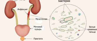
The liver and intestinal mucosa produce xanthine oxidase, an enzyme that breaks down purines into uric acid, which dissolves in the blood, enters the kidneys, where it is filtered and excreted from the body. Up to 70% of the total supply of uric acid is excreted in the urine (approximately 12-30 g per day). The remaining 25-30% enters the gastrointestinal tract, where one part of it is absorbed by intestinal bacteria, and the other part is excreted along with the stool.
An increase in uric acid in the urine (hyperuricemia) can provoke the formation of salt crystals (potassium, sodium) in the sediment (urate). The more urates accumulate in the body, the higher the risk of developing pathology - uraturia. This condition is typical for many serious diseases (gout, urolithiasis, etc.).
Also, an increase in the concentration of uric acid leads to a change in the internal acidity of the body. This condition is observed in diabetes mellitus (impaired glucose metabolism), lactic acidosis (increased lactic acid levels), etc.
Uric acid (UA) analysis
Uric acid is a substance whose content in the body is the most important marker of the state of purine metabolism. In healthy people, uric acid levels may increase due to increased consumption of foods containing purines - fatty meats, offal, beer.
A pathological increase in uric acid indicates cell damage due to the use of certain medications, widespread malignant tissue damage, severe atherosclerosis, and cardiovascular diseases.
If the concentration of uric acid in the blood exceeds the norm, there is a high probability of developing gout, which is also called “the disease of kings” (occurs due to the consumption of expensive fatty foods). Yes, this is that same hard lump on the foot in the area of the big toe.
Therefore, the level of uric acid in the body is the main marker in the initial diagnosis of gout. The analysis must be periodically taken by all patients with this pathology for subsequent monitoring of the course of the disease over time.
What is uric acid
Uric acid is a nitrogen-containing substance that is produced as a result of the body's processing of purine bases by cells.
By removing uric acid from the body, we also get rid of excess nitrogen. In healthy people, purines are produced by the body through the natural process of cell renewal, and are also slightly supplied through food.
During the normal functioning of all organs and systems, the breakdown of purines produces uric acid, which, after processing in the liver, is transported with the blood to the kidneys. There it undergoes filtration, and 70% of urea is excreted in the urine, and 30% is sent to the gastrointestinal tract, where it is disposed of in feces.
An increase in the concentration of uric acid in the blood is observed with massive cell destruction, a genetic predisposition to increased production of the substance, and kidney disease.
A condition in which the concentration of uric acid in the blood increases is called hyperuricemia. Uric acid is excreted from the body primarily through urine, so an increase in the concentration of the substance in the blood indicates impaired renal function.
If the body cannot cope with the utilization of uric acid, it accumulates in the blood in the form of sodium salts. They crystallize, which provokes the development of urolithiasis.
Increased levels of uric acid in the body over a long period of time lead to gout. This is a pathology characterized by the deposition of uric acid crystals in the joint fluid, which leads to inflammation and damage to the joints. If nothing is done, the disease will progress. Uric acid crystals will accumulate in organs and soft tissues, causing gouty damage to the kidneys and other structures of the body.
Crystallization of uric acid at its increased concentration is caused by the fact that this substance has extremely low solubility. It is noteworthy that hyperuricemia itself is not a separate pathology. Doctors consider it as a trigger risk factor for metabolic disorders, as well as a symptom of a number of diseases.
It is important to understand that the level of uric acid will be different for each person. It depends on age, gender, cholesterol levels, alcohol consumption and other factors.
Also, when interpreting test results, it should be taken into account that in children the level of uric acid in the blood will be significantly lower than in adults. The same applies to women in relation to men. The levels of uric acid in the blood of both sexes become equal only after 60 years.
Interpretation of uric acid analysis. Reference values.
| Age | Uric acid, µmol/l |
| children under 14 years old | 120 — 320 |
| women > 14 years old | 150 — 350 |
| men > 14 years old | 210 — 420 |
Indications
The prerequisite for the analysis is the main symptom of uraturia: brown or dark red color of urine against the background of excess urate. Assessment of the quality of the metabolic process of uric acid in the body.
- Identification of systemic disorders that accelerate or slow down the secretion of uric acid;
- Diagnosis of gout and choice of treatment tactics for the disease;
- Family history of gout (to diagnose early and asymptomatic increases in uric acid concentrations);
- Carrying out a complex of rheumatic tests to determine the etiology (cause) of inflammation and damage to the joint;
- Determination of kidney dysfunction, degree of complications;
- Diagnosis of urolithiasis;
- Carrying out a set of kidney tests to diagnose kidney diseases;
- Diagnosis of pathologies of the cardiovascular system (assessment of the likelihood of complications, general prognosis);
- Diagnosis of metabolic disorders and related diseases (diabetes mellitus, anorexia, obesity, etc.);
- Monitoring the patient during the treatment of oncological pathologies (radiation and chemotherapy), against the background of which there is massive cell destruction and the release of uric acid;
- Development of a diet plan;
- Monitoring the patient’s condition during long-term alcohol abuse;
- Monitoring uric acid levels in patients after prolonged fasting (diet, fasting, vegetarianism, hydrotherapy, etc.);
- Diagnosis and monitoring of therapy for chronic polycythemia (blood disease with increased red blood cell volume).
The interpretation of the analysis results is carried out by specialists: nephrologist, urologist, pediatrician, general practitioner, therapist, etc.
Microscopy of urine sediment (NICON)
What are urinary sediment salts?
Urine is a solution of various salts, the precipitation of which is determined by the urine reaction (pH) and a number of other factors. The presence of salt crystals in urine sediment primarily indicates a change in the urine reaction to the acidic or alkaline side.
Thus, in acidic urine, uric acid, oxalates and urates often precipitate, while in alkaline urine crystals of ammonium urate, tripel phosphates, amorphous phosphorus salts, lime carbonate, etc. can often be found. The nature of nutrition is of great importance.
Indications for the purpose of analysis:
- diseases of the urinary system;
- suspected diabetes mellitus;
- assessment of the toxic state of the body;
- assessment of the course of the disease;
- monitoring the development of complications and the effectiveness of treatment;
- Persons who have had a streptococcal infection (tonsillitis, scarlet fever) are recommended to take a urine test 1-2 weeks after recovery.
Urine is a solution of various salts, which can precipitate (form crystals) when the urine stands. Low temperature promotes the formation of crystals. The presence of certain salt crystals in the urinary sediment indicates a change in the reaction towards the acidic or alkaline side. Excessive salt content in urine contributes to the formation of stones and the development of urolithiasis. At the same time, the diagnostic value of the presence of salt crystals in urine is usually small. When and what kind of crystals appear in a general urine test?
Uric acid and its salts (urates):
- highly concentrated urine;
- acidic reaction of urine (after physical activity, meat diet, fever, leukemia);
- uric acid diathesis, gout;
- chronic renal failure;
- acute and chronic nephritis;
- dehydration (vomiting, diarrhea, fever);
- severe inflammatory-necrotic processes;
- malignant tumors;
- leukemia;
- cytostatic therapy;
- lead poisoning;
- in newborns.
Hippuric acid crystals:
- eating fruits containing benzoic acid (blueberries, lingonberries);
- diabetes;
- liver diseases;
- putrefactive processes in the intestines.
Tripelphosphates, amorphous phosphates:
- alkaline urine reaction in healthy people;
- vomiting, gastric lavage;
- cystitis;
- Fanconi syndrome, hyperparathyroidism.
Calcium oxalate (oxaluria occurs with any urine reaction):
- eating foods rich in oxalic acid (spinach, sorrel, tomatoes, asparagus, rhubarb, potatoes, tomatoes, cabbage, apples, oranges, strong broths, cocoa, strong tea, excessive consumption of sugar, mineral water with a high content of carbon dioxide and organic salts acids);
- severe infectious diseases;
- pyelonephritis;
- diabetes;
- ethylene glycol poisoning;
- oxalosis or primary hyperoxalaturia (genetic deficiency).
Neutral phosphate lime:
- arthritis and arthrosis of rheumatic etiology;
- Iron-deficiency anemia;
- chlorosis.
Leucine and tyrosine:
- severe metabolic disorder;
- phosphorus poisoning;
- destructive liver diseases;
- pernicious anemia;
- leukemia
Cystine:
- congenital disorder of cystine metabolism - cystinosis;
- cirrhosis of the liver;
- viral hepatitis;
- state of hepatic coma
- Wilson's disease (congenital defect of copper metabolism).
Xanthine : Xanthinuria is caused by the absence of xanthine oxidase.
Uric acid levels
| Patient age | Uric acid in urine, mmol/day (with a normal diet) |
| Up to 1 year | 0,35 – 2,0 |
| 1 – 4 years | 0,5 – 2,5 |
| 4 – 8 years | 0,6 – 3,0 |
| 8 – 14 years | 1,2 – 6,0 |
| Over 14 years old | 1,48 – 4,43 |
Factors of influence
Increase the research result:
- insufficient volume of daily urine, violation of collection and storage rules;
- long-term use of medications or their large doses: atophan;
- cortisol;
- allopurinol;
- cytostatics;
- salicylates;
- vitamins;
- ibuprofen;
- paracetamol;
- lithium salts;
- methotrexate;
- prednisolone, etc.;
Lower the analysis result:
- drugs: thiazide diuretics;
- glucocorticoids;
- insulin;
- furosemide;
- pyrazinamide, etc.;
Uric acid in daily urine
Uric acid in urine is uric acid from the blood filtered through the kidneys.
Synonyms Russian
Purine-2,6,8-trione, product of purine base metabolism, trihydroxypurine, 2,6,8-trioxypurine, heterocyclic urea ureide.
English synonyms
Urine Uric Acid, Urine Uric Acid Quantitative (24-Hour), Uric Acid in urine.
Research method
Enzymatic method (uricase).
Units
mmol/day (millimoles per day).
What biomaterial can be used for research?
Daily urine.
How to properly prepare for research?
- Eliminate alcohol from your diet the day before donating urine.
- Do not eat spicy, salty foods, or foods that change the color of urine (for example, beets, carrots) for 12 hours before the test.
- Do not take diuretics 2 days before the test (as agreed with your doctor).
- Avoid physical and emotional stress during the collection of daily urine (during the day).
General information about the study
Uric acid is formed as a result of cell renewal and also enters the body with food. Most of it leaves the body in urine, less in stool. With excessive formation of uric acid, its concentration in the urine can increase significantly, and if the kidneys are unable to filter blood in normal volumes, it can decrease.
Consistently high levels of uric acid cause the formation of uric acid crystals in the joint cavity. This painful pathological condition is called gout. If left untreated, uric acid crystals inside joints and adjacent tissues can form deposits that protrude on the surface of the body as hard bumps.
Persistently high levels of uric acid in the urine can lead to stone formation.
Uric acid, which is dissolved in the blood, is delivered to the kidneys, where, after filtration, it is excreted in the urine. If the body produces too much uric acid over a long period of time or does not eliminate it well enough, a person may experience problems urinating, fever, chills, fatigue, and joint pain.
A condition in which the level of uric acid in the urine is elevated is called hyperuricosuria. This can cause kidney stones to form, blocking the normal flow of urine in the kidney tubules, ureter, and bladder.
What is the research used for?
- To assess uric acid metabolism.
- To identify disorders affecting uric acid production.
- To determine the severity of kidney damage.
When is the study scheduled?
- If necessary, find out the cause of the formation of kidney stones.
- When monitoring the condition of patients with gout.
What do the results mean?
Reference values: 1.48 - 4.43 mmol/day.
Causes of increased concentration of uric acid in urine:
- eating large amounts of food rich in purine bases (meat, especially offal),
- gout (increased production or insufficient excretion of uric acid),
- urolithiasis disease,
- polycythemia vera (excessive production of blood cells),
- Lesch-Nyhan syndrome (increased uric acid synthesis),
- Wilson–Konovalov disease,
- viral hepatitis,
- sickle cell anemia,
- malignant neoplasms with metastases, multiple myeloma, chronic myeloid leukemia (uncontrolled cell growth and division),
- Fanconi syndrome (decreased tubular reabsorption of uric acid due to a defect in tubular development).
Reasons for low concentration of uric acid in urine:
- chronic kidney diseases, such as chronic glomerulonephritis,
- xanthinuria (little uric acid is formed due to xanthine oxidase deficiency),
- lead intoxication (due to a pronounced decrease in kidney function),
- chronic alcoholism,
- folic acid deficiency.
What can influence the result?
Falsely inflated results are contributed to by:
- stress and intense physical activity,
- injuries,
- beta-blockers, caffeine, vitamin C, large doses of acetylsalicylic acid, calcitriol, asparaginase, diclofenac, isoniazid, ibuprofen, indomethacin, piroxicam, paracetamol, lithium salts, mannitol, mercaptopurine, methotrexate, nifedipine, prednisolone, verapamil.
A falsely low result can be caused by:
- allopurinol, glucocorticoids, imuran, contrast agents, vinblastine, azathioprine, methotrexate, spironolactone, insulin, small doses of acetylsalicylic acid, furosemide, ethambutol, pyrazinamide.
Important Notes
- 20-25% of people with nephrolithiasis develop hyperuricosuria.
Also recommended
- Serum uric acid
- Urea in serum
- Serum creatinine
- Urea in urine
- Creatinine in urine
Who orders the study?
Therapist, rheumatologist, gynecologist, hepatologist, oncologist, nephrologist.
Reasons for increased values (hyperuricemia)
Eating disorder:
- the predominance of meat products and alcohol in the diet;
- consumption of foods and drinks that acidify cells: fermented milk products, wine, citrus juices, kvass, etc.;
- the use of sweeteners (xylitol, fructose, sorbitol), which activate the formation of uric acid in the liver.
A significant number of cells die in the body as a result of:
- gout;
- pneumonia (pneumonia);
- anemia (hemolytic and pernicious);
- psoriasis (non-infectious skin lesion);
- rhabdomyolysis (destruction of muscle cells);
- polycythemia vera;
- oncological processes: leukemia (blood cancer);
- myeloma (plasma cell cancer);
- lymphoma (malignant neoplasms in the lymphatic system);
The acid-base balance of the blood decreases when:
- energy deficiency in the body as a result of fasting;
- oxygen starvation of tissues;
- ketoacidotic (diabetic) coma;
- carbon monoxide intoxication;
- lactic acidotic coma as a result of increased lactic acid in the serum.
Disorders of uric acid metabolism with:
- hypothyroidism (hypersecretion of thyroid hormones);
- cirrhosis of the liver (changes in the structure of its tissues);
- glycogenosis enzyme deficiency (hereditary defects in enzyme production);
- Lesch-Nyhan syndrome (hereditary increase in uric acid synthesis in children).
Impaired production of uric acid and its filtration in the kidneys as a result of:
- renal failure (regardless of severity);
- complicated pregnancy: gestosis (late toxicosis);
- eclampsia (increased blood pressure in pregnant women);
Main symptoms of hyperuricemia
Hyperuricemia is an increase in uric acid in the urine. The main symptoms of this condition:
- Pain, burning and discomfort when urinating;
- Feeling of incomplete emptying of the bladder;
- Chills and fever;
- Increased fatigue and weakness;
- Aches in the joints;
- Pain and discomfort in the kidney area (may indicate the formation of stones), etc.
Complications of hyperuricemia
Gout is the most common disease that occurs due to increased levels of uric acid in the urine. The main manifestation of gout is inflammation of the joints (arthritis), caused by the deposition of salt crystals in it.
Another complication of hyperuricemia is renal failure due to the accumulation of toxic metabolic products in the kidneys. Lack of treatment for this pathology for a long time can lead to the need for dialysis (extrarenal blood purification) and kidney transplantation.
Kidney stone disease: features and clinical picture
This disease affects about 15% of the world's population. It is characterized by the formation of kidney stones. Formations may consist of various mineral deposits. Small stones can be removed from the body unnoticed by a person. Large stones can block the ureter, causing severe pain. The reasons for the development of kidney stones are currently unknown. Risk factors include genetic predisposition, gout, excess calcium, metabolic disorders, and insufficient fluid intake. Stones cause inflammation of the mucous membrane, provoke secondary nephritis and other diseases. Often the disease occurs in a latent form for a long time. Stones form, but the person does not experience any health problems. Symptoms appear when the stone begins to move. In this case, the person experiences pain in the back, stomach and side. Pain may spread to the groin. Urine appears brown and red. Urination hurts. Symptoms of kidney stones also include nausea and frequent urination. If the disease is complicated by the presence of infection, fever and chills may occur. With small stones, the disease manifests itself as recurrent renal colic. After a while the calculus comes out. Between attacks, a person experiences dull pain in the lower back. The pain may worsen with heavy lifting, walking, or for no apparent reason. The appearance of pus in the urine indicates the presence of an infectious process. Prolonged lower back pain that cannot be relieved is a reason for hospitalization. A purulent infection can lead to the development of serious complications, so the patient should be under constant medical supervision. In some cases, surgical treatment is indicated.
Lowering values
A decrease in the concentration of uric acid (hypouricemia) occurs with kidney dysfunction (inability to perform a filtering function).
- Celiac disease (gluten protein intolerance);
- Wilson-Konovalov disease (hereditary disorder of copper metabolism, which leads to liver damage);
- Fanconi syndrome (the kidneys are unable to retain uric acid in the body);
- Xyntinuria (lack of an enzyme involved in the synthesis of uric acid);
- Oncological processes: Hodgkin's disease (oncology of lymphoid tissue);
- bronchogenic carcinoma (lung cancer);
- multiple myeloma (plasma cell cancer);
Gout: general information
Men are more susceptible to the disease; women are less likely to have it. Excess uric acid is deposited in the joint in the form of needle-shaped crystals. The joints of the big toes are most often affected first. The disease is chronic, manifested by acute recurrent attacks. Usually the attack begins at night. It is characterized by an acute inflammatory process, which is accompanied by high fever. The duration of the attack is on average 10-15 days. After its completion, the function of the joint is restored, pain disappears, and the person feels healthy until the next attack occurs. The diagnosis is made based on the clinical picture, results of laboratory and instrumental studies. A complete cure is impossible. Therapy relieves symptoms and reduces the frequency of attacks. Thanks to treatment, a person can return to normal life. It must be borne in mind that for gout, diet is an important part of treatment. Dishes and foods that contain a large amount of purines (meat soups, fatty fish, spinach, coffee, legumes, alcohol, etc.) are excluded from the diet. Another important point is compliance with the drinking regime. Without drug therapy and dietary nutrition, gout leads to disability. The frequency of attacks increases. New joints are involved in the pathological process.
Preparing for analysis
- diuretics;
- aspirin;
- antipyrine;
- furagin;
- vitamin B, etc.
2-3 days before the analysis, you must stop using diuretics, and also remove diuretic foods and drinks from your diet (watermelon, tea, etc.);
2 days in advance, exclude from the diet vegetables and fruits that can affect the color of urine (beets, blueberries, carrots, etc.)
per day:
- It is not recommended to eat too spicy, salty and fatty foods;
- It is prohibited to consume alcoholic beverages and energy drinks, smoking, including electronic cigarettes;
- It is advisable to avoid overheating the body (staying in the sun, including visiting a solarium, hygiene procedures in a bathhouse/sauna).
Urine testing is not recommended during menstruation.
Rules for collecting biomaterial
- at 6-7 o’clock in the morning the bladder is emptied (in the usual way, into the toilet);
- after this, a thorough hygienic shower of the genitals is performed using boiled or purified water;
- during the day, all daily urine is collected in a clean container (2-3 liter jar) with a lid, with the last urination being carried out at 6 o’clock in the morning the next day;
- during urine collection, the container with biomaterial must be stored in the refrigerator at a temperature of 4-8°C;
- after collecting the last portion of urine, the contents of the jar must be mixed and, having previously measured the total volume (daily diuresis), pour about 40 ml into a sterile container;
- The following information must be indicated on the container: patient’s name;
- age;
- date of collection of material;
- daily diuresis in ml (total volume of urine per day);
The entire volume of urine is not required for analysis!
Other urine tests
- Uric acid in urine
- Urea in urine
- Creatinine in urine
- Rehberg's test
- Blood in urine
- Free cortisol in urine
- Protein in urine
- General urine test during pregnancy
for>at>on>for>for>