The small intestine is the part of the intestine that is located between the large intestine and the stomach. Its function is difficult to overestimate: it carries out a significant amount of food processing processes, ensuring the absorption of nutrients. An X-ray of the small intestine makes it possible to determine the state of this anatomical formation and identify a number of disorders characteristic of it. The procedure can be performed using contrast and has a high diagnostic value. It is a relatively safe non-invasive diagnostic method that has become widespread due to its simplicity and accessibility.
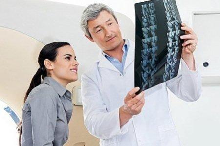
You can undergo diagnostics of the small intestine at the CELT multidisciplinary clinic. Our specialists will help identify the cause of the ailment, determine the localization of the pathological focus and provide all the data necessary to the attending physician.
X-ray of the small intestine - 6,000 rubles.
15-20 minutes
(procedure duration)
Types of X-ray of the small intestine
Modern radiology offers different techniques for performing radiography. Their choice is made on an individual basis, taking into account the clinical picture and patient characteristics.
| Types of research | Their distinctive features |
| Radiography | Aimed at linear visualization of the small intestine on film or digital media. It is highly accurate and informative. |
| X-ray | Aimed at studying an organ in the process of its functioning. It is distinguished by its duration and relatively high radiation exposure to the patient’s body. |
| CT scan | Allows you to obtain a three-dimensional image of the diagnosed organ. It has a high radiation dose and price. |
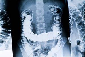
Most often, diagnosis of the small intestine is carried out using contrast, due to its anatomical features. Being hollow, it is poorly visualized on the image, so the use of contrast agents is advisable for better visualization. The process can use so-called double contrast. This is how an X-ray of the small intestine is performed with barium, after which air is used.
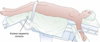
Conditions for performing fluoroscopy of the gastrointestinal tract
X-rays are taken at intervals of twenty minutes to an hour. The process may take several hours. The subject periodically needs to change position on the table (lying, standing, on his side) and wait from half an hour to an hour for the contrast to move to the next section of the digestive canal.
Depending on the areas studied, there are several types of fluoroscopy of the stomach and intestines :
- irrigoscopy - x-ray of the colon (the suspension is administered by enema);
- fluoroscopy – images of the upper gastrointestinal tract (pharynx, esophagus, stomach, first part of the small intestine);
- esophagography – pictures of the esophagus, pharynx.
During intestinal fluoroscopy, general photographs and targeted ones are taken from different angles for a better examination of pathologies or disorders of the examined area. A suspension of pure barium sulfate is used as a contrast.
Contrast in intestinal fluoroscopy
X-ray diagnostics can be conventional (contrast), with double and air contrast.
- The patient takes part of a pre-prepared barium suspension orally. To better distribute the solution throughout the gastric mucosa, the patient’s abdomen is thoroughly massaged.
- Double contrast - taking the solution through a perforated tube. X-ray of the intestines and stomach with 2nd contrast allows you to study in detail the disturbances in the relief of the organ lining.
- Air contrast - taking a solution of crystalline soda instead of barium.
Indications and contraindications for TC radiography
Indications
Diagnostics is indicated in cases where the patient complains of the following clinical manifestations:
- pain symptoms localized in the abdominal area and not going away for a long time;
- frequent vomiting;
- symptoms of anemia;
- painful bloating due to the accumulation of excess gases;
- constipation for a long time;
- diarrhea mixed with blood or pus.
Contraindications
The procedure is used with caution if the patient is pregnant or breastfeeding, for patients who are in serious condition, for patients under 15 years of age and for those patients who have received a high dose of radiation over the past year. When using radiocontrast agents, the list expands significantly. It is added to patients who have individual intolerance to the components of the contrast agent or suffer from:
- severe renal and liver failure;
- intestinal obstruction;
- blood clotting disorders;
- diabetes mellitus in severe forms;
- rapidly progressing ulcerative colitis;
- tachycardia.
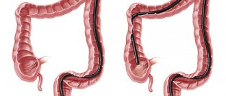
X-ray of the intestine
13.07.2020
Today, the most common in quantitative terms are diseases of the gastrointestinal tract. There are a large number of diagnostic techniques to identify them. X-ray examination is one of them. When examining the intestines using fluoroscopy , a contrast agent is used, in most cases it is barium sulfate. X-ray examination of the intestine has its own unique name - irrigoscopy. Carrying out irrigoscopy helps to identify a number of pathological processes in the colon. Using this type of diagnostics, a specialist determines with maximum accuracy whether the colon lumen meets the standards, the elasticity of the intestinal , and the relief of the mucous membrane. Any deviations from the norm in functional terms will also be detected during an X-ray of the intestine .
The most common reasons for performing an X-ray of the intestine include bleeding from the rectum , mucous or purulent discharge, severe pain in the anus, and regular bowel movements (constipation or diarrhea). Irrigoscopy can be prescribed as an additional study, often this happens in cases where the results of previous diagnostic procedures are of little information value. It should be noted that intestinal x-rays are contraindicated in pregnant women, during breastfeeding, in severe general condition of the patient, in cases of suspected violation of the integrity of the intestinal , in a number of inflammatory pathological processes, for example, ulcerative colitis.
Carrying out fluoroscopy of the intestines requires special preliminary preparation. To obtain maximum informative results, it is necessary that the intestinal be completely cleared of food debris. In this regard, the diagnostic procedure is usually prescribed in the morning, and the patient should follow the doctor’s regarding nutrition for several (3-4) days before the procedure. First of all, you will have to exclude products that contribute to gas formation. It is necessary to completely avoid alcohol these days. On the eve of the diagnostic procedure, it is recommended to perform a forced cleansing of the body. Moderate activity laxatives or a cleansing enema will help with this.
Consider taking an X-ray of the intestine . The procedure can take three quarters of an hour. Initially, the radiologist performs a survey fluoroscopy . The patient is placed on a special couch and the specialist takes a picture. The patient must then assume the position recommended by the radiologist and a contrast agent is injected. Contrast is administered through the anus. During the diagnostic process, the specialist may ask the patient to change position several times, this helps to better distribute the contrast agent in intestines and thereby ensures optimal information content of the study. In some cases, they resort to filling the intestines with air, this is also done through the anus. Air is necessary to straighten the intestinal . At the end of the contrast-enhanced diagnosis, the patient must have a bowel and return for a final image to reflect functional compliance with normal bowel . X-ray of the intestine is a painless and harmless procedure. It is necessary to take into account that any X-ray diagnostics, including irrigoscopy, must be carried out exclusively as prescribed by a doctor . Only a specialist should interpret the diagnostic results.
Published in Diagnostics and examinations of Premium Clinic
How is TC radiography performed using contrast?
One of the most common techniques using contrast involves the use of barium. It allows you to assess the condition of not only the small intestine, but also the stomach along with the esophagus, since the barium mass passing through them makes them radiopaque. The procedure includes the following:
- The patient assumes a standing position in front of the x-ray machine and undergoes a standard x-ray;
- The radiologist asks him to drink the barium suspension in small sips and lie down on the X-ray table in half an hour;
- The shooting is carried out as the diagnosed parts of the gastrointestinal tract are filled with contrast. In this case, the patient lies on his back, left and right side, turning at certain intervals at the command of the diagnostician.
On average, this procedure lasts up to two hours, after which the patient must immediately eat in order to restore the body’s energy balance.
Symptoms that are referred for fluoroscopy of the gastrointestinal tract
X-ray examination of the intestine and upper tract is mandatory for :
- long-term heartburn;
- symptoms of the acute stage of the disease (blood in the stool, persistent vomiting);
- sudden weight loss, diarrhea;
- lack of appetite;
- pain in the chest and abdomen;
- suspected tumors;
- ulcers;
- ingestion of solid foreign objects.
In some cases, fluoroscopy of the stomach and intestines is performed or not, depending on the expected benefit of the procedure. Contraindications to the examination are perforation, intestinal obstruction and pregnancy.
However, in conditions of high threat to the patient, radiography of the intestines and stomach can be carried out in compliance with special requirements. The same applies to cases of obstruction, when the patient is scheduled to undergo perforation surgery.
Advantages of carrying out X-rays of the chest cavity at the CELT clinic
We have our own diagnostic center with a full range of equipment necessary to conduct research in a wide variety of areas. Modern X-ray equipment not only makes the procedure safe, but also takes it to a new quality level. Our patients can count on:
- affordable prices;
- accurate diagnosis;
- the procedure is performed by radiologists with over 15 years of experience;
- use of effective safe means;
- use of modern gentle technologies.
You can find out our prices and make an appointment by calling in advance.