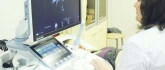
Pelvic ultrasound during early pregnancy is the only safe and non-invasive diagnostic method that allows you to assess the health of the expectant mother and fetus. The procedure will help to confirm the fact of successful fertilization in the early stages, exclude incorrect localization of the embryo and, assess the characteristics of the course of pregnancy, find out the exact timing of gestation. is available to all patients of the International Hemostasis Clinic, where appointments are conducted by experienced specialists of the highest category. With us you can undergo all types of diagnostics and receive high-quality medical care. The study is carried out using highly sensitive ultrasound equipment. Diagnostic results are provided immediately upon completion of the procedure.
Indications for ultrasound during pregnancy

Ultrasound diagnostics are performed to confirm pregnancy, assess the condition of the genital organs, see how the child is developing, examine amniotic fluid, and check the condition of the placenta.
There are several methods for performing ultrasound: when the sensor is inserted into the vagina (transvaginal diagnosis), the examination is carried out from the anterior wall of the abdomen (transabdominal method). The first option is considered more accurate.
How to prepare?
The day before the ultrasound, it is necessary to exclude from the diet foods that increase gas formation in the intestines - legumes, cabbage, yeast baked goods, brown bread, soda, etc. As prescribed by the doctor, additional medications for flatulence are taken.
Before a transabdominal ultrasound, drink at least a liter of still liquid to ensure your bladder is full at the time of the procedure. In this case, the uterus moves closer to the abdominal wall, and the doctor sees a clearer picture.
Before a transvaginal ultrasound at home, perform a toilet of the external genitalia. Immediately before the procedure, empty your bladder - this will make you more comfortable during the examination - and refresh the perineum with a damp cloth.
Diagnosis of the course of pregnancy and the condition of the fetus using ultrasound in the first trimester:
- accurate – allows you to accurately determine normality and pathology;
- informative – provides the necessary data for correct management of pregnancy, taking into account the individual characteristics of the mother and child;
- painless - does not require drug pain relief;
- proven safe for mother and child;
- does not require violating the integrity of the skin and mucous membranes, so there is no risk of infection;
- does not require special training;
- carried out in 20 minutes;
- The results are ready within 10 minutes after the procedure and can be used for medical purposes.
In addition, paid ultrasound of the 1st trimester in our clinic in St. Petersburg is affordable for a wide range of patients. If the screening results reveal a deviation from the norm, the doctor prescribes a more detailed examination.
There are the following routine ultrasound examinations during pregnancy:
- Ultrasound examination in the first trimester - done at 11-14 weeks. The procedure is necessary to exclude ectopic pregnancy, assess embryo development (heartbeat, motor activity, etc.), and determine the gestational age. The yolk sac and umbilical cord are also checked. They look to see if there are any pathologies in the mother’s genital organs.
- In the second trimester - approximately 20-24 weeks , they can be carried out at earlier stages if the pregnant woman is prescribed a second biochemical screening. Diagnostics allows you to assess the dynamics of development and growth of the embryo: the length of the femur, abdominal parameters, and head circumference are assessed. During the second examination, the blood flow between the pregnant woman and the fetus is examined (Doppler measurements are performed), the cervix is measured, as well as the length of the cervical canal. Now it is already possible to accurately determine the sex of the child (if the fetus does not turn away) and assess the possible threat of premature birth.
- In the third trimester - an ultrasound is performed at 30-34 weeks in order to see the maturity of the placenta (if it has aged ahead of schedule, premature birth is possible), to check the condition of the child and its location. The examination helps to identify blood flow disorders and defects in the functional state of the fetus.
This is a list of routine ultrasound examinations approved by the Ministry of Health, however, there may be more of them, it all depends on the condition and well-being of the pregnant woman, as well as the child.
Doctors in this field
Indications for examination
If there is no regular menstruation, you need to take a rapid pregnancy test. Even if such a test shows a negative result, but your period still does not occur, it is worth visiting a gynecologist, who, after examination and medical history, will give a referral for an ultrasound scan. Pelvic ultrasound during pregnancy is performed for the following indications:
- The delay lasts more than 10 days, while the woman complains of discomfort in the abdomen, spotting brown discharge, not similar to menstrual discharge.
- Previously, the patient had an ectopic or frozen pregnancy, or an early miscarriage.
- Surgical intervention on the uterus, including caesarean section.
- Irregular menstrual cycle.
Also, an ultrasound of the pelvis during early pregnancy is indicated; if there is a delay, the test is positive, but the size and features of the uterus upon palpation do not tell the specialist about the onset of pregnancy. Ultrasound diagnostics will confirm or refute the specialist’s fears and show whether successful implantation of the fertilized egg has occurred and its location.
Ultrasound is mandatory for women who have previously had surgery on the uterus. The procedure will help assess the condition of the scar, confirm the correct implantation of the fertilized egg, and eliminate complications.
What is examined during an ultrasound during pregnancy
In the first trimester, the location of the ovum is diagnosed: uterine or ectopic pregnancy. The collar area is examined; if the area on the back surface is thicker than the norm, then this may be a genetic pathology. The pregnant woman is then sent for consultation with a geneticist. Ultrasound examination can detect Down syndrome. At 10-14 weeks, the amount of amniotic fluid must be checked. If there is not enough, the diagnosis is oligohydramnios, and if there is too much, polyhydramnios is diagnosed. The color of the water is examined; cloudiness indicates the presence of infection. The uterus is checked: wall thickness, tone, presence of myomatous nodes.
A second ultrasound is necessary to identify possible abnormalities in the development of fetal organs. This examination can detect facial defects, such as cleft lip. The structure of the heart is studied and the heart rate is calculated. The structure of the placenta is examined; if there are deviations, placenta previa can be diagnosed. The doctor must check whether the umbilical cord is entangled.
At the last scheduled examination, the fetus is examined for the presence of pathologies: heart defects, developmental delay syndrome. The degree of maturity of the placenta is determined and the height and weight of the child are determined.
Minimum terms for determining pregnancy
After fertilization, global changes begin to occur in the expectant mother’s body, which affect the functioning of internal organs and general well-being. In the first day after conception, the egg continues to actively divide, moving along the fallopian tube into the uterine cavity. This trip lasts 4 – 6 days. A set of cells is already implanted in the uterus. After the implantation of the fertilized egg, a woman may feel a nagging discomfort in the lower abdomen. Many people are concerned about implantation bleeding in the middle of the cycle. This is a characteristic symptom indicating conception.
If you suspect pregnancy, you need to do a hCG test, which will show a reliable result already on the 2nd - 3rd day of the delay. It is impossible to diagnose pregnancy by ultrasound during this period, because the size of the fertilized egg is microscopic - 0.2 mm. However, from the first day of implantation, the embryo begins to produce human chorionic gonadotropin - a specific hormone, the increase of which indicates successful implantation and the development of pregnancy.
2 weeks after conception, when the embryo has already grown to 1 mm, the woman notices a delay in menstruation. However, even at this stage it will be difficult to determine pregnancy by ultrasound - the fertilized egg is not yet visualized. 3 weeks after conception, the embryo grows to 4 mm. During this period, the neural tube, brain and spinal cord are formed, the heart begins to beat, and the placenta is formed. The third week after conception is equal to five full obstetric weeks. It is at this time that you can see the embryo on an ultrasound and evaluate its development. However, this possibility depends on various factors:
- Absence of pathologies of the reproductive system of the pregnant woman. If a cyst, polyps, or malignant tumor has formed on the lining of the uterus, the diagnostician may confuse the fertilized egg with a disease, so it will not be possible to diagnose pregnancy.
- High sensitivity and resolution of the ultrasound scanner. Modern equipment with high sensitivity will help determine pregnancy in the earliest stages. The experience of the specialist conducting the diagnostics plays an important role. An experienced specialist will be able to distinguish a tumor or polyp from a fertilized egg and make the correct diagnosis.
Pelvic ultrasound detects pregnancy starting from the first day of the last menstrual cycle. The possibility of visualizing the ovum appears in the fifth week - this is the beginning of the first trimester. However, gynecologists advise performing an ultrasound no earlier than 10–11 weeks. This is the most optimal period when it is possible not only to confirm pregnancy, but also to identify congenital abnormalities in the development of the fetus.
Service cost*
| NAME | PRICE (Kolomenskaya) | PRICE (Vidnoe) |
| Ultrasound examination of the pelvic organs (comprehensive) (on the device Voluson, Mindray, Logiq) | 2650 rubles | 2650 rub. |
| Ultrasound examination of the pelvic organs (comprehensive) 1st trimester of pregnancy | 2000 rubles | 2000 rubles |
| Ultrasound examination of the pelvic organs (comprehensive) screening of the first trimester of pregnancy (on the device Voluson, Mindray, Logiq) | 2550 rubles | 2550 rubles |
| Ultrasound examination of the pelvic organs (comprehensive) screening of the second trimester of pregnancy (on the device Voluson, Mindray, Logiq) | 3400 rubles | 3400 rubles |
| Ultrasound examination of the pelvic organs (comprehensive) III trimester of pregnancy (on the device Voluson, Mindray, Logiq) | 2900 rubles | 2900 rubles |
| Ultrasound examination of the pelvic organs (comprehensive) III trimester of pregnancy with Dopplerometry (on the device Voluson, Mindray, Logiq) | 6800 rubles | 6800 rubles |
| Duplex scanning of the fetal heart and blood vessels (using Voluson, Mindray, Logiq) | 2500 rubles | 2500 rubles |
| Ultrasound examination of the fetus (on the device Voluson, Mindray, Logiq) | 950 rubles | 950 rubles |
| Ultrasound examination of the fetus (determination of sex) (on the device Voluson, Mindray, Logiq) | 1100 rubles | 1100 rubles |
| Ultrasound examination of the fetus in 3D mode (15-36 Weeks) with recording on disk (on a Voluson, Mindray, Logiq device) | 4850 rubles | 4850 rubles |
| Ultrasound examination of the fetus in 3D mode during multiple pregnancy with recording on disk (on a Voluson, Mindray, Logiq device) | 6000 rubles | 6000 rubles |
| Ultrasound examination of the fetus in 3D mode with recording on disk with Doppler ultrasound (on a Voluson, Mindray, Logiq device) | 6300 rubles | 6300 rubles |
We accept payment:
*Attention! The indicated prices are provided as reference information and do not constitute a public offer. Check current prices by phone and directly in clinics.
To make an appointment for an examination or ultrasound examination, please call:
8 – Kolomenskaya 8 or – Vidnoe.
You may also be interested in:
Kidney ultrasound
Ultrasound of the thyroid gland
Ultrasound of the liver

How is ultrasound performed and is such a procedure important?
During the entire pregnancy, 3 examinations, or screenings, are performed. Screening is a procedure that identifies patients with an existing anomaly or pathology during pregnancy.
The first screening includes genetic testing. It can detect malformations and defects in fetal genes. If complex pathologies are detected, additional tests may be performed or the pregnancy may be terminated. This diagnosis is carried out from 11 to 14 weeks of the gestational process, and the gestation period is specified.
The second examination is carried out from 20 to 24 weeks of pregnancy. At this time, specialists can determine the sex of the fetus and exclude the presence of most congenital pathologies, because all its organs and systems are formed by this time.
During the third diagnostic procedure, the state of the “uterus-placenta-fetus” system is checked, the gestational age and the weight at which the child will be born are determined. The presentation of the fetus and the relationship between the size of the child and the patient’s birth canal are determined. All this helps determine how obstetric care will proceed and whether surgical delivery will be necessary.
What should I do if for some reason I missed a scheduled ultrasound?
If an ultrasound procedure was missed, it should be done as soon as possible. The timing of ultrasound diagnostics is set for special reasons - in the early stages you can see whether the fetus is developing correctly, begin therapy or terminate the pregnancy with the least harm to the patient’s body.
The patient must save all the results of ultrasound examinations. This will allow the specialist to understand how the pregnancy is progressing, the embryo is developing, and also to identify emerging pathologies in time.