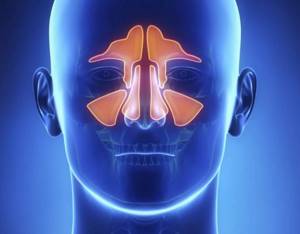
The paranasal sinuses (paranasal sinuses) are air cavities in the bones of the skull that communicate with the nasal cavity. In order to most accurately diagnose the condition of the paranasal sinuses, computed tomography (CT) is used.
Author:
- Chuprikov Roman Sergeevich
ENT pathology expert
5.00 (Votes: 1)
The paranasal sinuses (paranasal sinuses) are air cavities in the bones of the skull that communicate with the nasal cavity. The mucous membranes of these cavities are sensitive to the effects of viral microorganisms and often become inflamed due to colds and infectious diseases. In order to most accurately diagnose their condition, computed tomography (CT) is used.
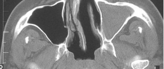
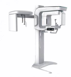
Computed tomography is a detailed examination using x-ray waves. During one rotation of the scanning system, the tomograph produces several slices at once, which reduces the procedure time and ensures maximum clarity of the information received. Based on the results of the procedure, the doctor receives high-quality images that allow a detailed study of the condition of all anatomical structures of the sinuses.
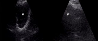
Compared to MRI, CT is more suitable for studying the paranasal sinuses, since its action is based on X-ray radiation. It most accurately visualizes bones and hollow organs, which include the sinuses, compared to MRI, which is more suitable for studying soft tissue.
Sinuses: description, where they are and what they are
Evolution made sure that the soft tissues of the brain were reliably protected by the bones of the corresponding section.
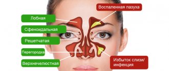
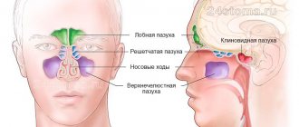
However, the outer side is more fragile and thin, and also consists of many elements. The design includes air pump cavities, which are known to most as sinuses or sinuses. These parts are connected by several passages to the nasal cavity, and are divided into four groups depending on their location:
- The frontal or frontal is located above the eyes.
- Maxillary - under the eyes in the upper part of the jaw.
- Ethmoidal - separates the cranial cavity from the nose, behind which, in fact, it is located in the ethmoid part of the bone.
- Sphenoid or sphenoid - inside the skull, behind the ethmoid bones and sinuses.
On the inside, the sinuses are covered with epithelium with a certain type of cells that produce mucus. Also inside there are special cilia that actually move this secretion to the immediate nasal cavity. The mucus passes through the small 1–2 mm openings of the ethmoid labyrinth. They move at a speed of 1 cm per minute.
Content
- Functions of the sinuses
- CT scan of the sinuses: what is it?
- When is such a diagnosis prescribed? What does it show?
- Preparation for the procedure
- How is it carried out and how is it done?
- How are the results interpreted?
- Indications for the procedure
- Contraindications to the procedure
- Where to get a CT scan of the sinuses?
Functions of the sinuses
Unfortunately, scientists have not come to a clear conclusion about what these organs are needed for. However, there are several most likely theories. First of all, the sinuses provide shockproof protection in case of injury, and also protect the more sensitive parts of the face (roots of teeth and eyeballs) from cold and hot air flows during inhalation and exhalation. It is also assumed that it is the sinuses that warm the inhaled air, since they block the flow.
It is likely that the cavities are an additional sensory organ that provides the brain with information about the environment. Also, some researchers suggest that the organ studies microorganisms that pass through inhalation.
Another important function of the sinuses: distributing the weight of the facial part of the skull. The shape of these areas is important because the facial muscles are attached to them. The organ is also responsible for vocal resonance, because there is an indirect connection with the pharynx.
CT scan of the sinuses: what is it?
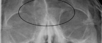
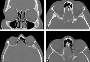
Computed tomography of the nasal and paranasal sinuses is one of the types of scanning of the human body, in this case the facial part. Based on the results, the patient receives an x-ray, which the doctor examines. Thus, it is possible to identify various pathologies and outline a further treatment plan.
The image is taken while the space inside the face is filled with air. The beams of the device illuminate this area, which is absolutely painless.
This is the most informative way to see the patient’s problem. Since liquids or air, as well as various pathologies, are quite clearly visible. This method is much more effective than conventional X-ray equipment, which at most can only detect sinusitis. When choosing an MRI or CT scan of the sinuses, preference is given to the latter, because only with the help of tomography can one identify how tumors grow and how close other areas are located.
CT or MRI
An alternative option for examining the paranasal tract is MRI. These two methods complement each other. CT allows one to determine the pathology of mainly bone tissue and determine the presence of cracks that cannot be seen with MRI. In such situations, CT is a more accurate examination method and is carried out in fine steps.
MRI is prescribed by a doctor to study mucous and soft tissues, namely to identify a tumor and determine its size. It allows you to find malignant tumors, as well as other complications caused by skull injuries. MRI is a more accurate and reliable method in such matters, but its cost is 2 times higher than CT. Before starting the examination, you should consult your doctor.
When is such a diagnosis prescribed? What does it show?
Doctors do not immediately resort to this method. Typically, a CT scan is prescribed if antibiotic therapy has not produced any results within a certain time. Therefore, in such cases, the doctor needs to find out in more detail the patient’s condition, which is facilitated by this procedure. Also, CT scanning of the nose and paranasal sinuses is prescribed in the following cases:
- diagnosis of sinusitis or cyst;
- find out the exact location of the anatomical features of the cavities;
- how the tumor or inflammatory process changes;
- for assessing cavities that are filled with liquid;
- how thick is the shell;
- for traumatic births.
Often such a study is needed before surgery. As a rule, this concerns endoscopic, corrective or plastic surgeries. Using this analysis, the doctor determines how effective the treatment was and, if necessary, adjusts the course.
Preparation for the procedure
CT is performed with or without contrast. Depending on this, preparation varies. If there is no dye, the patient is not required to do anything; they can come at any time and make a diagnosis. But when using contrast, the patient must first do a creatinine test to identify a possible allergy.
It is necessary to inform the specialist about the medications that are currently being taken and allergic reactions to certain pathogens. If you have serious illnesses: heart disease, asthma, diabetes, kidney or thyroid problems, you should also notify your doctor about them. Eating should occur no earlier than 1 – 2 hours before the examination.
It is better to take a medical card with your medical history, other pictures and test results, etc. to the examination. The study is carried out only as prescribed by the doctor, so do not forget this paper.
Preparatory process
X-ray examination does not require special preparation. Before starting, the patient must remove dentures, jewelry, metal objects and any magnetic media from pockets.
If you need to use contrast agents, you should take a blood test to check your creatinine level. You can drink and eat food before a CT scan if it is performed without the use of contrast agents.
The radiation dose from CT is minimal, but this study should not be overused. It is recommended to do it once a year; all other cases are discussed with your doctor.
How is it carried out and how is it done?
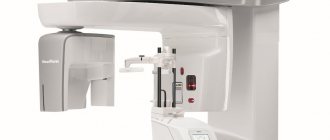
Tomography of the paranasal sinuses in St. Petersburg practically does not differ from clinic to clinic. More precisely, the course of the procedure itself is similar. It all depends on the quality of the devices and the qualifications of the doctors who will subsequently interpret the results.
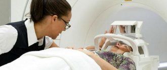
The mechanism affects the human body with x-rays of varying degrees, which makes it possible to distinguish tissues by density. During the scan, a radiation tube moves around the patient and a set of detectors measures the amount of radiation absorbed. A special computer program that displays the data transmits a cross-sectional image of the body on the monitor.
When examining children, the operator adjusts the size and dose of radiation. The patient is usually positioned on his back to ensure maximum immobility. If contrast is administered, the patient may experience a slight burning sensation or metallic taste that will quickly subside.
How are the results interpreted?
Specially trained personnel should be well versed in what CT scans of the sinuses show. As a rule, these are radiologists who know how to “read” the results of the images that are displayed on the screen of the device. Before the procedure, he calculates the required dose based on the patient’s data. The employee also sets up the tomograph and prepares the patient, because during the examination itself the patient remains alone in a special room.
A doctor with the necessary skills to interpret images is required to issue a conclusion on the day of the study. The document states that the sines correspond to normal values. The size of the sinuses, various deformations that violate the integrity of the tissues, the thickness of the mucous membrane, the presence of foreign objects or neoplasms, their type, the position of the nasal septum, fluid filling, etc. are described.
How does the procedure work?

Examination of the brain and other areas of the head is carried out in the following order:
- the patient lies down on a special table, the neck is placed on a comfortable stand, and the head is secured with straps;
- the table slides into a special chamber equipped with an X-ray tube that produces radiation;
- The sinus scanning process lasts several minutes. The more revolutions the sensor makes, the more detailed the area is shown;
- The image obtained as a result of scanning is displayed on the monitor screen;
- During the procedure, the patient should not move, as this will affect the quality of the image.
A radiologist interprets the results obtained. Ultimately, the patient receives an image, a written report and digital media with a recording of the study. This data will help prescribe treatment. A repeat study is carried out to monitor the development of pathologies, evaluate treatment methods, and in case of inaccurate analysis results.
Indications for the procedure
A CT scan of the paranasal sinuses is prescribed by a doctor only in certain cases. Since the patient must be irradiated during the procedure, doctors try not to abuse this method and first find out the patient’s condition in simpler ways. Especially if the person has already taken several x-rays before. The procedure is prescribed for the following indications:
- traumatic brain injuries;
- constant headaches or stable temperature 37 – 37.2 for several weeks;
- if you have previously had maxillofacial surgery;
- if tumors are suspected in the nasopharynx or bones of the facial part of the skull with probable growth into the sinuses.
For chronic sinus diseases, such as sinusitis, sinusitis, frontal sinusitis, this procedure is periodically prescribed so that the ENT can check the course of the disease and, if necessary, prescribe new treatment. Also for various suspicions of certain types of diseases, formations (neurioma, angioma) or injuries, etc. CT scan is required.
Contraindications to the procedure
Tomography is contraindicated when performing a procedure with a contrast agent if there is a possibility of a severe reaction. There are also a number of diseases for which it is undesirable to do a CT scan. As a rule, these include asthma and various allergies. If the patient’s condition is serious, the process may result in shock, respiratory or cardiac arrest, as well as convulsions.
In case of serious diseases of the liver and kidneys, especially with insufficiency, and the thyroid gland, CT is prohibited. Pregnant and lactating women also cannot undergo the procedure. The main risk is an additional dose of ionizing radiation, so if similar procedures have been performed previously, it is worth limiting the amount of radiation.
Indications and contraindications

Before a CT scan of the nasopharynx, tell your doctor about your pregnancy or allergies to iodine-containing contrast
If, as a result of conventional radiography, the diagnosis is in doubt or requires clarification, it is reasonable to perform a CT scan of the nasopharynx. The study is carried out as a preoperative preparation to assess important anatomical landmarks and their variations. We list the complaints for which CT scanning is indicated:
- trismus (inability to open the mouth normally due to muscle spasms);
- bloody discharge from the nose (from one nostril);
- persistent otitis;
- unexplained nosebleeds;
- persistent rhinorrhea of unknown cause;
- nasal pain, stuffiness;
- adenoid type of face;
- hearing impairment;
- palpable (enlarged) regional lymph nodes;
CT scans of the nasopharynx and larynx are often performed in patients to monitor the stage of the tumor process of already diagnosed and treated nasopharyngeal cancer. Symptoms for which diagnosis is justified:
- difficulty eating or swallowing;
- feeling of a foreign body in the throat;
- growth of lymph nodes;
- cough;
- discomfort (numbness) in the lower part of the face;
- hoarseness/change in voice quality;
- pain in the neck, ears or jaw.
Contraindications to native CT are childhood and pregnancy; contrasted computer scanning is supplemented by hypersensitivity reactions to radiopharmaceuticals and kidney disease with an increase in creatinine levels. For patients with severe obesity (more than 150 kg), closed CT is technically impossible, since the volume of the body exceeds the size of the tomograph tunnel.
Where to get a CT scan of the sinuses?
If you want to have a CT scan of the nasal cavities in St. Petersburg, then you should carefully choose a clinic. Be sure to check the availability of all licenses that indicate the quality of the device, as well as the competence of the doctors. Employees, as a rule, explain all the features of the technique during consultation. These must be people who have the appropriate medical education and have been trained in this type of equipment.
We offer to do a CT scan of the nasal sinuses in an x-ray, where responsible employees will conduct the selected examination in compliance with all safety standards. The staff will also decipher the results and provide all the necessary information. The medical complex offers high-quality care during the procedure, which is completely painless and with increased comfort. We also have an adequate pricing policy.
Sign up for a study by phone
+7 (812) 332-52-54
Advantages
Tomography of the paranasal sinuses provides much more information than ultrasound or radiography. In a CT scan, scanning is performed layer by layer, which allows you to obtain high-quality images with the possibility of reconstruction in various planes.
Other advantages of performing the procedure in our medical center:
- The study lasts 10 minutes - maximum information about the condition of the paranasal sinuses can be obtained in a minimum time period.
- The description is checked by the chief physician of the medical center.
- You can sign up for a convenient time from 9-00 to 21-00.