When do you need to take a General blood test without leukocyte formula (capillary blood)?
- To assess general health (routine medical examinations, screening examinations, etc.);
- For diagnostics at the first stage of a wide range of diseases that may manifest themselves as changes in the characteristics of peripheral blood;
- Diagnosis of anemia, polycythemia along with the study of hemoglobin and hematocrit;
- Monitoring ongoing drug treatment for patients suffering from bleeding or chronic anemia;
- Determination of the activity of erythrocyte formation processes in the bone marrow.
Detailed description of the study
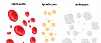
Blood is a multicomponent structure that is part of the internal environment of the body. It consists of plasma, which contains a suspension of formed elements - red blood cells, platelets, leukocytes and some others. This study is carried out to assess quantitative blood parameters. During a general blood test, the number of blood cells and cells of different types is determined, which gives an idea of their functioning, and, accordingly, the state of the body:
During a general blood test (CBC) without a leukocyte formula, the following parameters are assessed:
- number of leukocytes (white blood cells, WBC);
- number of red blood cells (RBC);
- hemoglobin level (hemoglobin content, Hb);
- hematocrit (hematocrit, Hct);
- mean erythrocyte volume (MCV);
- average hemoglobin content in erythrocytes (MCH);
- mean erythrocyte hemoglobin concentration (MCHC);
- platelets (platelet count, PC).
The main indicators that are included in the table of results of a general blood test:
Leukocytes (WBC, White Blood Cells)
Leukocytes, also called white blood cells or white blood cells, are a population of diverse but keyly similar immune system cells that are produced in the bone marrow. They are capable of active movement, capture and absorption of foreign particles and cells of their own body. In the first case, leukocytes provide protection from microbes and other foreign particles, in the second - maintaining the constancy of the internal environment of the body, ensuring the timely destruction of old or defective cells of the body's own.
Their number increases in the presence of inflammation - the degree of increase in the number of leukocytes indicates the severity of the infectious process.
CBC allows you to estimate only the total number of leukocytes. In order to assess the population composition, structure and structure of leukocytes, it is necessary to additionally undergo a leukocyte formula analysis. If a general blood test shows abnormalities, the doctor may prescribe a more detailed option.
You can also take a blood test with leukocyte formula and ESR at the Hemotest Laboratory.
Hemoglobin (Hb, Hemoglobin)
Hemoglobin is a protein that contains iron. Its main function is to transport oxygen to cells and remove carbon dioxide from them. The normal limits for it are well known, which vary somewhat depending on gender and age: in men, its content is, on average, higher than in women, and in children of the first year of life, a slight decrease in its content in the blood is normal. The combination of iron in hemoglobin with oxygen gives the blood a bright scarlet hue.
A decrease in hemoglobin levels beyond normal values is one of the types of anemia. This disease is also called “anemia”. There are several types of anemia depending on the causes, but with any of them the amount of hemoglobin per unit volume of blood will be reduced.
The most common cause of anemia is iron deficiency in the diet. In addition, anemia can occur due to blood loss and impaired absorption of iron in the gastrointestinal tract. The cause may also be too intense destruction of erythrocytes (red blood cells) or disruption of their formation. To clarify the diagnosis, additional tests will be required. A complete blood count can be a starting point in the diagnosis and treatment of anemia.
Hematocrit (Ht, Hematocrit)
Hematocrit is a numerical indicator of the ratio of the volume of blood cells, 99% of which are red blood cells, to the total blood volume. However, the statement is true only in cases where the size of red blood cells is normal. If the number of normal-sized red blood cells increases, the hematocrit also increases. In cases of large or small red blood cells, this is not always the case. For example, with iron deficiency, red blood cells decrease and the hematocrit will be reduced, but the number of red blood cells per unit of blood may be normal or even slightly higher.
Hematocrit increases with dehydration, lung and heart diseases, and while in high mountains. On the contrary, a decrease in hematocrit is observed with anemia of various origins, with liver diseases, with the consumption of large amounts of fluid or the administration of large volumes of fluid intravenously.
Red blood cells (RBC, Red Blood Cells)
Erythrocytes (red blood cells) are the formed elements of blood, not entirely correctly called “cells”. They are indeed formed from precursor cells, but upon maturation they lose their nucleus - one of the key features of a living cell. In this way, they provide themselves with the opportunity to implement a highly specialized function: transporting oxygen to the cells of the body and removing carbon dioxide from them. These are the most numerous formed elements of blood, which have the shape of a biconcave disc. Their entire volume is occupied by hemoglobin in order to effectively transport oxygen to cells and remove carbon dioxide and metabolic products from them.
The number of red blood cells is a very important indicator of health. CBC allows you to identify deviations from the norm and prescribe additional studies to clarify the diagnosis and identify the causes of changes in the number of red blood cells, including:
Erythropoietin is a hormone that is synthesized in the kidneys and stimulates the formation and maturation of red blood cells. With a lack of oxygen - hypoxia, it is synthesized more intensely. The number of red blood cells may decrease due to a lack of this hormone.
Vitamin B12, folic acid and sufficient iron are necessary components for adequate hemoglobin synthesis and red blood cell formation.
Red blood cells live in the bloodstream for 120 days, after which they are destroyed in the spleen. Excessive destruction of red blood cells can also lead to a decrease in their number. Determining the number of red blood cells, together with assessing the hemoglobin content, establishing hematocrit and characterizing red blood cells, makes it possible to diagnose the type of anemia and its causes with a high degree of probability. Based on this data, it is possible to select the most effective treatment.
Color Index (CPU)
The numerical indicator of hemoglobin saturation of red blood cells indicates the amount of hemoglobin per red blood cell.
MCH (Mean Cell Hemoglobin, average hemoglobin content in red blood cells)
Numerical expression of the average hemoglobin content in one red blood cell. Together with MCV it is used for the differential diagnosis of anemia.
MCV (Mean Cell volume, average volume of red blood cells)
Numerical indicator of the average volume of red blood cells. Normally it has no diagnostic significance, but is necessary for the differential diagnosis of anemia in combination with MCH. If the range of red blood cell volumes is large, or there are many red blood cells of a changed shape, then averaging the indicator will not be so informative, but differences in the size and structure of red blood cells indicate the need to pay close attention to this: this is not the norm.
MCHC (Mean Cell Hemoglobin Concentration, mean hemoglobin concentration in red blood cells)
An indicator that shows the average concentration of hemoglobin in red blood cells. A sensitive indicator of changes in hemoglobin synthesis processes - a possible cause of iron deficiency anemia, thalassemia, and some hemoglobinopathies.
Platelets (PLT, Platelets)
Platelets are the formed elements of blood. Like red blood cells, they are formed from precursor cells, but they are not cells: they do not have a nucleus or other important intracellular structures, so their lifespan is only about a week.
The loss of cellular characteristics is due to the high specialization of these formed elements: almost their entire volume is filled with granules, which contain substances involved in the processes of blood coagulation and platelet aggregation. With the help of special receptors, platelets detect a violation of the integrity of blood vessels, form a clot there and stimulate local blood clotting to stop bleeding.
An increase in the number of platelets can occur as a result of taking certain medications, indirectly indicating malignant neoplasms, some inflammatory and infectious diseases. Some types of cancer, malaria and bronchial asthma can reduce their levels. Their levels may also be reduced as a result of disruption of their formation from progenitor cells in the bone marrow or due to increased consumption during wound healing. The main symptom of a low platelet level is bleeding, up to life-threatening conditions: from the appearance of bruises even with light pressure and bleeding gums, to internal hemorrhages.
Rules for collecting biomaterial
Determination of amino acids and acylcarnitines in blood using tandem mass spectrometry
Blood is collected on a standard filter card (No. 903), which is used to screen newborns for phenylketonuria. Blood can be either capillary (from a finger) or venous. It is necessary to saturate the selected area on the filter well! The filter card must clearly indicate the full name, who and where the patient was referred from, date of birth, date of sample collection and telephone number of the attending physician. The sample is dried for 2-3 hours at room temperature (heating and exposing the sample to direct sunlight is unacceptable) and is sent to the laboratory with a completed application form. It is advisable to attach an extract from your medical history.
Determination of oxysterols in blood plasma (Niemann-Pick disease type C)
To determine oxysterols in blood plasma, strict adherence to the rules for transporting biomaterial is necessary. Collect 5-7 ml of the patient’s blood in a test tube with an EDTA preservative (usually a purple cap), carefully invert the tube several times to mix with the preservative, but do not shake to avoid hemolysis. Clearly and legibly sign the patient's Last Name, First Name, and the date of blood collection. Deliver the test tube with blood within 24 hours at a temperature of +2-+8 degrees Celsius, under no circumstances should it be frozen! Transport blood in a thermos with food ice (1-2 ice cubes) or in a container with a cold pack.
If blood cannot be delivered within 24 hours, additional manipulations with the blood must be performed in the laboratory where it is collected. After mixing with the preservative, the tube must be centrifuged for 10 minutes at 1000 g. After centrifugation, collect the plasma (supernatant) into an Eppendorf tube (two tubes, if possible), without touching the sediment! Next, the blood and tubes with plasma must be frozen and possibly stored at -20 degrees Celsius. Such samples must be sent frozen.
Be sure to send to the laboratory a completed clinical phenotype registration card or an extract from the medical history. A special map can be downloaded in the “Documents” section
Isofocusing of transferrins (diagnosis of congenital disorders of glycosylation)
2-3 ml of the patient's blood is collected either into a tube WITHOUT PRESERVATIVE (usually a red cap) or into a tube WITH HEPARIN (usually a green cap). The contents of the tube must be mixed, but not shaken, to avoid hemolysis; the tube must be closed and labeled. Transport blood in a thermos with food ice (1-2 ice cubes). Blood must be delivered to the laboratory no later than 24 hours after collection. Blood should not be frozen! It is advisable to attach an extract from your medical history.
If blood cannot be delivered within 24 hours, it is necessary to perform additional manipulations with the blood in the laboratory where it is collected. After mixing with the preservative, the tube must be centrifuged for 10 minutes at 1000 g. After centrifugation, collect the serum/plasma (supernatant) into an Eppendorf tube (if possible, two tubes), without touching the sediment! Next, the serum/plasma must be frozen and possibly stored at -20 degrees Celsius. Such samples must be sent frozen.
This analysis is possible on the material of blood stains on a filter card. (The rules for collecting blood on the filter are specified in the “General Recommendations” section)
Determination of ceruloplasmin (diagnosis of Wilson-Konovalov disease and Menkes disease)
2-3 ml of the patient's blood is collected in a test tube without a preservative (usually a red cap). The contents of the tube must be mixed, but not shaken, to avoid hemolysis; the tube must be closed and labeled. Transport blood in a thermos with food ice (1-2 ice cubes). Blood must be delivered to the laboratory no later than 24 hours after collection. Blood should not be frozen. It is advisable to attach an extract from your medical history.
If blood cannot be delivered within 24 hours, it is necessary to perform additional manipulations with the blood in the laboratory where it is collected. After mixing with the preservative, the tube must be centrifuged for 10 minutes at 1000 g. After centrifugation, collect the plasma (supernatant) into an Eppendorf tube (two tubes, if possible), without touching the sediment! Next, the blood and tubes with plasma must be frozen and possibly stored at -20 degrees Celsius. Such samples must be sent frozen.
Determination of very long-chain fatty acids, cholesterol isomers (diagnosis of peroxisomal diseases, Smith-Lemli-Opitz syndrome)
5-7 ml of blood from the patient and parents (on an empty stomach) is collected in a test tube with a preservative (EDTA). The contents of the tube must be mixed, the tube must be closed and signed. It is recommended not to eat bananas, nuts, chocolate and cheese 3 days before the test. Transport blood in a thermos with food ice. Blood must be delivered to the laboratory no later than 24 hours after collection.
If blood cannot be delivered within 24 hours, it is necessary to perform additional manipulations with the blood in the laboratory where it is collected. After mixing with the preservative, the tube must be centrifuged for 10 minutes at 1000 g. After centrifugation, collect the plasma (supernatant) into an Eppendorf tube (two tubes, if possible), without touching the sediment! Next, the blood and tubes with plasma must be frozen and possibly stored at -20 degrees Celsius. Such samples must be sent frozen.
Important Notes
Material for research
Children under 7 years of age: venous blood/capillary blood (for special indications).
Children over 7 years of age and adults: venous blood.
Capillary blood collection for research is carried out only for children under 7 years of age (for special indications).
According to GOST R 53079.4-2008, indications for taking capillary blood are possible in newborns, in patients with very small or hard-to-reach veins, with large-area burns, and in severely obese patients.
References
- Nazarenko G.I., Kishkun A.A. Clinical assessment of laboratory research results // M.: Medicine - 2006. - 544 p.
- Tatkov O.V., Stupin F.P. General blood test. Information collection // M.: Publishing solutions. — 2021. — 72 p.
- Fauci, Braunwald, Kasper, Hauser, Longo, Jameson, Loscalzo Harrison's principles of internal medicine, 17th edition, 2009
- Mosby's Diagnostic and Laboratory Test Reference 14th Edition, 3251 Riverport Lane
- St. Louis, Missouri 63043: Elsevier Publishing Hall 2019
Preparing to give a clinical finger blood test to a child
A clinical blood test is NOT taken on an empty stomach; you can eat 2 hours before the test; it is advisable to drink a small amount of water before the procedure.
For infants, blood can be drawn 1-2 hours after feeding; it is advisable to give the child water to drink before the procedure. Material for a clinical blood test for newborns is taken from the heel or big toe.
It is recommended to take a finger prick blood test before an x-ray examination.


