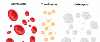
Natural causes of increased erythrocyte sedimentation rate
If the ESR is elevated, this can be caused by various reasons, but these are not always malignant tumors.
- In women, ESR increases during menstruation, as well as during pregnancy. The absolute norm for a pregnant woman will be 44-46 mm/hour, but remember that at the beginning of pregnancy, ESR, on the contrary, is reduced.
- The cause of increased subsidence can be stress, excessive tension and emotional stress.
- This indicator also increases with allergic reactions of the body.
- Various reactions, inflammation due to cuts or splinters are also the cause.
- Pay attention to the medications you take - sometimes the ESR increases because of them.
The reasons given above are not the most serious. If we are talking about natural processes in a woman’s body, there is absolutely no need to worry. It’s another matter when the ESR increases due to internal inflammation, which can develop into a dangerous disease.
Therefore, it is so important not to resort to self-medication, and find out from your doctor the reason for the high rate.
Clinical and laboratory features during pregnancy
Pregnancy
is the period of time during which the fetus develops inside a woman's uterus, ending with the birth of the child.
During pregnancy, numerous physiological changes occur to meet the needs of the growing fetus and the mother's body, which adapts to them. Most of these changes begin soon after conception and continue until late pregnancy. Physiological adaptation is reflected in changes in the values of laboratory parameters. Some of the changes are well known, for example, a decrease in hematocrit and hemoglobin, creatinine; others, on the contrary, are known to a lesser extent and therefore their observation in the analysis result form can lead to incorrect interpretation.
The cardiovascular system
Changes in the functioning of the cardiovascular system occur among the very first. Their profound restructuring begins already in the early stages of gestation. The main events are physiological vasodilation and fluid retention in the body. Peripheral vasodilation leads to a decrease in vascular resistance and an increase in cardiac output, an increase in heart rate, and an increase in venous pressure.
Blood pressure decreases in the first and second trimesters, but rises to levels in non-pregnant women in the third trimester.
Water balance
Low blood pressure during pregnancy leads to activation of the renin-angiotensin aldosterone system, resulting in increased release of antidiuretic hormone. There is a tendency to retain water and sodium, and the likelihood of edema formation increases.
Hematological changes
Pregnancy is accompanied by stimulation of hematopoietic processes. There is a general increase in plasma, red blood cell (RBC) counts, and total circulating blood volume. Plasma volume increases during normal pregnancy. An increase of 15% occurs in the first trimester; in the second trimester, this trend continues, reaching a maximum at 32 weeks. By this time, the plasma volume has increased exactly by half.
The number of red blood cells increases, but occurs more slowly compared to plasma, making the blood more dilute and leading to the “physiological anemia” of pregnancy. There is a decrease in hemoglobin and hematocrit and mean erythrocyte hemoglobin content (MCH). The maximum decrease in hemoglobin levels is observed at 32–34 weeks of pregnancy.
During pregnancy, the volume of red blood cells changes. During the first 8 weeks of pregnancy, MCV decreases, by week 16 it returns to normal values, as in non-pregnant women, and then MCV increases.
White blood cell (WBC) levels increase by an average of 20%. The following changes are noted in the leukocyte formula: the absolute number of neutrophils (band and segmented) increases, the number of lymphocytes decreases.
The platelet count (PLT) changes ambiguously. Pregnancy is associated with increased platelet turnover.
An increase in blood volume is accompanied by an increase in ESR. In the first trimester, the ESR value is 15 mm/hour, in the second - 25 mm/hour, in the third - 40 mm/hour.
Hemostasis indicators
Throughout the entire period of pregnancy, the body prepares for the upcoming blood loss, so certain changes occur in the hemostatic system.
Changes in the blood coagulation system during pregnancy lead to a physiological state of hypercoagulability or increased susceptibility to thrombosis. In the third trimester, coagulation activity is twice as high as normal. The procoagulant activity of the hemostasis system increases, on the other hand, the activity of the fibrinolysis system decreases.
The concentrations of factors VII, VIII, IX, X, XII and von Willebrand factor increase.
Factor XI values decrease to 60–70% of those in non-pregnant women.
Fibrinogen levels increase significantly - up to 50%.
Levels of protein S and antithrombin III gradually decrease during pregnancy, while protein C activity remains unchanged.
Plasma fibrinolytic activity decreases throughout pregnancy but returns to normal within one hour after delivery.
Thrombin is generated as the gestational age increases. The D-dimer value increases throughout pregnancy. Only 3–5 days after delivery, the D-dimer value returns to its original values.
A shortening of APTT is observed in the second and third trimester and is associated with an increase in the activity of procoagulants in the blood. In the third trimester, a shortening of prothrombin time is observed.
Carbohydrate metabolism
Pregnancy is a diabetogenic condition because it is associated with the development of insulin resistance. An increase in the level of estrogen and progesterone in the initial stages leads to hypertrophy of pancreatic cells that secrete insulin. As a result, insulin secretion and tissue sensitivity to it increase in the early stages.
In the second trimester, insulin resistance begins to appear, reaching a peak in the third trimester. This results from the secretion of contrainsular hormones: human placental lactogen, growth hormone, progesterone, cortisol and prolactin. These hormones cause a decrease in the sensitivity of peripheral tissues to insulin, especially in adipose tissue and skeletal muscle, by interfering with insulin receptor signaling.
Insulin levels during pregnancy increase during fasting, after eating.
Fasting glucose levels often decrease due to:
- increasing storage of tissue glycogen reserves;
- increased use of peripheral glucose;
- decreased liver glucose production;
- glucose absorption by the fetus.
Insulin resistance and relative hypoglycemia lead to an increase in the process of lipolysis - the formation of fats, which primarily allows the use of fat as fuel, preserving glucose and amino acids for the fetus. The placenta allows the transfer of glucose, amino acids and ketones to the fetus, but is impermeable to large lipids. If a woman's endocrine pancreatic function is impaired and she cannot overcome the insulin resistance associated with pregnancy, she develops gestational diabetes.
During pregnancy, physiological transient glycosuria is often observed, which is associated with an increase in the glomerular filtration rate and an increase in the permeability of the renal tubular epithelium.
Protein metabolism
Throughout pregnancy, a woman needs more protein, since anabolic processes prevail over catabolic processes.
There is a physiological decrease in blood albumin; dilution of the blood also contributes to a decrease in the proportion of albumin.
The proteinogram in the first and second trimester has the following features: a decrease in albumin levels, a slight increase in the a-2 and b-1 globulin fraction. In the third trimester, there is a sharp increase in the a-1 globulin fraction.
And due to increased protein metabolism, a positive nitrogen balance is observed. Due to an increase in GFR by 75%, creatinine and urea levels decrease. A decrease in creatinine occurs mainly in the first and second trimesters, when intensive growth of the uterus is observed. The level of urea falls due to increased protein utilization, especially pronounced in the third trimester.
Lipid metabolism
The general effects of altered lipid metabolism during pregnancy are the accumulation of fat reserves in the mother's body in the first half and increased mobilization of fats in the second half of pregnancy.
Hypercholesterolemia is caused by increased production of sex steroid hormones, changes in metabolism in the liver and adipose tissue. Elevated triglyceride values provide the mother with energy needs. An increase in LDL cholesterol is associated with an increase in progesterone, in addition, LDL cholesterol is a source of placental progesterone. Increased concentrations of estrogen during pregnancy cause an increase in total cholesterol, LDL cholesterol and triglycerides.
Regional fat deposition in the mammary glands and subcutaneous fat is associated with increased conversion of carbohydrates into fats under the influence of insulin.
Mineral metabolism
Pregnancy causes an increase in the need for iron by 2-3 times for the synthesis of hemoglobin, for the production of certain enzymes. The need for folic acid increases 10–20 times, the need for vitamin B12 doubles.
The third trimester marks the peak demand for calcium.
During pregnancy, the concentration of total calcium in the blood serum decreases due to a decrease in the proportion of blood albumin, but the level of ionized calcium remains unchanged.
Dangerous causes of increased ESR
- The very pathology of the behavior of red blood cells may indicate a blood disease - leukemia, anemia, etc.
- A malignant formation or purulent infectious accumulation (for example, an abscess) may also be detected in the body.
- An increased ESR also indicates acute intestinal, bacterial infections, and inflammatory diseases of the gastrointestinal tract in general.
- An increase in indicators also indicates an excess of toxins in the body or poisoning.
The body could have suffered great blood loss under various circumstances, or suffered from tuberculosis or rheumatism. Then in the tests, in addition to an increased ESR, platelets may be observed.
As you can see, there are many reasons for increased ESR, but this does not always mean the development of pathology or a serious disease. The main thing in this case is to find out the main problem with your doctor, and only then can you understand whether you need treatment or not.
How is ESR determined?
The expectant mother needs tenderness and support.
To calculate whether the subsidence rate of pregnant women in the 2nd trimester is high or low, it is necessary to undergo appropriate diagnostic procedures. The most reliable data today are considered to be ESR data using the Westergren method. Techniques for determining the ESR coefficient according to Panchenkov are also used. The latter suggests using sodium citrate (5%) as an anticoagulant substance that prevents blood clotting. Westergren also suggests using sodium citrate, however, in a much lower concentration (3.8%).
Medical diagnostic practice shows that the method for determining ESR is considered more indicative and reliable with a lower concentration of sodium citrate. Therefore, the Westergren technique is recognized as the most preferable method. To conduct the study, patients donate blood from a finger prick.
Preparing for analysis
To obtain the most accurate research results, it is necessary to prepare for diagnosis.
- Blood is donated only on an empty stomach in the morning; the pregnant woman should not eat or drink for about 8 hours before the biomaterial is taken. Only drinking water is allowed.
- On the eve of the study, the patient should exclude fried and fatty foods, spicy foods or seasonings from the diet, otherwise such a diet may distort the ESR standards.
- Approximately two days before the test, start following a gentle diet with boiled or steamed lean, light food, which will help avoid distortion of the results.
- It is also recommended to give up physical activity such as running, training, etc., about a day before the diagnosis.
- Before taking blood, you need to sit quietly for at least a quarter of an hour, calm down and restore your normal state, otherwise the study may erroneously show that the ESR rate is increased.
- In addition, the use of certain medications such as cortisone, etc., greatly affects the diagnostic results. Therefore, if any hormonal or other medications are prescribed for mommy, the doctor should be informed about this.
Blood tests are prescribed quite often during pregnancy, so mothers should take pre-procedural preparations more seriously.
Decoding the results
Only a specialist can decipher the result.
The erythrocyte sedimentation rate has a specific decoding that differs in patients of different ages and gender. For example, for women under 30 years of age, the normal ESR coefficient is about 3-15 mm/h. For patients over 30 years of age, these figures are already 8-25 mm/h.
For women in an interesting situation, the normal erythrocyte sedimentation rate begins to increase from the first or second week of gestation. Experts associate the fact that the ESR rate is elevated with changes in the biochemical composition of the blood typical for pregnant women.
In what cases does ESR not need to be treated?
Elevated ESR can also occur due to poor nutrition, if the diet is dominated by unhealthy foods high in fat, as well as an excess of spicy foods.
The level of ESR is also affected by a lack of vitamins in the body. In this case, the doctor may even prescribe a special therapeutic diet.
If you have recently had surgery, increased subsidence will be normal.
Therefore, if you notice a deviation from the norm in your blood test, do not rush to sound the alarm, and treatment without knowing the cause will not be beneficial.
ONLINE REGISTRATION at the DIANA clinic
You can sign up by calling the toll-free phone number 8-800-707-15-60 or filling out the contact form. In this case, we will contact you ourselves.
If you find an error, please select a piece of text and press Ctrl+Enter
Effect of pregnancy on liver function
The liver during pregnancy is in a state of high functional tension. However, in healthy women during the physiological course of pregnancy, the functioning of the liver is not impaired.
Noted:
- slight increase in liver size in the absence of pronounced histological changes;
- there is a decrease in the antitoxic function of the liver;
- the level of protein in the blood serum decreases, by the time of childbirth it can reach 60 g/l;
- the result of changes in the composition of serum proteins is an increase in ESR;
- blood clotting and fibrinolysis changes. These changes help increase blood clotting ability.
Why does ESR increase?
The erythrocyte sedimentation reaction coefficient is not constant and can change even throughout the day. Typically, during the daytime this indicator is slightly increased, while at other times this value decreases. Experts note the existence of many pathological and physiological factors that can provoke an increase in the sedimentation rate of red blood cells in pregnant girls. The main reasons for a decrease in ESR include different situations.
- A marked increase in fibrinogen levels, reaching maximum values around the third trimester. This substance is responsible for blood clotting. In general, such an increase is quite natural for pregnant women, because in this way the body prepares for unexpected bleeding, which can complicate pregnancy.
- Vegetarians also experience an increase in ESR values. During pregnancy, plant food is not enough for the full development of the fetus. Mothers must consume animal protein, otherwise the baby may have serious health problems.
- Autoimmune pathologies that are accompanied by connective tissue damage, for example, rheumatoid arthritis or rheumatism, etc.
- Against the background of anemia in pregnant women, an increase in ESR also occurs. Maternal red blood cells, which carry oxygen to the baby, quickly wear out and are destroyed. And new ones do not have time to be fully produced, because the consumption of vitamins and minerals increases significantly.
- Pathologies of endocrine origin that provoke metabolic disruptions. Such pathologies include diabetes, hypo- or hyperthyroidism and other hormonal disorders.
- ESR increases with sore throat or pneumonia, syphilis or tuberculosis, osteomyelitis and arthritis.
- Also, an increase in the erythrocyte sedimentation reaction is observed in strokes and liver pathologies, heart attacks and kidney diseases.
- Frequent blood transfusions, large blood losses, and bone fractures also provoke upward deviations in ESR.
But even if the ESR rate increases to 45-50 mm per hour, you should not wait in fear for terrible and deadly diagnoses. The gynecologist will most likely refer the pregnant woman for additional diagnostic examination, which will help detect possible inflammatory lesions. Sometimes elevated ESR levels indicate metabolic disorders or infectious pathologies accompanied by tissue decay or malignant processes.


