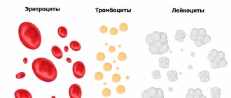
Reticulocytes (Retic count) are nuclear-free cells. Their formation occurs in the bone marrow. They then enter the bloodstream. Under the influence of erythropoietin, these cells are converted into red blood cells. The latter play an important role in the life of organs and systems. The lifespan of red blood cells is approximately three months. After this, they are destroyed and replaced by new cells. The function of red blood cells is to deliver nutrients and oxygen to the cells for which they are necessary.
Indications for the purpose of analysis
Young red blood cells, reticulocytes, are formed during the process of differentiation and division of stem cells. In fact, these are the precursors of adult red blood cells with a nucleus, mitochondria, remnants of ribonucleic acids and a number of other organelles. The shape is round, the color is pinkish with a bluish tint. They are formed within 1-2 days, and then mature in the general bloodstream for about another 3 days, after which they are transformed into red blood cells. The norms of reticulocytes for men and women are different; in newborns they are much higher.
A blood test shows the total number of reticulocytes in the blood, their ratio as a percentage. Laboratory testing can determine the adequacy of red blood cell production and bone marrow activity.
During normal functioning of the body, the number of red blood cells circulating in the bloodstream is almost always the same. And their average life expectancy is within 120 days. Obsolete red blood cells are broken down in the spleen, new ones are formed in the bone marrow. This continuous process is controlled by a special hormone produced by the kidneys, erythropoietin. When the oxygen level drops, the secretion of erythropoietin increases, the hormone enters the bone marrow through the bloodstream, where, under its influence, the production of young red blood cells is activated. After the volume of produced red blood cells reaches normal values, the secretion of erythropoietin decreases.
When hemolysis (destruction of red blood cells) or deterioration in their production by the bone marrow, anemia develops. The cause of this pathology can also be bleeding, since the body, in order to compensate, increases the secretion of red blood cells and, accordingly, the indicators of reticulocytes in the blood increase.
Determination of reticulocytes in a blood sample is used:
- To confirm the suspected type of anemia;
- To determine the activity of the process of production of red blood cells by the bone marrow;
- To identify the severity of anemia. Additionally, a general blood test is required, showing the erythrocyte index, total volume of erythrocytes, hemoglobin concentration and hematocrit value.
The patient is referred for advanced laboratory diagnostics if general tests show signs of a decrease in hemoglobin, red blood cells and other values indicating possible anemia. Symptoms of anemia, in turn, may include:
- Pale or yellow skin;
- Fast fatiguability;
- Dyspnea;
- Weakness;
- Hair fragility and loss.
Chronic blood loss is indicated by the appearance of blood in the feces.
Reticulocyte tests are also prescribed for patients with iron deficiency anemia, cancer pathologies, lack of folic acid or vitamin B12 during a course of treatment. In this case, the study allows you to determine how effective the prescribed therapy is and, if necessary, adjust it.
Reticulocytes in the sample are also counted if there are more red blood cells in the blood compared to the norm. This is necessary to assess the functioning of the bone marrow.
Counting reticulocytes in a blood smear (quantities)
What are reticulocytes?
Reticulocytes are young forms of erythrocytes (precursors of mature erythrocytes), containing a granular-filamentous substance, revealed by special (supravital) staining. Reticulocytes are detected both in the bone marrow and in the peripheral blood. The maturation time of reticulocytes is 4-5 days, of which within 3 days they mature in the peripheral blood, after which they become mature erythrocytes. In newborns, reticulocytes are found in greater numbers than in adults.
The number of reticulocytes in the blood reflects the regenerative properties of the bone marrow. Their counting is important for assessing the degree of activity of erythropoiesis (production of red blood cells): when erythropoiesis accelerates, the proportion of reticulocytes increases, and when it slows down, it decreases. In the case of increased destruction of red blood cells, the proportion of reticulocytes may exceed 50%. A sharp decrease in the number of red blood cells in the peripheral blood can lead to an artificial increase in the number of reticulocytes, since the latter is calculated as a percentage of all red blood cells. Therefore, to assess the severity of anemia, the “reticular index” is used: % reticulocytes x hematocrit / 45 x 1.85, where 45 is a normal hematocrit, 1.85 is the number of days required for new reticulocytes to enter the blood. If the index is < 2, it indicates a hypoproliferative component of anemia; if > 2-3, then there is an increase in the formation of red blood cells.
Indications for the purpose of analysis:
- diagnosis of ineffective hematopoiesis or decreased production of red blood cells;
- differential diagnosis of anemia;
- assessment of response to therapy with iron, folic acid, vitamin B12, erythropoietin;
- monitoring the effect of bone marrow transplantation;
- monitoring of erythrosuppressor therapy.
- Preparation for the study: the study is carried out on an empty stomach or at least 2 hours after
- the last meal, when taking blood, it is necessary to avoid prolonged compression of the vein with a tourniquet.
When does the number of reticulocytes increase (reticulocytosis)?
- Posthemorrhagic anemia (reticulocyte crisis, increase 3-6 times).
- Hemolytic anemia (up to 300%).
- Acute lack of oxygen.
- Treatment of B12-deficiency anemia (reticulocyte crisis on days 5 - 9 of vitamin B12 therapy).
- Therapy of iron deficiency anemia with iron preparations (8 - 12 days of treatment).
- Thalassemia.
- Malaria.
- Polycythemia.
- Tumor metastases to the bone marrow.
When does the reticulocyte count decrease?
- Aplastic anemia.
- Hypoplastic anemia.
- Untreated B12 deficiency anemia.
- Metastases of neoplasms to bone.
- Autoimmune diseases of the hematopoietic system.
- Myxedema.
- Kidney diseases.
- Alcoholism.
Preparing for the fence
Venous blood is considered the best biomaterial for research, since:
- When capillary blood is collected, some of the blood cells are deformed and destroyed, resulting in microscopic blood clots appearing in the test tubes. Their formation makes the results unreliable, so repeated sampling is required;
- Innovative vacuum systems are used to collect blood from a vein, making the procedure instant, virtually painless and eliminating infection.
However, it is also acceptable to use capillary blood for analysis. According to GOST, its collection is mainly permissible in newborns, in patients with difficult access to a vein, with severe obesity and with extensive burns in which it is impossible to take venous blood.
It is possible to obtain the most reliable result only if the patient follows all the rules for preparing for the collection:
- For at least 24 hours before the collection, it is necessary to completely avoid the consumption of alcoholic beverages, fatty and spicy foods;
- You cannot eat at least 2 hours before taking the test, but drinking water (only still) is permissible;
- 30 minutes before the collection, you should try to eliminate physical and psycho-emotional stress; Stop smoking for 30-60 minutes before the test.
The doctor issuing a referral for diagnostics must be warned about the medications you are taking, as some medications may affect the results of the study.
Features and advantages of the technique
Venous blood is considered the best material for laboratory research:
- To ensure the quality of the test result, it is necessary to donate venous blood (unless the child has special indications for taking capillary blood). When blood is taken from a finger, the blood cells are deformed, and some of the red blood cells are destroyed, forming microscopic clots in the test tubes. In this case, it is impossible to conduct research; in this case, repeated collection of biomaterial is required.
- In venous blood, blood cells are not destroyed, microscopic clots are formed much less frequently. That is why capillary blood is used only in children in isolated cases.
- Thanks to modern technologies, the procedure for collecting venous blood is painless and safe even for small children, since completely closed disposable BD vacuum systems are used, which eliminate infection and meet all international standards.
- Taking blood from a vein takes a few seconds.
Interpretation of the obtained indicators
Qualified doctors interpret the tests. However, it is advisable for the patient to know the norm; this will allow timely attention to deviations and contact the doctor for an advanced diagnosis.
Reticulocyte percentage table (RET%)
| Patient category | Age | Reference (acceptable) values |
| Children | Newborns (up to 2 weeks) | 0,15 — 1,5 % |
| From 2 weeks to a month | 0,45 — 1,4 % | |
| From month to 2 | 0,45 — 2,1 % | |
| From 2 months to 6 | 0,25 — 0,9 % | |
| From 6 months to 24 months | 0,2 — 1 % | |
| From 2 years to 6 years | 0,2 — 0,7 % | |
| From 6 years to 12 years | 0,2 — 1,3 % | |
| Women | From 12 to 18 years old | 0,12 — 2,05 % |
| Over 18 years old | 0,59 — 2,07 % | |
| Men | From 12 to 18 years old | 0,24 — 1,7 % |
| Over 18 years old | 0,67 — 1,92 % |
In children under 12 years of age, the reticulocyte rate is the same for both sexes. With the onset of puberty, the values begin to differ. The concentration of young red blood cells increases as you rise to higher altitudes, as the body adapts to the reduced amount of oxygen at high altitudes. An increased number of young reticulocytes is detected in pregnant women and smoking patients.
Important! An increase or decrease in reticulocytes cannot make a diagnosis. However, deviation from normal values is a reason for prescribing additional examinations.
Reticulocytes
Reticulocytes are young red blood cells formed in the bone marrow and found in small quantities in the blood. They are a transitional form between the precursors of red blood cells in the bone marrow and adult red blood cells, which are found in large numbers in the bloodstream.
Synonyms Russian
Reticulocyte count, reticulocyte count, reticulocyte index.
English synonyms
Retic count, reticulocyte index, corrected reticulocyte, reticulocyte count.
Research method
Flow cytometry.
What biomaterial can be used for research?
Venous, capillary blood.
Units
*10^9/l (10 in st. 9/l), % (percent).
How to properly prepare for research?
- Eliminate alcohol from your diet for 24 hours before the test.
- Do not eat food 2-3 hours before the test (you can drink clean still water).
- Avoid physical and emotional stress 30 minutes before the test.
- Do not smoke 30 minutes before blood collection.
General information about the study
Reticulocytes are young red blood cells (erythrocytes). They are formed in the bone marrow when stem cells differentiate and divide to become adult red blood cells through the reticulocyte stage, gradually losing their nucleus and decreasing in size.
Newborns have more reticulocytes than adults.
Most red blood cells are already fully mature when they leave the bone marrow and enter the bloodstream, but 0.5-2% of those circulating in the blood are reticulocytes, which turn into adult red blood cells within two days. This test shows the number and percentage of reticulocytes in the blood and reveals the adequacy of red blood cell production by the bone marrow and the degree of its activity.
The body tries to maintain approximately the same number of circulating red blood cells; the normal lifespan of each of them is about 120 days. In this case, old red blood cells are destroyed in the spleen, and new ones are formed in the bone marrow. This process is regulated by erythropoietin, a hormone produced in the kidneys. In response to decreased oxygen levels in the blood, the kidney produces erythropoietin, which is then transported by blood to the bone marrow, where it stimulates the formation of red blood cells. As the red blood cell count increases, the production of erythropoietin in the kidneys decreases.
If red blood cells are destroyed (hemolysis) or their synthesis in the bone marrow is disrupted, anemia occurs. Also, its development is facilitated by the loss of red blood cells due to bleeding - then the body increases the formation of red blood cells in the bone marrow and the number of reticulocytes in the blood increases.
Damage to the bone marrow (eg, from a tumor, chemotherapy, or ionizing radiation) results in decreased production of red blood cells and the number of reticulocytes in the bone marrow and peripheral blood.
Insufficient production of red blood cells leads to a decrease in their circulation in the bloodstream, the amount of hemoglobin and the ability to carry oxygen. Accordingly, fewer reticulocytes are formed.
In turn, the number of reticulocytes and red blood cells increases with more active bone marrow function. This can occur for a variety of reasons, such as increased production of erythropoietin, a chronic disorder that increases the number of red blood cells (polycythemia vera), or smoking.
What is the research used for?
- For differential diagnosis of types of anemia, as well as to determine the activity of the formation of red blood cells in the bone marrow.
- To determine the severity of anemia, a general blood test should be performed along with this study, which evaluates the number of red blood cells and red blood cell indices, hematocrit and hemoglobin concentration.
When is the study scheduled?
- With a decrease in the total number of red blood cells, hemoglobin and other signs of anemia or suppression of bone marrow function. The main symptoms of this are: pale skin, increased fatigue, shortness of breath, blood in the stool (a sign of chronic blood loss).
- The analysis is also prescribed to monitor the treatment of patients with iron deficiency, vitamin B12 or folic acid deficiency, renal failure, and cancer.
- If the red blood cell count is elevated (to assess bone marrow function).
What do the results mean?
Reference values
- Reticulocytes,% (RET%)
| Floor | Age | Reference values |
| Female | Less than 2 weeks | 0,15 — 1,5 % |
| 2 weeks – 1 month | 0,45 — 1,4 % | |
| 1-2 months | 0,45 — 2,1 % | |
| 2-6 months | 0,25 — 0,9 % | |
| 6 months – 2 years | 0,2 — 1 % | |
| 2-6 years | 0,2 — 0,7 % | |
| 6-12 years | 0,2 — 1,3 % | |
| 12-18 years old | 0,12 — 2,05 % | |
| Over 18 years old | 0,59 — 2,07 % | |
| Male | Less than 2 weeks | 0,15 — 1,5 % |
| 2 weeks – 1 month | 0,45 — 1,4 % | |
| 1-2 months | 0,45 — 2,1 % | |
| 2-6 months | 0,25 — 0,9 % | |
| 6 months – 2 years | 0,2 — 1 % | |
| 2-6 years | 0,2 — 0,7 % | |
| 6-12 years | 0,2 — 1,3 % | |
| 12-18 years old | 0,24 — 1,7 % | |
| Over 18 years old | 0,67 — 1,92 % |
- Reticulocytes (absolute count, RET#)
| Floor | Reference values |
| Female | 17 - 63.8 *109/l |
| Male | 23 - 70 *109/l |
- Immature reticulocyte fraction (IRF)
| Floor | Reference values |
| Female | 3 — 15,9 % |
| Male | 2,3 — 13,4 % |
- Low fluorescence reticulocyte fraction (LFR): 83 - 97%.
- Average fluorescence fraction of reticulocytes (MFR): 2.9 - 15.9%.
- High fluorescence fraction of reticulocytes (HFR): 0 - 1.7%.
In a healthy person, the number of reticulocytes is generally stable. When the red blood cell count or hematocrit decreases, the percentage of reticulocytes may be elevated relative to the total red blood cell count—an artificially high reticulocyte count. So, for a more accurate assessment of the severity of anemia in such cases, it is better to use the reticulocyte index - determination of the absolute number of reticulocytes. This compares the patient's hematocrit with a normal hematocrit level.
The reticulocyte index is calculated by the formula: percentage of reticulocytes x hematocrit (an indicator characterizing the degree of blood thinning) / 45 x 1.85. In this case, an index of less than 2 will indicate a decrease in the activity of red blood cell production, and an index of more than 2-3 will, on the contrary, indicate an increase.
It must be remembered that the reticulocyte level reflects recent bone marrow activity.
Causes of increased reticulocyte levels
- Bleeding. If bleeding occurs, the reticulocyte level will rise after 3-4 days, which will indicate an attempt by the bone marrow to increase the production of red blood cells in order to replace their losses. With chronic blood loss, the level of reticulocytes will be persistently increased.
- Hemolysis (the rate can increase up to 300% of normal) is the destruction of red blood cells inside the body. It can occur for various reasons: due to a hereditary defect of red blood cells, as a result of the appearance of antibodies to one’s own red blood cells, or toxic effects in malaria.
- The result of anemia treatment. An increase in reticulocytes can be used as a criterion for the effectiveness of treatment of anemia, as well as the adequacy of the selection of the dosage of an iron supplement for iron deficiency anemia (an increase in reticulocytes occurs on the 8-12th day). When treating B12-deficiency anemia, the so-called reticulocyte crisis occurs on days 5-8, which indicates the adequacy of the prescribed treatment.
- Inflammatory processes.
- Bone marrow cancer or metastases of other tumors to the bone marrow.
- Polycythemia (increased hemoglobin and red blood cells) of any origin.
- Restoration of bone marrow function after chemotherapy or radiation therapy.
- Taking erythropoietin.
- If the bone marrow is unable to adequately produce red blood cells in response to increased demand, the number of red blood cells may be normal or slightly low, although it will eventually decline.
- If a patient is anemic and the reticulocyte count does not increase, this means that there is some degree of bone marrow dysfunction and/or erythropoietin deficiency.
Reasons for low reticulocyte levels
- Iron-deficiency anemia.
- B12 deficiency anemia or folate deficiency anemia.
- Alcoholism.
- Myxedema is a decrease in thyroid function.
- Aplastic anemia (consistently reduced reticulocyte levels - poor prognosis).
- Kidney diseases.
- Tumor involvement of the bone marrow or metastases of other tumors to the bone marrow.
- Chemotherapy or radiation therapy.
- Chronic infections.
- Uremia.
- Taking carbamazepine or chloramphenicol.
What can influence the result?
- In individuals who rise to high altitudes, the concentration of reticulocytes increases as their body adapts to the reduced oxygen concentration.
- In smokers and pregnant women, reticulocyte levels may be elevated.
- Antipyretics and levodopa increase the level of reticulocytes, while azathioprine, chloramphenicol, methotrexate, and sulfonamide drugs, on the contrary, decrease them.
Important Notes
- The reticulocyte level allows us to more accurately suggest what exactly is happening, but in itself does not provide grounds for making a single diagnosis. But it can help determine further examinations or be used to monitor therapy.
- Previously, reticulocyte counting was done manually under a microscope. This method is still sometimes used, but automatic counting provides much more accurate results.
Also recommended
- Complete blood count (without leukocyte formula and ESR)
- Leukocyte formula
- Ferritin
- Serum iron
- Transferrin
- Erythropoietin
- Bilirubin indirect
Who orders the study?
Therapist, hematologist, pediatrician, infectious disease specialist, surgeon, gynecologist.
Reasons for increasing the value
An upward deviation of reticulocytes may indicate:
- Massive bleeding;
- Hemolysis. The destruction of red blood cells may be due to antibodies to red blood cells, the toxic effects of malaria, or a congenital defect of red blood cells;
- Inflammatory processes in the body;
- Malignant bone marrow lesion or metastases in it;
- Taking erythropoietin;
Reticulocytes increase after radiation and chemotherapy, indicating restoration of bone marrow function. An analysis carried out during the period of monitoring the treatment of patients with anemia confirms the effectiveness of the prescribed medications. An increase in reticulocytes should be observed approximately 10-12 days from the start of therapy (for anemia) and 6-8 days when treating B12-deficiency anemia.
Reasons for the decrease in value
Deviations of the analysis from the norm in the direction of decreasing indicators are observed when:
- Anemia – iron deficiency, aplastic and B12 deficiency;
- Alcoholism;
- Myxedema. The disease is characterized by decreased thyroid function;
- Chronic infectious processes in the body;
- Uremia;
- Malignant neoplasms in the bone marrow;
- Carrying out chemotherapy and radiation exposure;
- Chronic kidney pathologies.
Reticulocytes can also decrease while taking chloramphenicol and carbamazepine.


