Which X-ray machine should I buy? The answer to this question must be thoughtful and balanced, because we are talking about quite expensive equipment, the purchase of which requires the assessment of many characteristics and criteria.
First, you need to understand what type of X-ray unit will suit the needs of your particular medical and diagnostic institution.
Secondly, determine the significant parameters by which it will need to be selected.
Thirdly, decide whether it is worth overpaying for the additional functionality of the equipment.
Since the decision to purchase an x-ray is not made every day, it is difficult to “keep your finger on the pulse” and keep abreast of modern decisions in this matter. How much does its price affect the quality of an X-ray machine? Is it enough to equip an outdated film X-ray with a digitizer and save money, or is it better to “fork out” for a modern digital X-ray machine, which will increase the throughput of the X-ray room and reduce the radiation dose to a safe minimum?
To answer all these questions, it is important to consider all the main criteria for choosing an x-ray diagnostic system.
Types of X-ray machines
The first thing you need to decide on when purchasing an X-ray machine is the research profile and the location of the device.
All X-ray installations can be divided according to the method of installation and operation into:
- stationary
- mobile or mobile
The use of stationary X-ray systems is intended exclusively in specially designed and equipped rooms. Their power starts from 40 kW.
They, in turn, are divided into X-ray systems
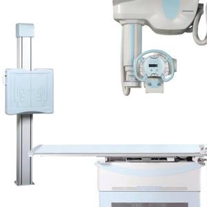
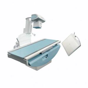
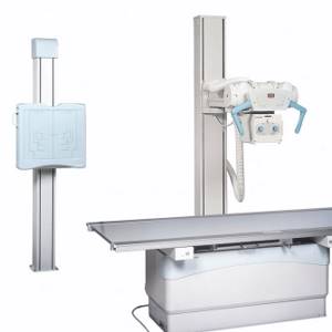
- for 2 workstations - the most popular stationary x-rays, including an x-ray tube, a table and an image stand, as well as a generator. The X-ray tube can be either ceiling-mounted or floor-mounted, the imaging table is designed for a “lying” position, and the stand is designed for a “standing” position.
- for 3 workstations - tele-controlled rotary tables-tripods - are distinguished by the highest price and unsurpassed functionality: all types of radiographic and fluoroscopic examinations become available to you.
- U-arm X-ray machines, often called “yoke” and suitable for small spaces
|
|
| X-ray machine for 2 workstations RADspeed with ceiling tube suspension | X-ray complex Shimadzu SONIALVISION G4 |
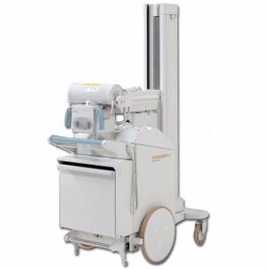
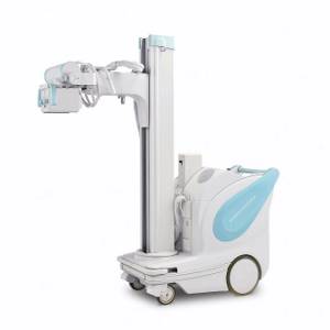
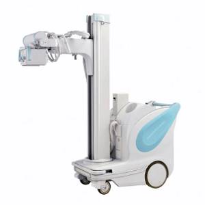
Mobile X-ray devices are often called ward or mobile. Their power starts from 12.5 kW and does not exceed 32 kW. Ward X-ray is the optimal solution if there are restrictions on the installation of stationary equipment.
|
|
|
| Mobile X-ray unit without motor drive MobileArt Eco | Ward X-ray unit MobileART Evolution | Mobile X-ray machine MobileDaRt Evolution |
Ultrasound examination (ultrasound)
X-ray is not the only way to “look inside” the human body; another technology is ultrasound. Some animals, such as bats, use sound waves to orient themselves in space. People have also learned to use waves to solve some problems, including in medicine. A picture of the internal organs can be obtained by sending a sound wave into the human body and monitoring its return. The computer helps process the results and present them in the form of a three-dimensional picture.
The main advantage of this research method is safety.
. Ultrasound can be performed even on pregnant women; in addition, ultrasound devices are mobile and can easily be placed in the patient’s room to monitor the condition of organs and blood flow in real time.
However, ultrasound cannot provide a high-definition image, so the use of this method of research is limited, for example, gastrointestinal diseases cannot be diagnosed using ultrasound.
Specialized X-rays
There is a classification by area of application:
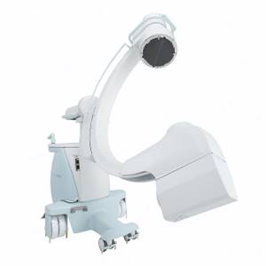
- surgical C-arms
- mammographs
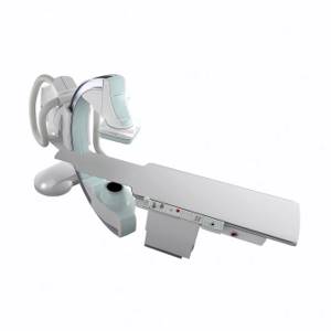
- angiography machines
- dental x-rays
|
|
|
| Mobile C-arm X-ray surgical system OPESCOPE ACTENO | Digital mammograph Planmed Clarity 3D | Angiographic system BRANSIST alexa |

Digital and film x-rays
Digital X-ray machines are increasingly replacing outdated film models. These new generation diagnostic systems allow you to obtain images of the highest quality in a matter of seconds, without wasting time and money on developing images, and conveniently store them in digital format.
What is the difference between a digital x-ray machine? In a DIGITAL method of processing anatomical structures that are obtained through exposure to X-ray irradiation.
Features of digital x-ray systems:
- high quality radiographs, excellent detail, resolution and contrast
- the ability to instantly take pictures
- the ability to conveniently store, as well as edit and process images
- low cost of research: no costs for film and reagents
- no need for a developing room
- environmental safety due to the elimination of the photo development stage
- minimal radiation exposure
- increasing the capacity of the X-ray room
Digital X-ray machines differ in the type of detector :
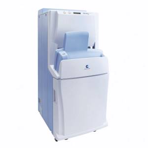
- CR systems with a digitizer (digitizer), using the principle of phosphor sensitivity. Imagine an ordinary X-ray machine, but instead of a film cassette, there is a special CR format cassette. After the photo is taken, it is removed from the device and placed in a digitizer - a reading device. After reading, the digital image is sent to the laboratory assistant’s workstation, and the cassette is cleaned and can be reused.
- Flat panel detector DR systems , which use semiconductor flat panel detectors. Moreover, a standard stationary X-ray machine for 2 workstations can be equipped with one wireless flat-panel detector, which will have to be moved from the table to the image rack, or with two separate ones - for the table and for the rack. Immediately after the image is taken, the image goes to the laboratory assistant’s station, bypassing digitization through a digitizer, which not only reduces the research time, but also generally guarantees greater reliability of the system.
The flat panel detector requires especially careful handling, because very sensitive to any damage and shock! Its price is the vast majority of the cost of the entire DR system.
If your clinic is already equipped with an analog (film) X-ray device, and the budget is limited, then to switch to digital technology it is enough to purchase a CR system or DR system based on a flat panel detector.
|
|
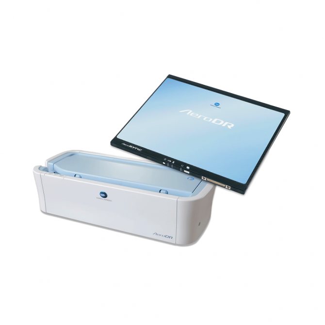
| Digitizer Konica Minolta Regius 210 | Konica Minolta AeroDR Digital Flat Panel Detector |
Digital diagnostics of pulmonary diseases
Digital diagnostics has great capabilities, and therefore the image is more informative. For example, with a digital X-ray you can even find out who the picture belongs to - a man or a woman, since the results show shadows of the mammary glands in the form of contours.
Doctors receive information about the correctness of the study - how deep the breath was and whether the patient was motionless at the time of filming.

Photo of healthy lungs
When assessing the results of digital fluorography, doctors consider the following positions using x-ray irradiation:
- soft tissues;
- integrity of bone elements;
- location of the trachea;
- the presence of calcifications;
- contours of the heart;
- pulmonary fields.
The study is carried out in an anteroposterior projection. The images are informative and make it possible to assess or identify the presence of pathology in the lungs.
Generator power
What do you need to know about such an important characteristic as generator power? The higher it is, the shorter the exposure time, and therefore the lower the radiation dose, and in some cases the higher the quality of the resulting image.
This advantage is especially valuable when examining heavier patients!
The highest generator power is typical for stationary X-rays, for which this figure ranges from 40 kW to 80 kW.
The most popular are x-rays with a generator power of 50 kW. This power is sufficient to carry out most of the necessary research.
What is important to consider? When choosing the generator power, it is necessary to take into account the power of the X-ray tube foci, which together determines the operating power of the system.
Purpose of the equipment
The X-ray machine is designed to generate Ro-radiation for further use for medical purposes without invasive intervention. X-ray equipment is used for diagnostic and therapeutic purposes. The operating principle of X-ray allows it to be used to treat a number of diseases using bremsstrahlung radiation. There are devices for superficial, intracavitary, medium and deep therapy.
A diagnostic X-ray machine is designed to examine patients to identify pathologies of internal organs. It is used for research:
- stomach and duodenum;
- gallbladder and bile ducts;
- colon;
- chest organs;
- spine and parts of the peripheral skeleton;
- abdominal cavity;
- female reproductive organs;
- mammary gland;
- teeth and others.
Generator type
Do not miss such an important characteristic as the type of generator. It is this criterion that determines the high contrast and resolution of the images, as well as the safety of the diagnostics performed.
Modern X-ray systems are equipped with high-frequency generators with a small pulsation of the anode voltage. It is precisely such technical innovations that make it possible not only to minimize the radiation dose to the patient, but also to increase the wear resistance and durability of the X-ray tube.
Where to look for such technological solutions? For example, in the line of Japanese X-rays Shimadzu, which are famous for their almost ideal rectangular pulses, providing unsurpassed quality of the resulting X-ray images.
|
|
| Digital X-ray diagnostic complex Shimadzu Flexavision F3 | X-ray complex Shimadzu Flexavision HB |
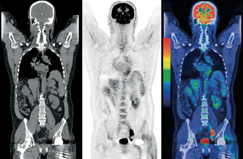
Hybrid Imaging Techniques
Probably, science would not be science if it did not constantly move forward and try to create something new from the old. So, doctors began to combine different scanning methods to obtain even more detailed and high-quality images. PET and SPECT are combined with CT, MRI complements PET. Such experiments are not cheap, but can sometimes help make decisions about further treatment for the patient.
X-ray tube
When studying the characteristics of an X-ray tube, pay attention first of all to the effective focus sizes: the lower the tube focus size, the more the image clarity increases and the operating power decreases.
The indicator of the anode’s heat capacity, which directly affects resource intensity, is also important. The high thermal capacity of the anode means improved wear resistance of the X-ray tube.
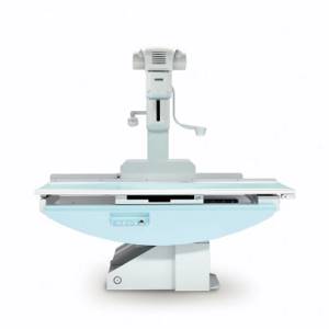
The X-ray tube of a stationary device for 2 workstations can have a floor or ceiling mount, with the first being the most popular due to the simplicity and ease of installation, as well as the absence of significant restrictions on ceiling height and room area. But it is worth remembering that when purchasing an X-ray with a floor-mounted tube and a large patient flow in the future, it is important to pay attention to models with a reinforced floor stand.
A tube with a ceiling mount always means a higher price for the device and its installation, but this option is distinguished by both high reliability and ease of use. If the high price and size of the room allow it, then ceiling mounting of the tube is recommended.
| |
| RADspeed X-ray machine with floor mounted tube column | RADspeed X-ray machine with ceiling-mounted tube |
Determining the price of an X-ray machine
What affects the cost of an X-ray machine?
- Class determined by the type of X-ray machine. If this is digital equipment, then it will cost more than analog. Entry-class X-ray machines – about 3-5 million rubles. If you choose the middle class, the price will be higher - from 5 to 14 million rubles. Premium class X-ray machines will cost an average of 14 million rubles.
- Location. A stationary one is more expensive than a mobile one.
- Functionality. The wider the functionality, the more expensive the installation.
Picture table
Check the maximum permitted load on the imaging table: a load of 200 kg is considered optimal, but as an option, if necessary, you can increase the load to 290 kg or more.
It will be useful to have a “lift” option, which allows you to move the surface of the imaging table vertically at a level of 50-85 cm from the floor.
In our catalog you will find X-ray machines from global manufacturers Shimadzu and Ziehm, as well as mammographs from the Finnish company Planmed. Our managers will help you choose the right model and tell you about its characteristics, as well as provide all the necessary assistance with the delivery and installation of equipment.