The following recommendations will be general for magnetic resonance imaging:
- It is strictly forbidden to undergo an MRI if there are any metal parts in the patient’s body that are attracted by a magnet . Under the influence of the field, they can heat up and move, causing tissue damage and burns. For this reason, MRI is not performed for patients with wire braces, metal vascular staples, and other “add-ons”;
- It is strictly forbidden to undergo an MRI if you have piercings , jewelry or other metal accessories on the body . This will not only lead to malfunction of the device, but can also cause harm to health;
- The study is prohibited for patients with a pacemaker, hearing aid, insulin pump, implanted cardioverter-defibrillator and other medical devices , since radio waves can affect and disrupt the operation of these devices;
- You cannot take a mobile phone or other electronic devices , as well as plastic bank cards, into the treatment room. Exposure to a magnet can lead to serious damage to gadgets;
- Due to the special loading capacity of the MRI machine table, there are weight restrictions : - for the Siemens Symphony MRI machine (1.5 T), located in Penza , the weight should not exceed 110 kg , the patient’s waist circumference should not exceed 120 cm; — for the MRI machine Philips Intera MR tomograph (1.5 T), located in Syzran , the weight should not exceed 130 kg , the patient’s waist circumference should not exceed 120 cm;
- Immediately before the diagnosis, you need to wear spacious and comfortable clothes that do not contain any metal fittings;
- When using contrast for examination, it is necessary to check for allergies to the components of the administered medication;
- Pregnancy during the 1st trimester. If the patient is pregnant or may be pregnant, she must inform the doctor conducting the study. MRI is much safer than computed tomography and radiography because This method does not use ionizing radiation (X-rays), but it does cause some heating of the body, so they try not to conduct this type of research in the first 3 months of pregnancy unless absolutely necessary. In the second and third trimester, MRI is considered safer, but if it is possible to postpone the scan until after childbirth, wait until the end of pregnancy;
- Mental inadequacy or serious condition of the patient;
- Due to the fact that the technique is carried out in a limited space, MRI cannot be done in the presence of claustrophobia and other neurological pathologies that have a similar clinical picture;
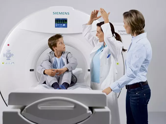
- The procedure is safe for children and can be performed on patients if the child behaves adequately and calmly;
How to properly prepare for an MRI procedure?
Before an MRI, you should refrain from using decorative cosmetics. This is because mascara and blush may contain metal particles. They will not harm your health in any way, but may interfere with the examination. As a result, images of the area of interest are unclear.
Magnetic resonance imaging is performed by appointment. It is recommended to arrive at the medical facility 15-20 minutes before the start of the diagnosis. This time is required for registration. If necessary, the patient can ask the doctor clarifying questions if any arise. Bank cards and other valuables will have to be left in the locker room.
It is advisable to put on a medical gown before the examination. It is unacceptable to take into the office:
- Metal jewelry, including watches.
- Gadgets.
- Bank cards.
If the patient remains in his clothes during the examination, they should be as loose and comfortable as possible. It should not have metal fasteners or decorative elements. It is better for the patient to wear a loose robe provided by the clinic staff.
Taking metal objects with you to magnetic resonance imaging is prohibited not only for the person undergoing the study, but also for the person accompanying this person.
Preparation for MRI examination of the abdominal organs and retroperitoneal space
Magnetic resonance imaging of the pelvic organs should be performed on an empty stomach, preferably in the morning. The last meal should be no later than 6-8 hours before the start of the study.
The day before the test, you must exclude from your diet coarse fiber (cabbage, other vegetables and fruits), legumes, carbonated drinks, brown bread, and dairy products. To eliminate increased gas formation, take activated carbon at the rate of 2 tablets per 10 kg of body weight or Espumisan. 30-40 minutes before the start of the study, take 1-2 No-Spa tablets (to relax muscles).
Rules for preparing for a diagnostic study
- Passport.
- Referral for research (if you did not leave it at the reception).
- Discharge summaries, descriptions and conclusions of ultrasound, CT, MRI, mammography, X-ray and scintigraphic studies (for the last 2-3 months), if any (it is advisable to make copies of them). Along with the conclusions, it is necessary to submit CDs (preferably copies) with recorded studies.
- REPLACEMENT OF FOOTWEAR (SLIPPERS).
- DISPOSABLE CUP (for receiving contrast agent and water).
- DRINKING WATER (without dye and gases) - 1-2 liters .
- Toilet paper (napkins).
- DVD - R or DVD - RW format disc for recording PET/CT studies, if necessary. Recording the study to disk is optional!
If for some reason you cannot come for the study on the specified day, be sure to notify us as early as possible (preferably at least one day before the study) by calling the registration desk.
The date and time of the study is announced when registering for it or is specified by YOU by phone within the time frame specified by the registrar.
The time that the registrar tells you is not the individual start time of your study, but the general registration time for the shift in which your study takes place. Next, at the Center, groups of patients are selected for the study. The order of the study in groups depends on the height and weight of the patients, the scope of the study, the severity of their condition, the presence of diabetes mellitus and other factors. The group usually includes 3-4 people, the groups follow each other with a gap of 10-15 minutes.
SEQUENCE OF PET/CT STUDIES
Upon arrival at the Center for the study, you must contact the PET Center reception directly and present your passport and available medical documents. Patients without a doctor's referral are not served !
Place your outerwear and outdoor shoes in the cloakroom for visitors, put on a change of shoes and wait for a call for research.
First of all, patients are taken to the internal cloakroom of the laboratory, where they can change into a change of clothes (if necessary) and leave things in a personal locker.
Next, in the treatment room, patients’ height and weight are measured, an “Agreement to Perform a PET/CT Study” is drawn up, and blood glucose levels are monitored. If the glucose levels correspond to acceptable values, the patient is given an intravenous catheter and he is escorted to a special room where he awaits the administration of the radioactive drug.
The procedure for administering the drug is safe and painless, and only in rare cases may it be accompanied by a feeling of heat. After an intravenous injection of the drug, before the start of the study, you must drink part of the liquid you brought with you (0.5 - 0.7 l) - the quality of the diagnostic images depends on this. At this time, the patient should be in a calm, relaxed state (otherwise the test results may be distorted). Optimal: close your eyes, don’t read, don’t listen to music, don’t talk, try not to move. Empty your bladder after visiting the bathroom, trying to move smoothly and avoid sudden movements. When using the toilet, you must flush the toilet twice with a full flush tank. Men are advised to sit down when urinating to avoid splashing urine.
At the end of the waiting period (lasting about 60 minutes), the patient must completely empty the bladder (even if there is no urge to urinate), after which the nursing staff will accompany him to the PET/CT procedure.
The examination is carried out in a supine position with the arms placed above the head in a hand lock. If this pose is not possible, alert staff. It is permissible to place the hands along the body near the buttocks or under them, but not on top of the body.
During the entire study, the patient must strictly follow the instructions of the staff of the diagnostic department: lie still, try not to cough, do not talk, and do not breathe deeply and shallowly.
The duration of the study is individual and ranges from 20 to 60 minutes. If it is necessary to conduct an additional delayed examination (40 - 60 minutes after the initial examination), you will be informed separately. In some cases, a contrast agent is used to improve the accuracy of a CT scan, which improves visualization of blood vessels or intestines. Its administration is also painless.
Upon completion of the study, the patient is accompanied to the exposure room - a rest room, where he stays for up to 1-2 hours to reduce the level of radioactivity in his body.
Subsequently, the patient on call undergoes radiation control and changes clothes in the wardrobe. At the same time, he is given a reminder about reducing radiation exposure and restrictions for the near future.
During their stay in the relaxation rooms, as well as on the territory of the PET center, patients are monitored by video surveillance .
Hospital clothing (if you used it) is returned to the medical staff.
The total time spent in our center for the study is 4-6 hours!
ATTENTION!!! Description of PET/CT examination is associated with the processing and interpretation of a large number of diagnostic images and the analysis of medical documents. In this regard, it is acceptable to issue a PET/CT examination report 2-5 working days from the date of its completion. The conclusion of our specialists will be pasted into your outpatient card and transferred to the office or department that referred you (or sent by e-mail to your dispensary). The readiness of the study results should be confirmed by calling the PET Center registry a few days after the study. When the registrar notifies you that the report is ready, you come for a consultation with the doctor who referred you. Please check with your clinic about the procedure and need to make an appointment for a consultation with your doctor.
A disk with a recording of the study in DICOM format (if you ordered it) is issued immediately after the study.
Preliminary consultations based on the research data are not provided until the final answer is provided.
Registration staff do not provide advice on the conclusion of the study.
PET/CT REGISTRATION. Tel.: 8 (017) 389-96-84
Preparation for MRI examination of the pelvic organs
An hour before the examination, you need to drink 2-3 glasses of water. MRI of the pelvic area is performed when the bladder is low or moderately full.
The day before the test, you must exclude from your diet coarse fiber (cabbage, other vegetables and fruits), legumes, carbonated drinks, brown bread, and dairy products. To eliminate increased gas formation, take activated carbon at the rate of 1 tablet per 10 kg of body weight or Espumisan.
It is advisable to empty your bowels. 30-40 minutes before the start of the study, take 1-2 No-Spa tablets (to relax muscles).
For women, the study is recommended to be carried out on days 5-14 of the menstrual cycle ; it is possible to conduct the study in the second phase of the cycle, but not during menstruation .
List of documents that the patient must have with him for MRI
Magnetic resonance imaging is performed for various reasons. It could be:
- Previously diagnosed disease.
- Suspicion of the presence of pathologies in the body, lack of convincing data on adverse changes based on the results of other examination methods.
- A disease for which therapy has already been performed (to evaluate its results).
In all of these cases, the patient has a doctor’s opinion and data from other examinations. You must take all available documentation with you. This will greatly simplify the work of the doctor performing functional diagnostics. It will allow you to draw up a more informative and detailed conclusion based on the results of magnetic resonance imaging.
For example, the treating specialist can give an opinion confirming the presence of oncology, indicate the location of the tumor and its size. The availability of data on previous examinations and therapy for the tumor will allow us to understand how effective the treatment was and whether there are complications after it.
It is advisable to show your medical documentation to the doctor before the examination. This will make it possible to choose more effective diagnostic tactics.
Tomography of various organs is done only if there are no contraindications to this procedure.