What does an ultrasound of the stomach show?
The content of the article
The anatomical location, contents and presence of a gas bubble in the stomach greatly complicates visualization of the organ. However, ultrasound is a rational diagnostic method, since it allows one to evaluate the terminal and output sections, and lesions are most often concentrated in them.
On an ultrasound of the stomach you can clearly see:
- minor and major curvature;
- part of the body of the stomach;
- gatekeeper cave;
- gatekeeper channel;
- pyloric sphincter - the area of transition into the duodenum;
- part of the duodenum.
During the procedure, the doctor evaluates the size of the organ, its shape and size, location, wall thickness, echostructure, motor function, as well as the presence of deformations and foreign objects.
Ultrasound of the stomach is often included in ultrasound of the abdominal cavity. In this case, the doctor sees other organs of the gastrointestinal tract.
What stomach diseases can be detected by ultrasound?
Sonography of the stomach allows you to identify many diseases at an early stage of development:
- diaphragmatic hernia;
- gastritis, peptic ulcer;
- gastroesophageal reflux;
- swelling of the walls;
- malignant lymphoma;
- varicose veins of the stomach;
- diffuse neoplastic wall thickening;
- malignant and benign neoplasms;
- congenital and acquired pyloric stenosis - narrowing of the pyloric outlet;
- carcinoma;
- lack of delimitation of the wall into normal layers;
- aberrant tumor vessels;
- cystic formations;
- inflammation of the internal cavity of the organ;
- mesenchymal tumors.
Indications
Timely ultrasound examination will help maintain health and avoid various serious consequences. Ultrasound of the stomach is indicated not only for certain obvious pathologies, but also when a number of alarming symptoms appear:
- prolonged heartburn, nausea, vomiting, frequent belching;
- frequent pain of various types in the gastrointestinal tract;
- disturbance of the digestive process, loss of appetite, bloating;
- gastritis, ulcer;
- congenital anomalies of organ development;
- suspicion of cancer;
- polyps;
- pyloric stenosis, intestinal obstruction;
- suspicion of embryonic anomalies;
- severe discomfort in the abdominal area in a pregnant woman.
There are a number of separate indications for performing ultrasound of the stomach in children:
- frequent bronchitis;
- asthma;
- causeless increase in temperature;
- dry cough;
- excessive regurgitation in infants;
- abdominal pain;
- diarrhea, constipation, change in stool character;
- causeless nausea and vomiting.
Even healthy people are recommended to undergo an ultrasound of the stomach for preventive purposes. Moreover, all types of ultrasound can be done without a referral from a gastroenterologist or therapist, on your own initiative.
Research results
An ultrasound of the esophagus and stomach may reveal signs of the following diseases in a child:
- gastritis;
- hypertrophic pyloric stenosis is a common cause of high intestinal obstruction in infants.
There are other pathologies that can be determined using this method: congenital gastric dystonia, polyps, gastric lymphoma, stones and foreign bodies.
Ultrasound with a water-siphon test can detect disorders of gastric motility and reflux.
How to prepare for the procedure
Compliance with all recommendations regarding preparation for an ultrasound examination of the stomach will give the most accurate and high-quality result. Preparation is carried out in several stages and consists of the following:
- 2-3 days before the procedure
. A diet aimed at reducing gas formation in the stomach and intestines is required. To do this, avoid drinking carbonated drinks, fresh vegetables and fruits, herbs, legumes, sweets, dairy and yeast products, rye bread, fresh juice, black tea and coffee. Also avoid alcohol and smoking. It is recommended to form your diet from boiled meat, fish, cottage cheese, soft-boiled eggs and porridge cooked in water. If necessary, you need to take enzymes that improve the digestion process (mezim, festal, etc.). A day before the procedure, it is necessary to cleanse the body with the help of sorbents (activated carbon, smecta, etc.), and for people suffering from stool disorders, with the help of laxatives. - On the eve of the procedure
. The study is carried out strictly on an empty stomach. As a rule, it is prescribed in the morning, so the last meal should be a light dinner 10-12 hours before the ultrasound. For children, fasting should last 6-8 hours, and for infants 3-3.5 hours, while their stomach is examined immediately after feeding. - Before the procedure
. Skip breakfast and drinks. It is not recommended to even brush your teeth, as there is a possibility of toothpaste getting into your stomach.
Pathology detected: should it be rechecked?
Ultrasound of the stomach and intestines is very informative, but it is impossible to make a diagnosis based on the data obtained. If problems are detected, the patient undergoes additional examination. The most popular methods for examining the gastrointestinal tract include:
- FGDS. This is an endoscopic method that allows you to see bleeding, tumors in the stomach and intestines.
- Probing. It involves taking the contents of the stomach for further laboratory testing.
- Gastropanel. This is an innovative method, according to which the patient is drawn from a vein, and a possible ulcer, atrophy, or cancer is detected using certain markers.
- CT scan. They take cross-sectional images in different projections and identify the location of tumors, hematomas, hemangiomas, etc.
- MRI. This is the most expensive and effective research method. Allows you to visualize not only the organ itself, but also nearby lymph nodes and blood vessels.
- Endoscopy. Used when collecting material for biopsy.
- X-ray. Reveals incorrect location of the stomach and intestines relative to other organs, pathology of shape, and various neoplasms.
- Parietography. Translucent the walls of the stomach and intestines thanks to the injected gas.
- Laboratory tests (blood, urine, stool tests).
After undergoing additional diagnostics, the doctor decides on treatment methods. It is important to understand that treatment of the gastrointestinal tract cannot be done in a “mono” mode - it is always a set of measures related to restoring health and preventing relapses and complications. You can also monitor the quality of treatment using ultrasound, comparing previous results of a gastrointestinal examination with new ones.
If you find an error, please select a piece of text and press Ctrl+Enter
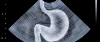
How is an ultrasound of the stomach performed?
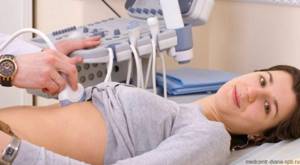
Diagnosis of the stomach using ultrasound is carried out transabdominally - through the abdominal wall using an external sensor. The patient lies on the couch with his back, on his side, or takes a semi-sitting position and exposes his stomach. The doctor applies a special gel to the epigastric area to improve the passage of ultrasound waves, and then scans, slowly moving the device over the area being examined.
Ultrasound of the stomach is carried out in 3 stages:
- 15 minutes before the examination, the patient needs to drink 1 liter of non-carbonated liquid (for children - 0.5 liters). This helps straighten the stomach and allows you to diagnose all possible pathological changes.
- The patient again needs to drink a small amount of liquid so that the doctor can diagnose the stomach filled with it by examining changes in the internal organs.
- After 20 minutes, the doctor conducts a second examination to determine the rate of gastric emptying and the motor function of the organ.
In some cases, for better visualization, the doctor may use a contrast agent, for example, Ekhovist-200, diluted with 0.5 liters of carbonated water.
Ultrasound machines for intestinal examination
The intestines are examined using two types of sensors: transabdominal (through the abdominal wall) and endorectal. To study the colon, a 2D device is sufficient, which produces a flat two-dimensional image. Such an examination already provides reliable information about the patient’s health status. The endorectal method is more informative because the sensor is inserted into the anus and examines the organ from the inside.
The doctor decides which sensor to choose depending on the patient’s complaints. In special cases, both methods are used.
- In 15% of cases, the transabdominal sensor “does not see” the rectum, as well as the area of the anal canal. The endorectal method is not possible with stenosis of the terminal gastrointestinal tract (abnormal narrowing).
- The endorectal probe usually examines the distal parts of the rectum. A rectal examination requires preparation.
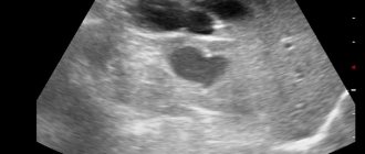
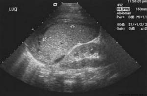
Interpretation of ultrasound of the stomach: normal parameters of the organ
A qualified specialist, knowing the normal indicators, can simply determine pathological changes in the stomach. Normally, sections of an organ have a round or oval shape - in the form of a ring with an echo-positive central part and an echo-negative rim. The walls of the stomach are uniform and consist of 5 layers. The removal of a glass of liquid from the stomach occurs in approximately 20 minutes, and the primary withdrawal of liquid is normally about 3 minutes.
Normal values for the thickness of parts of the stomach
| Stomach in the pyloric region | up to 8 mm |
| Stomach in the proximal zone | up to 6 mm |
| Muscle layer | 2 – 2.5 mm |
| Slime layer | up to 1.5 mm |
| Submucosa | up to 3 mm |
| Lamina mucosa | up to 1 mm |
Normal echogenicity values
| Outer serous membrane | high |
| Muscle layer | low |
| Slime layer | high |
| Submucosa | average |
| Lamina mucosa | low |
| Stomach wall | varying degrees of echogenicity of the layers |
Interpretation of pathological indicators visible on ultrasound
| Pathology | Signs on ultrasound |
| Impaired gastric emptying | maximum distension of the stomach with liquid; lack of peristalsis (not always); internal echostructure may vary from anechoic to hyperechoic |
| Swelling of the wall | the thickness of the stomach wall exceeds 7 mm; homogeneous wall structure; hypoechogenicity, uniform thickening |
| Diffuse neoplastic wall thickening | the walls of the stomach are hard, inelastic, with low echogenicity; boundaries between layers are not detected; there are no organ contractions |
| Aberrant tumor vessels | a thickened wall with low echogenicity is detected due to the penetration and accumulation of tumor particles in it |
| Lack of differentiation of the wall into normal layers | the lumen in the stomach is significantly narrowed due to obliteration |
| Congenital hypertrophic pyloric stenosis | thickening of the pyloric muscle ring; slow removal of contents from the stomach; increasing organ mobility and antiperistalsis |
| Acquired pyloric stenosis | frigidity of the pyloric wall; anechoic structure, homogeneous pinpoint echo structure, or coarse internal echoes |
| Varicose veins of the stomach | the outer wall of the stomach is anechoic or cystic thickened in some areas; blood flow from the liver and portal-systemic blood flow were identified |
| Peptic ulcer | the stomach wall is thickened; crater-shaped depressions are visible on the inner surface |
| Benign tumor | formation of a round shape; has smooth edges and low echogenicity; no metastases |
| Malignant tumor | the wall of the stomach is thickened unevenly (round or lobed); the gastric outlet is narrowed; the layers of the stomach are blurred, the contours are uneven |
| Carcinoma | polypoid: lobular formation; circular: a round or oval-shaped formation with an echogenic center, inside of which there is air and mucus. Possible detection of metastases |
| Malignant lymphoma | appears similar to diffuse lymphoma or carcinoma |
| Mesenchymal tumor | benign: no more than 6 cm, vessels are not identified; malignant: more than 6 cm, tumor vessels, possible metastases |
| Cystic formations | formations of 2 layers: echogenic internal mucous and hypoechoic muscular external |
Tests and studies that complement intestinal ultrasound
As mentioned above, intestinal ultrasound is not 100% confirmation of a particular diagnosis, although in many ways the method is informative and accurate. Depending on the preliminary diagnosis, in addition to ultrasound, the patient is prescribed:
- Capsule examination
. The patient swallows a capsule with a sensor inside, which conducts video surveillance and transmits the image to the monitor screen. The method allows you to see areas inaccessible to the endoscope. Significant advantages also include the absence of trauma (the intestinal walls are not scratched) and radiation (unlike X-rays).
The disadvantages of the capsule technique include the low prevalence of capsule examination, because the method was first tested in the USA in 2001, and today it is still not widespread. Its cost is very high, and this limits the circle of clients. Other disadvantages include the inability to conduct a capsule study in case of intestinal obstruction, infections, and peritonitis. The method has age restrictions associated with the peculiarities of peristalsis.
- Colonoscopy
. This is an endoscopic method that allows you to examine the internal mucous membrane for polyps, colitis, tumors, Crohn's disease, inflammation and other pathologies. The disadvantage of this method is the risk of intestinal trauma and perforation (punctures of the walls). Colonoscopy also does not see tumors between the intestinal walls. - Irrigoscopy
. This is a special method aimed at identifying hidden tumors located between the inner and outer lining of the intestine. In addition, the method, unlike colonoscopy, sees areas on the folds of the intestine and its remote areas.
Irrigoscopy involves the introduction of a liquid solution of barium sulfate through the anus, which allows a clear contrast image to be obtained upon contact with air. The advantages of irrigoscopy are the ability to examine structural changes in tissue (scars, diverticula, fistulas). The method is used for diarrhea or constipation, mucus in the intestines, pain in the anus.
Examination of the stomach with abdominal ultrasound
Ultrasound examination of the abdominal organs
is a modern non-invasive diagnostic procedure that is absolutely safe for the patient’s body, but has a high level of information content.
This research option is often chosen by the doctor as the primary one, since it is the most accessible, has minimal training requirements, and can be carried out urgently. Also, the wide popularity of abdominal ultrasound
is ensured by
the price
, which, unlike many techniques, is low.
When is an abdominal ultrasound prescribed and what can the doctor see?
This method, in addition to the stomach, allows you to visualize many vital organs, therefore it has a wide range of indications. A general practitioner, gynecologist, urologist, nephrologist, gastroenterologist and surgeon can refer a patient for this procedure. The main indication for ultrasound diagnostics is pain of any kind in the abdominal area. There are no contraindications for diagnostics.
Complaints, the appearance of which requires an urgent ultrasound of the abdominal cavity:
- severe abdominal pain;
- feeling of heaviness in the area of the right hypochondrium;
- bitter taste in the mouth;
- aversion to fatty foods;
- yellowing of the tongue;
- detection of a tumor or other neoplasm in the peritoneum.
Ultrasound easily visualizes internal organs, and hollow organs can also be seen if they are filled with fluid. The main obstacles to a quality examination are air and fat - these features are important to consider when preparing for the procedure.
- Liver
. Ultrasound allows you to determine focal changes in liver tissue, such as adenomas, cancer, cysts. In addition, diseases such as hepatitis or cirrhosis can be seen using ultrasound. - Gallbladder and its ducts
. Ultrasound clearly identifies the following deviations from the norm: defects associated with the abnormal structure of the gallbladder (kinks, constrictions); chronic and acute cholecystitis; disturbances in the functioning of the bile ducts; cholelithiasis; polyps; tumors, both benign and malignant. - Pancreas
. Ultrasound diagnostics can reveal: acute, chronic pancreatitis; structural anomalies; tissue changes caused by diabetes; cysts, cancer and other neoplasms. - Spleen
. An organ that is difficult to diagnose, and therefore the doctor will ask the patient to take a certain position. Ultrasound allows you to identify malformations, heart attacks, mechanical damage, tumors and estimate the size of the organ. - Stomach.
In addition, the study covers organs such as the bladder, prostate gland in men and uterus in women. Abdominal ultrasound can visualize the retroperitoneum and kidneys.
How is an abdominal ultrasound performed?
The procedure is simple and painless for the patient and usually takes about half an hour. It is important to pay attention to the need to prepare for an ultrasound, which consists of completely emptying the intestines of gas, which is achieved by following a diet.
The technique involves examining the patient in the “lying on his back” position; sometimes it is necessary to turn on his side and hold his breath. If the patient has deviations in the position of the organs, then the examination is carried out in the “sitting/standing” position.
During an ultrasound, the doctor can evaluate the dimensions, position, shape, density of the structure of organs and the thickness of their walls, the ability of ducts and vessels to pass physiological fluids (bile, secretions, blood, urine), the presence of stones, and the condition of bile. Based on the results of the examination, the result is given in the form of photographs and their interpretation.
What are the advantages and disadvantages of ultrasound of the intestinal gastrointestinal tract?
Ultrasound diagnostics of the intestine is used for initial examination in case of suspected pathology, as well as in cases where the endoscopic method is contraindicated due to the patient’s health condition (perforation (damage) of the intestine, inflammatory process).
Ultrasound examination of the intestines has a number of advantages:
- The patient does not experience psychological discomfort.
- The doctor receives information about the size of the organ, its structure, thickness, number of layers, without penetrating inside the organs.
- Ultrasound allows you to examine the inflamed intestines and clearly sees the upper gastrointestinal tract.
- Peristalsis is visualized in real time and intestinal obstruction is determined.
- On an ultrasound of the intestines, the specialist will see even small compactions or changes in the echostructure of tissues.
- Ultrasound allows you to do screening (endorectal method), completely confirm or refute oncology.
Despite the large number of advantages, diagnosing this organ with ultrasound has some disadvantages, the main one of which is the impossibility of making an accurate diagnosis without additional examination.
Also, the disadvantages of the method include the following:
- Only functional disorders in the functioning of the organ are detected.
- Structural changes are determined without defining the parameters of the changes.
- It is not possible to assess the condition of the internal mucous surface; if structural changes are detected, colonoscopy is prescribed - an endoscopic method
Alternative studies: how best to examine the stomach
Ultrasound examination does not replace endoscopy and x-ray examination; to some extent it is even inferior in terms of information content, but at the same time it has many advantages compared to them:
- high information content of the study;
- speed of the procedure;
- absolute safety – has no complications and does not harm the body;
- painlessness and lack of discomfort during the procedure;
- visualization of the state of the organ in real time, the result is visible immediately;
- the ability to see even small structures;
- allows you to study the functions of the organ and use duplex scanning to identify reflux and blood supply to the stomach wall;
- the possibility of conducting research even for infants and pregnant women;
- X-rays visualize only one projection of the organ, which is why some pathologies can be overlooked;
- during endoscopy there is a risk of infection if the endoscope is not sterilized sufficiently;
- X-rays and endoscopy do not study the entire thickness of the stomach wall and deep layers.
A significant disadvantage of gastric ultrasound is the inability to take biopsy material and physiological fluids. This can only be done by endoscopy.