All internal and external systems of the human body are closely interconnected, especially when it comes to areas adjacent to each other - the brain and the cervical spine. Even minimal deviations in the degree of functionality of these areas entail disturbances of varying degrees of severity in other areas of the body. Any biological tissue needs nutrition and purification from waste products and toxins, including brain cells. The energy-supplying and oxygen-conducting function is performed by the vascular network. All important channels entering the brain structure pass through the neck, therefore, if pathology in the skull is suspected, a joint diagnosis with the adjacent cervical area is often prescribed.
Despite the large selection of diagnostic methods, specialists most often give preference to the safest and most accurate of them - MRI of cerebral vessels. A distinctive feature of this technique is the ability to set settings in the software that clearly show the vascular structures of the organ, bypassing adjacent sections that “interfere” with a detailed study. If an MRI examination of brain structures clearly displays all the “details” of an organ: gray matter at the cellular level, configuration and relief, then an MRI of the vessels of the head and neck focuses only on the blood pathways, and the soft fibers around them remain transparent in the images.
Unlike technologies that use radiation exposure (x-ray, computed tomography), nuclear resonance screening works on the principle of generating natural magnetic fields that do not damage living tissue and do not leave critical health consequences. Over more than 40 years of practical use of magnetic resonance devices, not a single case of harmful effects has been recorded.
Advantages of MRI of head and neck vessels over alternative methods
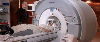
If you do not use an effective diagnostic technique in a timely manner, there is a high probability of making a medical error in making a diagnosis. It is impossible to determine by eye the presence of a hematoma or contusion disorder, the degree of thrombosis or destruction of the walls. Instrumental penetration, puncture sampling of biomaterial for biopsy, etc. – painful and traumatic methods that cause additional harm to the patient. Remote, hardware examination does not imply invasive penetration into membranes and organs. It is enough for a person to be in the field of action of a powerful magnet so that the response signature from the cells is recorded by the sensors of the installation and transmitted in the form of high-resolution images to the computer screen. The person does not experience any pain or discomfort at this moment.
In addition to non-invasiveness, MRI of vessels and arteries of the neck and cranial region has a whole list of advantages over other diagnostic techniques:
- absolute harmlessness to general health due to the absence of harmful ionizing radiation;
- simplicity and high speed of obtaining effective results;
- a small number of contraindications, which allows people of any age and pregnant women (from the second trimester) to undergo examination;
- the ability to identify the disease at the earliest stage of its onset;
- no prohibition on repeated access to tomography (if necessary);
- greater information content compared to ultrasound examination;
- lack of radiation exposure compared to X-ray and CT;
- high degree of hypoallergenicity of contrast-enhancing drugs.
When is MRI of the head and neck vessels prescribed?
Screening can be prescribed both as a primary examination and as a confirmatory method when other methods are ineffective. If classical methods are powerless or a specialist doubts the correctness of their results, the only one hundred percent method is MRI of brain vessels. Scanning is often required preoperatively to assess the patient's general condition and operability.
The proper nutrition of the systems located in the cranium depends on the correct functionality of the circulatory structures in the neck. If the cervical spine is damaged, an MRI of the vessels and arteries of the neck is required. The main indications for referral for MR screening are:
- regular pain, migraines of unknown etiology, dizziness and fainting;
- attacks of muscle strain, convulsions that cannot be purposefully controlled;
- loss of sensitivity in certain areas of the body, limbs, skin;
- loss of coordination, sense of reality;
- decreased sound perception and visual function;
- speech disorder;
- manifestations of mental disorders;
- suspicion of the development of a tumor process;
- “crunching” in the cervical region during physical activity, turns;
- old and new traumatic brain injuries;
- fractures and displacement of the cervical vertebrae;
- VSD, tinnitus;
- ischemic damage.
Also, an examination can be prescribed after surgery to monitor tissue regeneration, analyze the patient’s condition and relapse of pathology. Such control can be carried out repeatedly. Neurosurgery uses scanning as an accompanying visualization during surgery.
What is better: MRI or ultrasound of neck vessels
There is no clear answer to this question. Each method has its own advantages and disadvantages. By examining soft tissues using a tomograph, the diagnostician obtains more accurate data, but at the same time, if a person (especially a child) cannot lie still, ultrasound is the only correct solution. In terms of cost, an ultrasound of the cervical spine will be cheaper, but after the examination additional tests may be prescribed, while magnetic resonance imaging gives a final result.
What can be seen on an MRI of the vessels of the head and neck?
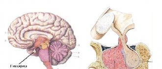
As mentioned above, MRI photographs of cerebral vessels clearly show the main arterial tracts and small capillary canals: circle of Willis, anterior, median and posterior cerebral tracts, basilar and vertebral basins, carotid canals, carotid basin. Any, even microscopic, pathology is noticeable when examined on numerous photo sections, combined into a multidimensional model. When performing MRI of vessels and arteries of the neck and intracranial cavity, the following abnormal conditions can be determined:
- intravascular atherosclerotic growths;
- degenerative processes as a result of TBI;
- foci of infection by bacterial, viral or fungal pathogens;
- examination of the consequences of stroke;
- tumor formations;
- stretching of the canal walls of an atherosclerotic nature;
- incorrect innate structure of the network;
- multiple sclerosis;
- exfoliating wall processes;
- thrombosis with plaques.
For pathologies occurring in the blood circulation of the neck, in addition to some of the described anomalies, pinching, damage to the walls as a result of injuries suffered by the patient, genetic changes and disorders, as well as neoplasms of various origins, cysts are diagnosed.
In what situations is an MRI of the neck indicated?
The most common situations in which research is necessary:
- suspicion of a neck tumor
- preoperative examination for an established tumor (for treatment planning), control examinations after surgical, radiation and chemotherapy treatment.
- suspicion of an infectious process
- swollen lymph nodes of unknown cause
- congenital structural abnormalities (for example, congenital neck cysts)
- neck injuries
- pathology of neck vessels
When is it prohibited to do MRI of head and neck vessels?
Like any medical procedure, MRI of vessels and arteries of the neck, as well as MRI of brain vessels, have a number of limitations based on individual indicators. Thus, the examination is prohibited for people who have:
- Implanted ferromagnetic elements in the form of metal implants and electronic life activity stimulators. Removable systems in the form of dentures or braces are also prohibited. They must be temporarily removed for the duration of the study. The presence of such inclusions in the body should be reported to the treating doctor in advance.
- Early pregnancy (up to 12 weeks). The ban is associated with the need to exclude any external influence on the actively developing organism of the unborn baby.
- Neurological and mental disorders leading to uncontrolled motor activity, spasms, tics, etc. The effectiveness of the examination directly depends on remaining still during screening. Otherwise, the images will be distorted and diagnosis will be impractical.
- Excess body weight. Standard hardware installations are designed for a weight category not exceeding 120-200 kg. Obese patients will not be able to physically fit inside a car with solid walls. In this case, there is an alternative option to use an open-configuration tomograph with a ring structure.
- Immune rejection to certain types of medications. This contraindication applies to cases where scanning is required using contrast enhancement. The solution is administered intravenously. If the patient is allergic to the components of the dye, the procedure in this mode is prohibited.
- Impaired functionality of the urinary system (with contrast). If the enhancing drug is not cleared from the body through the kidneys through urination in time, its retention leads to complete intoxication.
- Shock, coma, unbearable pain, uncontrollable fear. In these conditions, a session can only be scheduled using general anesthesia, which also has individual contraindications.
MRI of the neck arteries: features of the procedure
As a rule, in order for the diagnostician to be able to see each vessel, an MRI of the neck with contrast is prescribed. A special drug containing halidonium is administered intravenously immediately before the examination. The substance quickly spreads throughout the bloodstream and allows a more accurate assessment of the condition of large and small vessels. The duration of the procedure is from 30 minutes to an hour. It is prohibited to use contrast in cases where the patient may have an allergic reaction to this substance or has kidney dysfunction.
In general, tomography is contraindicated for:
- the presence of metal devices and devices in the patient’s body - pacemaker, implants, insulin pump;
- pregnancy (especially not recommended in the first half);
- mental illnesses, as a result of which a person cannot lie still.
Otherwise, MRI of the cervical spine with contrast is the same as the procedure without the use of this substance. The patient must remove jewelry, piercings, glasses, watches, and change clothes. Next, it is located on a retractable table, fixed with special belts and moved inside the circuit. During the examination, a person can talk through a microphone with the doctor and hear the answers.
Depending on what the MRI of the soft tissues of the neck shows, the patient may be prescribed a consultation with a phlebologist, endocrinologist, or oncologist.
How to prepare for MRI of head and neck vessels?
A special system of preparatory manipulations for MRI of vessels and arteries of the neck and cranium has not been developed, since it is not required. The exception is the planned event under the contrast protocol. For comfortable administration of the dye solution, it is necessary that the patient come to the session on an empty stomach. Otherwise, nausea and vomiting may occur. It is better to refuse your last meal 5-8 hours before the tomography.
There is also a list of recommendations that you should know about in advance:
- before the examination, you need to remove all metal objects from clothing and body, women should wash off their makeup (it also contains metal particles);
- prepare in advance documentation related to the disease (medical record, referral from a doctor, results of past studies);
- testing for allergic resistance to a contrast agent;
- women take a pregnancy test, even if you are sure you are not pregnant;
- If you have anxiety and fear, it is better to take light sedatives before the examination.
Stages of MRI diagnostics of head and neck vessels
Each nuclear resonance screening has a standard implementation plan. MRI of vessels and arteries of the neck is no exception. The procedure is divided into several stages:
- Preparatory stage. The patient undergoes registration, questionnaires and instructions, is invited to a separate room with equipment, and, if necessary, changes into comfortable clothes or disposable clinic underwear. Next, the employee helps to take a comfortable position on the retractable part of the tomograph, secures the patient with belts, and attaches the sensors.
- Installation inspection. The conveyor with the patient's body moves inside the device, and screening begins. During the process, the machine may move slowly and make loud noises. Multiple images of the study area are taken. The center staff is in the next room and monitors the progress of the diagnosis through a transparent partition. If you experience discomfort or complaints, you can contact the doctor via the speakerphone system and ask to pause the session.
- Waiting for examination results. Interpretation of results may take from an hour to two hours. In particularly complex clinical cases, the conclusion can be obtained only the next day.
Are blood vessels visible on MRI of the brain?
MRI without contrast shows only brain tissue. Occasionally, the sections contain minor fragments of large vessels that cannot be fully assessed. If the doctor suspects an aneurysm, atherosclerosis, angiopathy or other pathology, an examination of intra- and extracranial vessels is carried out using a contrast method.
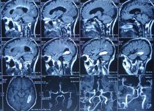
MRI scans of the brain and blood vessels
What to do after an MRI of the head and neck vessels?
As a result of the examination, the client is left with a detailed description of the identified pathologies and photographic images printed on film or transferred to electronic media.
After an MRI of the cerebral vessels, the patient must go to the attending physician, who will make a final diagnosis.
If necessary, the doctor will redirect you to a more specialized specialist who is competent in the detected disease.
If the quality of the images does not satisfy the doctor, a repeat scan may be ordered in a different mode.
What does an MRI of the neck show?
During scanning, the device takes a series of layer-by-layer images, and they can be taken in different projections, at the angle necessary for diagnosis. After the procedure, the doctor will combine the resulting images into one whole and receive a single three-dimensional model of the internal organs, on which blood vessels, bone and soft tissue, and lymph nodes are clearly visible.
Thanks to this examination it is possible:
- identify a hernia formed between the cervical vertebrae;
- see abnormal processes;
- identify neoplasm;
- determine the location of the injury and assess the consequences;
- see developmental abnormalities or identify changes in the thyroid gland.
Make an appointment now!
In some cases, when a brain examination is carried out, it is very important what an MRI of the neck lymph nodes shows, since more than one artery and vein pass through it, and the functioning of the brain directly depends on their “correct operation.”