According to medical indicators in the field of urology, one of the most common urological diseases is urolithiasis - urolithiasis.
One of the forms of this disease is the development of compactions (calculi) in the urinary system - in the kidneys. This pathology is called nephrolithiasis. The formation and growth of stones can be caused by various factors, ranging from congenital predisposition to alcohol abuse and physical inactivity.
The phenomenon of kidney stones has been known since ancient times. According to data obtained as a result of excavations, this pathology was discovered in Egyptian mummies.
- Nephrolithiasis at risk
- 01congenital predisposition
- 02Alcohol abuse
- 03Sedentary
In this article we will reveal what causes stones, and also consider the first manifestations of the disease, methods of determination, and what to do if a sad diagnosis is made.
What does "Kidney stones" mean?
Kidney stones on ultrasound, photo. Dense hyperechoic calcification is visible in the renal pelvis with an acoustic shadow.
The kidneys are a bigeminal paired organ that filters and removes metabolic products from the body. When exposed to external negative conditions, a disruption in the functioning of the organ may occur, causing salts dissolved in the urine to crystallize and adhere to the walls of the renal calyces (pelvis). Over time, these deposits turn into calculi - stone-like formations.
The disease manifests itself quite quickly and acutely.
- Cutting pain in the abdomen, lower back and internal organs can sometimes lead to loss of consciousness.
- In men and women, the symptoms are almost the same; they cannot be confused with another ailment.
- Nausea, gag reflex, high fever, sharp cutting pain in the abdomen are characteristic manifestations of the pathology.
Check your kidneys for stones in St. Petersburg
Preparation
For good visualization of all internal organs, it is necessary to thoroughly clean the intestines, the accumulation of gases in which can cause darkening of the images. To do this, 3-4 days before the x-ray, the patient is advised to follow a diet without certain foods (cabbage, legumes, black bread and others).
The day before the examination, you can take activated charcoal to reduce flatulence, and before the procedure, it is necessary to cleanse the intestines. X-ray of the kidneys is done on an empty stomach; a light dinner is possible the night before.
Why do kidney stones appear?
There are different assumptions explaining the formation of stones in internal organs. None of them can be called the most accurate and true.
Experts identify two stress factors that contribute to stone formation:
| Endogenous (internal) factors | Exogenous (external) factors |
| Hereditary predisposition | Consuming High Protein Foods |
| Increased absorption of calcium in the intestine | Prolonged hunger |
| Dysfunction of bone metabolism | Abuse of coffee and alcoholic beverages |
| Urinary system dysfunction | Indiscriminate use of antibiotics, hormones, diuretics and laxatives |
| Infectious and inflammatory processes | Physical inactivity |
| Deviation in the function of uric acid metabolism and purine metabolism | Living conditions: climatic influence, location |
| Disease of the parathyroid glands | Type of professional employment |
| Disorders of the digestive system | |
| Some cancers | |
| Forced bed rest for a long time (after serious injuries) |
In other words, the main catalysts that provoke an imbalance in the renal system and the formation of stones are:
- insufficient water consumption (ideally, the recommended amount of required fluid should be more than 2 liters per day);
- inactivity, sedentary work, lack of physical activity;
- consumption of coffee and caffeine-containing products in large volumes: such food increases the quantitative content of stone-forming substances (calcium, for example) in the urine;
- infectious diseases of the urinary organs;
- overweight;
- hard water, etc.
Indications for examination on a magnetic resonance imaging scanner
Reasons for referral for MRI may be as follows:
- dysfunction of the urinary system (problem with urine outflow);
- suspicion of the presence of a neoplasm;
- damage to the genitourinary organs;
- pathologies of a congenital or acquired nature.
In urology, doctors resort to the use of tomography to clarify the existing results of other types of diagnostics, in order to confirm the diagnosis, as well as as part of control measures in the postoperative period.
Residents of Rostov-on-Don and guests of the city can undergo this type of study in a diagnostic one. The clinic, equipped with a Philips Intera MRI-1.5 Tesla high-field closed tomograph, specializes in conducting MRIs covering all organs and systems.
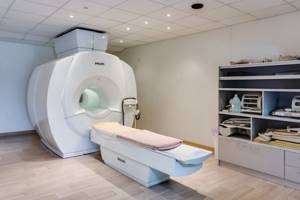
Modern tomograph installed in
Various types of kidney stones. Description
Before you begin therapy for urolithiasis, you need to know about the different types of stones that form. Under no circumstances should you diagnose and eliminate the pathology yourself. The chemical content of stones is determined only by medical examination. Stone is a dense formation made from a combination of organic substances and minerals.
Under no circumstances should you diagnose and eliminate the pathology yourself.
The type of kidney stones depends on their chemical composition. Let's take a closer look:
Oxalate stone
A high content of oxalic acid salts in the body with insufficient calcium intake can cause the development of hard black-gray lumps. If these formations are not identified in a timely manner, the pathology takes the form of a hard stone with a spiky structure. This is an oxalate stone, which can be detected using x-rays. Removing it from the organ is possible only surgically.
Kidney stones on x-ray, photo. Based on radiography, they can be confused with gall bladder stones; in order to answer this question, it is necessary to perform a CT scan or ultrasound.
Urate stone
Prolonged dehydration of the body leads to the fact that the volume of urine decreases and its acidity decreases. This phenomenon is observed in residents of countries with particularly hot climates, as well as in patients who have undergone chemotherapy or with certain types of cancer. Not loading the body with plenty of water can contribute to the formation of urate stones. A urine and blood test at an early stage of the disease will help prevent further development of stones.
Struvite stones
Struvite stones are considered one of the most dangerous types of stones due to their rapid growth in size, which can lead to more complex stones that are difficult to remove - the coral spiny subspecies. Struvite stones originate in the event of an infectious process in the ureters. An increased content of the urease enzyme in the urine leads to its breakdown and crystallization of salts, which outwardly resemble “coffin lids” (crystals can be detected by examining urine). To eliminate the deposits that have formed, the surgical method of ESWL is used - lithotripsy. To avoid relapse of the disease, the patient should be monitored even after surgery.
Cystine stone
People who have kidney pathology, or have had cases in their family, are predisposed to nephrolithiasis. A gene defect can cause an amino acid (cystine) to be unable to dissolve in urine, forming crystalline formations. As these forms grow and unite, the growth and development of cystine stone begins. The texture of the yellow-white stone is soft, smooth, and round in shape. It is difficult to detect with x-rays. Therefore, CT of the organ and intravenous urography are more suitable here. Crystals of cystine molecules can be detected through urine analysis. Unfortunately, even complete elimination of dense deposits will not provide relief from subsequent renal colic.
Examples of normal (on the radiograph on the left) and pathology (on the right) with excretory urography.
Number 1 on the left indicates contrasted calyces, number 2 – renal pelvis, 3 – ureters. On the right, blue arrows indicate sharply dilated calyxes. Also note the lack of contrast in the renal pelvis and ureter - all these are signs of urolithiasis. In this case, the situation is acute - to describe dilated calyxes, you need to use the term “calicoectasia” (from the Latin “calyx” - renal calyx and “-ectasia” - dilation). The term "hydronephrosis" is used for chronic, long-term conditions associated with increased pressure in the urinary tract; Unlike calicoectasia, hydronephrosis reveals atrophic changes in the renal parenchyma.
Examples of conditions that may reduce the validity of research. Excretory urograms.
On the left, a significant expansion of the renal cavity system is revealed. The bladder filled with contrasted urine is clearly visible. Note the gas-inflated loops of intestine that mask the right kidney. On the right, arrows indicate multiple calcifications in the pelvis, which can mimic stones in the ureter. Usually these are calcifications in the uterus or in the appendages (in women) or in the prostate gland (in men).
Contraindications to MRI of the ureter
Despite all the advantages of magnetic resonance imaging, there are conditions that do not allow this type of examination. The main contraindications are:
- the presence in the patient’s body of non-removable metal objects (prostheses, clips, plates, fragments and shot after wounds);
- existing implanted devices (cochlear implant, cardiac or neurostimulator);
- the patient’s weight category is over 130 kg and waist circumference is over 150 cm;
- pregnancy in the first trimester;
- children under five years of age;
- allergy to a medication used for diagnostics with contrast;
- the person being examined is intoxicated or under the influence of psychoactive drugs.
Minor contraindications include:
- late pregnancy and lactation;
- inability to keep the body still during the procedure;
Fear of being in a closed space and increased physical activity can be relieved with various sedatives as prescribed by the attending physician.
Preparing the patient for tomography
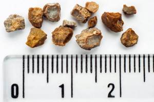
Classification of kidney stones by number and shape
Depending on how many “stones” are identified in the kidneys, doctors divide the phenomenon into single formations, multiple formations, as well as two-stone and three-stone formations. The size and weight of the stones vary from 0.1 to 15 cm or more, and from less than a gram to 2.5 kg or more, respectively. Often the growing stone takes the form of a cup-pelvical system (like a cast), with compacted thick ends. They are called coral. Stones can form in one or both kidneys. The variety of stone shapes is quite impressive: from shapeless to perfectly round.
results
The results of this precise diagnostic study in the form of a layer-by-layer image of the kidneys make it possible to identify any pathology in them. MRI is of particular importance in oncology: it detects tumors even up to 1 cm in diameter and determines their nature.
The doctor carefully analyzes the images obtained during the examination, and after about 2 hours writes a conclusion. It is given to the patient along with the images for presentation to the attending physician. In some medical institutions, the result is issued the next day.
Symptoms of urinary system disease
A person may not suspect for a long time that there is a foreign formation in the organs. The first symptoms of pathology, as a rule, appear only when the formed stone changes its location. More precisely, when it begins to move along the urinary tract. The severity and tangibility of pain depends on the shape and size of stone formations. Small stones (sand) do not always cause discomfort.
The first symptoms of pathology, as a rule, appear only when the formed stone begins to move along the urinary tract.
Here are the most basic signs of kidney problems:
- Varying degrees of pain, depending on the location of the stone in the organs
- The presence of blood, pus and other impurities in the urine
- Impaired urination, up to anuria (blockage of the urinary tract with kidney stones)
Dependence of symptoms on the size and position of stones
Stones can be detected by chance, but more often they are discovered when there are already complaints - during ultrasound or x-ray examinations. The severity of symptoms largely depends on the location of the stone in the urinary tract, its shape and size, and the presence of sharp edges. So, they can be localized:
1.In the renal calyx(es).
Moreover, even if the size of the formation is large, there may be no symptoms at all. A long-existing stone impedes the outflow of urine from the calyx and can provoke atrophy of the kidney parenchyma and its cystic degeneration.
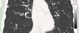
Computed tomography is a very valuable research method in identifying kidney stones. The blue arrow on the images indicates a large calcification located in the lower group of the calyces of the left kidney and (partially) in the pelvis. It can be seen that both the calyx and the pelvis are significantly dilated, in the lower parts the kidney tissue is thinned and atrophic, which indicates the duration of the disease.
Same patient, magnified CT image and 3D reconstructions. The stone is marked with an arrow.
CT scan of the kidneys with contrast, arterial-parenchymal phase.
A small (no more than 4-5 mm in diameter) dense calcification was detected in the lower calyx on the left. Such stones are more dangerous than large ones, because they can (when washed out and pass into the ureter) cause obstruction (blockage) of the urinary tract and provoke renal colic.
2.In the renal pelvis.
Large accumulations of calcium salts are usually localized here, their shape following the internal contours of the pelvis - the so-called coral stones. Coral kidney stones - what to do? First of all, you need to carefully analyze the CT results and contact a urologist to decide on surgery. The operation is necessary if there is a risk of complications, because if the lumen of the pelvis is blocked, signs of increased pressure in the urinary tract may occur, and hydronephrosis may subsequently develop (persistent dilation of the calyces and pelvis, often in combination with atrophy of its parenchyma). We will look at the complications in detail below.
Coral kidney stones, photo.
Computed tomography (CT). The formation is large in size, but clearly does not impede the outflow of urine - there are no signs of hydronephrosis or thinning of the parenchyma.
3.In the ureter.
When displaced downwards (from the renal pelvis to the ureter), painful sensations occur, the severity of which depends on the size of the stone and its shape, the presence of sharp edges. How to identify a stone in the ureter? Ultrasound can be used as a primary method, but CT is a more accurate method.
How to see a stone in the ureter.
On the contrast-enhanced CT scan of the pelvis, the arrow in the top row on the left indicates a small calculus at the ureteric orifice (where it enters the bladder), which causes dilatation of the pelvis (top left) and ureter (in the images in the bottom row). In this case, the condition is acute, the patient has an attack of renal colic, therefore, we are talking about ureteropyeloectasia (dilation of the ureter and pelvis), and not about hydronephrosis.
4.In the bladder.
Such stones usually have the shape of an “egg” or “ball”, measuring 10-20 mm in diameter - they are freely located in the cavity of the bladder (since they are formed in it), and usually do not cause any symptoms. If there are many of them and they occupy a significant part of the bladder volume, urination may be impaired.
Interesting diagnostic observation.
A large stone in the bladder cavity, identified during an X-ray examination of the hip joints (as an accidental finding). The images in the top row show an X-ray of the pelvis and an axial section (CT of the pelvis), a dense shadow in the form of an “egg” measuring 8x6 cm is visible. The image below shows the same stone after surgery. Its layered structure is clearly visible.
5.In the urethra.
Usually, stones formed in the upper parts of the urinary system pass through the urethra, with painful symptoms, and if the urethral wall is damaged, the urine becomes pink from the presence of blood - hematuria.
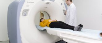
Progress of the procedure
The patient puts on disposable clothing (you can leave yours if there are no metal parts on it). The patient is placed on the table and moved inside the tomograph.
When using a closed-type device, an air supply is provided inside the tunnel, there is lighting and an intercom for two-way communication with the doctor during the procedure. If desired, the patient can use vacuum headphones or earplugs to get rid of the loud sound when the tomograph is operating.
The most important requirement is to remain still during the procedure. The clarity of the resulting images depends on this. If contrast is necessary, the marker is injected intravenously and the study continues. The average duration of the procedure is 30 minutes.
Medical diagnosis of kidney stones
For competent and complete treatment of urolithiasis, it is necessary to make a correct diagnosis. Before laboratory tests are carried out, all information about the possible causes of the pathology is collected from the patient’s words. Based on the examination, the tactics for further examination and treatment are determined.
For competent and complete treatment of urolithiasis, it is necessary to make a correct diagnosis.
In medicine, various methods for detecting kidney stones are used, depending on the period of relapse and the severity of the disease:
- Doing an ultrasound of the kidneys
- Ultrasound of the bladder
- Perform urography
- Take a urine and blood test
- Thanks to nephroscintigraphy, the degree of kidney dysfunction is determined
- Multislice CT allows you to determine the type and size of the formation
Get a kidney CT scan in St. Petersburg
An example of detection of renal calcifications on computed tomography
An example of detecting renal calcifications on computed tomography (small stones in the renal pelvis are circled). A stone at the mouth of the ureter on the left is also clearly visible (it is marked with a green arrow). A dilated left ureter (ureterectasia) is visible.
The blue arrow marks a large formation in the lower calyx of the left kidney, described in the CT scan as “an ovoid-shaped stone, with smooth edges, of a homogeneous structure.”
How is urography performed?
Before entering the X-ray room, the patient must take off his clothes, putting on a special robe and metal jewelry. Pictures are taken in a lying or standing position, as determined by the doctor. The examination time depends on the method, the use of contrast agents and the characteristics of the kidneys. The procedure is completely painless, and the main thing that is required of the patient is not to move for several seconds while the image is being taken.
The result of an x-ray is one or a series of images of the examination area. The images are saved on a disk (or other storage medium), and based on the images, radiologists will draw up a conclusion. It describes in detail the structure and condition of the kidneys, bladder and ureters, detected pathologies and their features.
Diagnosis of complications of urolithiasis
The presence of stones does not go unnoticed. If they exist for a long time, there is a high risk of developing various complications. For example, in all cases, even after the stone passes through the ureter, expansion of its lumen (ureterectasia) is observed for some time; expansion of the lumen of the pelvis (pyelectasia) and calyces (calicoectasia) can also be detected. This condition is temporary and goes away after the cause of the obstruction in the outflow of urine is eliminated.
If stones exist for a long time, there is a high risk of developing various complications.
If an obstruction to the outflow of urine exists for a long time, the expansion of the urinary tract becomes persistent and is combined with a decrease in the volume of the kidney parenchyma. This condition is called hydronephrosis. Hydronephrosis can be of varying degrees of severity - from minimal expansion of the collecting system to terminal (in which the kidney becomes like a thin-walled “bag” filled with fluid). Of course, such a kidney does not perform its function in any way.
Kidney CT. Severe right-sided hydronephrosis and hydroureter.
Cystic degeneration of the right pyelocaliceal system; normal kidney tissue is virtually invisible due to thinning and atrophy. On the right, the arrow indicates the cause of hydronephrosis - a stone in the lower third of the ureter, obstructing (blocking) its lumen.
Against the background of stagnation in the pelvicalyceal system (especially with concomitant diabetes mellitus), there is a high risk of infectious complications - pyelonephritis (and even pyonephrosis - a condition in which the kidney becomes a sac filled with pus), cystitis, urethritis.
Right-sided hydronephrosis due to urolithiasis, complicated by pyelonephritis.
The arrows in the images in the top and bottom row indicate dilated cups. Also note the sharp difficulty in the outflow of urine from the right into the excretory phase (10 minutes after the introduction of the ultravist into the vein) in the image below on the right. The perinephric tissue is cloudy and has a heterogeneous structure (due to edema and infiltration). Urine tests show leukocytes.
Why are kidney stones dangerous?
The table shows the possible consequences of nephrolithiasis.
| Possible pathology | Description |
| Chronic pyelonephritis | The first signs: cutting pain in the abdominal area, pulling, aching. The temperature rises sharply. Transition to the chronic stage is possible. |
| Acute pyelonephritis | Inflammation of the urinary system, which leads to swelling of urine into the kidneys. The first signs: renal colic, fever, weakness, intoxication. |
| Pyonephrosis | A type of purulent-destructive pyelonephritis. |
| Urethritis | Inflammation of the mucous tissue of the urinary tract of the urethra, that is, the urethra |
The best treatment is good habits
The most optimal and timely action to prevent kidney stone formation is your good habits:
- Drink clean water daily at least 2.5 liters per day
- Regular walks in the fresh air
- Maintaining personal hygiene rules
- Eliminate “heavy” foods and fried foods from your diet
Well, if stones have already been discovered in the kidneys, then it is necessary to strengthen all these actions. Additionally, follow these rules:
- drink only filtered, soft water;
- follow a strict diet designed specifically for your case;
- in cold weather, insulate the lumbar region and prevent hypothermia;
- avoid infectious diseases that are sexually transmitted;
- limit the consumption of coffee, tea, and caffeine-containing products;
- increase the consumption of diuretic drinks: cranberry juice, green tea, drinks made from decoctions of anti-inflammatory herbs;
- If you are overweight, strive to lose it.
Specifics of x-ray studies of the kidneys in children
This procedure is performed only in cases where the data obtained from ultrasound is insufficient to make a diagnosis. There are a number of features of the procedure:
- the child is given sedatives;
- the procedure is carried out in the presence of a doctor and sometimes an anesthesiologist;
- if the child is small, an adult (usually a parent) will be needed to help hold the child as still as possible;
- double cleansing of the intestines with an enema on the eve of the procedure is mandatory
What not to do if you have kidney stones
Self-medication is strictly forbidden. It was described above that even after foreign bodies are removed from the kidneys, pathological consequences may arise, which will only complicate the healing process of the affected organ.
Self-medication is strictly forbidden.
Diagnosis and treatment of urolithiasis should be carried out only in a clinical setting.
During the period of exacerbation and treatment of nephrolithiasis, it is recommended to limit, or better yet completely eliminate, the following foods:
- greens and vegetables rich in oxalic acid and vitamin C
- broths from meat, fish, mushrooms;
- chocolate, peanut butter, sweet and flour products, fruit jam;
- strong tea, coffee;
- carbonated drinks;
- milk drinks;
- hot spices and seasonings;
- sour fruits, berries;
- legumes