What is a colonoscopy?
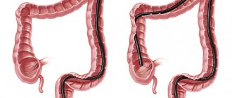
Colonoscopy (video colonoscopy) is a type of endoscopic examination during which all parts of the large intestine are examined, as well as a section of the small intestine - the ileum. An incomplete examination of the colon is called “rectoscopy” or “sigmoidoscopy”, in which only the rectum and part of the sigmoid colon are examined.
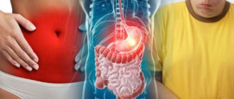
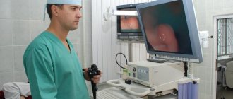
The purpose of the study is to detect any pathology of the colon. During the procedure, a very flexible, thin, completely non-traumatic endoscope with a built-in video camera at the end is inserted into the colon, allowing the doctor to evaluate the entire mucous membrane (inner surface) of the colon.
Colonoscopy is falsely associated with discomfort - many people follow unverified rumors and find dozens of what they think are “reasonable” reasons not to subject themselves to this, often the most important examination in their lives. Some people refuse to believe they are at risk for colon cancer. Some people are afraid of the pain that may occur during the study. Someone is afraid of the research results themselves - the unknown is scary!
However, none of these reasons are valid. What's more, colon cancer is one of the most preventable types of cancer, and a colonoscopy can truly save your or your loved one's life.
Only through colonoscopy can one detect and remove polyps (polypoid adenomas) of the rectum or the entire colon, which always degenerate into a malignant tumor. Colonoscopy also helps detect cancer at an early stage, which allows the disease to be completely cured in 90% of cases.
Tumors of the colon, unlike many others - for example, the stomach - always have a benign stage of development that lasts for years! And all you need to avoid tragedy is to perform a colonoscopy, get rid of polyps and return to your normal life in 1 hour!
Amazing, isn't it? It is worth taking advantage of the chance offered to us by nature and modern technology!
When is it necessary to have a colonoscopy?
Colonoscopy on a regular basis - once a year, is recommended for all patients over 45 years of age. If your family has had or has relatives diagnosed with colorectal cancer, then you must perform the first test no later than 25-30 years of age, and then follow the recommendations that you will receive at the end of the procedure.
You must also undergo a video colonoscopy if you are suspected of having one of the following diseases:
- Crohn's disease
- Ulcerative colitis
- Pseudomembranous colitis
- Ischemic colitis
- Solitary rectal ulcer
- Rectocele
- Diverticular disease
- Volvulus
- Colon tumor
Indications for colonoscopy
The main indications for colonoscopy, which we use in our clinical practice, may be one or more symptoms:
- Stomach ache
- Abdominal mass (on ultrasound, fluoroscopy or computed tomography)
- Unreasonable weight loss
- Iron deficiency anemia (especially in men of any age)
- Increased ESR (erythrocyte sedimentation rate)
- The appearance of blood from the rectum while maintaining the frequency and consistency of stools
- Monitoring patients with previously diagnosed colitis, Crohn's disease or ulcerative colitis
- Monitoring of patients with familial intestinal polyposis
- Monitoring patients after previous polypectomies
- Observation of patients with operated colon (inflammatory bowel diseases, familial polyposis, cancer)
- Increased levels of calprotectin in feces
- Prolonged diarrhea (especially during long-term antibiotic therapy)
- Examination of patients whose close relatives have been diagnosed with adenomas or colon cancer
- Presence of mutations in the BRCA 1 and BRCA 2 genes
- Preparing the patient for surgery for chronic bleeding hemorrhoids or anal fissure
Indications and contraindications for CT scanning
- routine diagnostics to exclude diseases of internal organs;
- screening for head injuries, headaches, fainting and lung cancer;
- convulsive syndrome, pupils of different sizes, neurological symptoms;
- mental status disorders, inappropriate behavior;
- suspicion of vascular damage, acute damage to a hollow or parenchymal organ;
- to control diagnostic or therapeutic procedures;
- for the purpose of the dynamics of the treatment process.
There are no absolute contraindications to CT scanning. However, taking into account the radiation exposure, it is necessary to rationally approach the issue of examining pregnant women and young children.
CT with contrast is contraindicated in patients:
- with an allergy to iodine-containing drugs;
- with severe renal failure;
- lactating women, but if there is a significant need, CT can be performed with the condition of interrupting breastfeeding for a period of at least two days.
You can do a CT scan in the cities: Zelenograd, Solnechnogorsk, Klin, Skhodnya, Andreevka, Istra, Khimki. You can find out more by phone
IMPORTANT: do not forget to bring all extracts, protocols or recordings (disks) of previous studies. This will allow you to assess the dynamics of the disease and determine the tasks for the radiologist.
Before a computed tomography scan with the introduction of a contrast agent, it is necessary to take a blood test for creatinine and urea. Without these tests, the patient is not allowed to undergo a study with a contrast agent.
Colonoscopy: preparation for the examination
For a colonoscopy to be successful, your colon must be thoroughly cleaned. Only in this case will the doctor be able to best assess the condition of the colon. Therefore, responsible preparation is required from you, which we recommend starting just 2 days before the test.
The main mistake patients make is taking special cleansing products without following a specific, “slag-free” diet. It is very important to exclude the following foods from the diet 2 days before the colonoscopy: salads, vegetables, legumes, fruits, “red” meat, sausage, rice, pasta. During preparation for the study, it is also necessary to exclude oral (by mouth) intake of activated carbon and iron supplements, prescribed, for example, for iron deficiency anemia.
During preparation for colonoscopy, patients should strictly avoid the introduction of suppositories into the rectum. Also, do not lubricate the anus with oil, cream or other fat-based substances. It is not allowed to take Vaseline oil or give cleansing enemas in addition to laxatives.
During preparation for colonoscopy, it is necessary to follow a diet with no fiber: broths, milk and fermented milk products, juices without pulp, tea, coffee and generally unlimited liquids (in the absence of therapeutic contraindications), a little chicken meat and “liquid” milk porridges are also allowed .
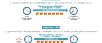
Only after this should you move on to taking “specialized” laxatives - “Moviprep”, “Pikoprep”, “Fortrans”. It is important to note that dividing their intake into 2 days (split system, described in the instructions for each drug), according to our observations, significantly improves the quality of bowel preparation for examination.
Endoscopist for gastroenterologist: the importance of colonoscopy in IBS
Dmitry Vyacheslavovich Zavyalov , candidate of medical sciences:
– The last talk on this evening's topic, irritable bowel syndrome. Presentation on the role of endoscopy in diagnosing irritable bowel syndrome. There is no doubt that endoscopic examination, colonoscopy, is the main method for diagnosing colon pathology in general. And, given that the diagnosis of IBS is an exclusion diagnosis, the role of colonoscopy here is undoubtedly high. However, a practicing physician, a clinician, has a number of questions when it comes to the problem of performing colonoscopy in this group of patients.
The first question is: in what cases is it still necessary to perform a colonoscopy in patients with suspected IBS? What results can we expect from a colonoscopy? Well, how to properly and efficiently prepare a patient for endoscopic examination in this group of patients?
In response to the first question, I would like to draw your attention to data published by the American Gastroenterological Association in 2009, which, from an evidence-based medicine perspective, determined when colonoscopy should be performed in this group of patients, patients with typical symptoms of IBS who do not have anxiety symptoms , and for those under fifty years of age, colonoscopy as a routine examination is not recommended. And at the same time, in patients over fifty years of age and who have symptoms of anxiety, an endoscopic examination is required to identify organic pathology and screen for colorectal cancer.
About the symptoms of anxiety. Today we have already discussed this issue. But I wanted to draw your attention to the fact that the first point is the presence of symptoms of irritable bowel syndrome at the age of fifty. This is in America. What about our practice? In our work, we tried to assess whether age, the recommended age of fifty years, is a reliable criterion, but in our conditions?
We carried out work in which we assessed the results of colonoscopy, which was performed in our department of the Yaroslavl Regional Oncology Hospital for five years on patients aged from forty to forty-nine years. So, in this group of patients, various pathological formations were identified in more than a quarter of cases. Of course, in most cases these were hyperplastic polyps. However, in almost a third of patients these were adenomatous polyps, almost half of which were so-called advanced adenomas - adenomas with a high risk of malignancy in the future. At the same time, some patients were diagnosed with colorectal cancer. First of all, these were patients aged from forty-five to forty-nine years. And, mostly, they were men.
According to our data, perhaps in Russia this is the age for starting screening, searching for precancerous pathology or early forms of cancer; perhaps this age for our conditions is less than fifty years, the age recommended in most American and European recommendations.
The next question is: what results do we expect from a colonoscopy? And here it is very important to determine what determines the quality of modern colonoscopy performed in this group of patients.
We present colonoscopy, the algorithm for performing colonoscopy in the form of such a pyramid, the tops of which are those new endoscopic technologies, those new endoscopic techniques that we now use, those technical approaches. The basis and foundation of this pyramid is high-quality and proper preparation of the intestines for examination.
I would also like to note the importance of a complete examination of the intestines. Even according to leading European and American clinics that screen for colorectal cancer and intestinal pathology in general, a total examination of the colon, examination of all parts of the colon, is carried out only in 90-95% of cases. That is, thus, part of the intestine remains unexamined. And in this clinical example, when we repeated a colonoscopy, did repeated studies on a patient whose right flank of the colon was not examined, we identified a carcinoid tumor in the area of the mouth of the appendix, which, in principle, can itself give certain clinical manifestations .
The next important point is examination of the terminal ileum. In this clinical example, we see an erosive process in the terminal ileum. Of course, such changes also give a certain clinical picture. And look how important it is when performing a colonoscopy for the endoscopist to examine this area and study it. The next clinical example is about Crohn's disease. Of course, when an endoscopist sees the typical manifestations, the endoscopic picture of Crohn’s disease (segmental lesions, deep slit-like ulcers), most often, there are no difficulties in diagnosis and in the endoscopic conclusion.
However, in the initial stages, in the so-called infiltration phase, changes in the intestine are minimal. And the only sign may be the presence of aphthous changes in the colon mucosa, as in this clinical case. And, unfortunately, endoscopists do not always correctly interpret these changes that they see when examining the colon.
The following clinical example is a patient undergoing colon examination, a forty-eight-year-old female patient who was referred for a colonoscopy with a preliminary diagnosis of IBS. And during the examination of the descending colon, we identified such a pathological area measuring only a few millimeters. And when we studied it in detail, we noticed the retraction in the center, and these changes seemed suspicious to us. We performed endoscopic resection of this area (unfortunately, I will not be able to demonstrate the video now). And during a morphological study of the removed specimen, the morphologist diagnosed “well-differentiated adenocarcinoma.”
Of course, a tumor of this size will not provide any clinical picture. However, you will agree how radically changes, firstly, the diagnosis of such a patient and her further fate and observation, fundamental changes in the life of this particular patient. And, in fact, the problem of missed pathology during colonoscopy is a worldwide problem. And so, our Spanish colleagues, Professor Firandes (00:06:40) published a large study in 2010, which included more than sixteen thousand patients, and showed that colon adenomas are equally often missed in both the left and right flank of the colon.
But colorectal cancer is even more common in the left flank and even in the rectum. New endoscopic techniques that are actively entering our lives may help change this situation. And one of these techniques, basic techniques, is chromoscopy - a technique for coloring the surface of the intestinal mucosa with special dyes. And in this example we see a practically unchanged intestinal mucosa. But when applying a special dye, we see a pathological formation marked with arrows, which was not visible during the initial examination.
Another clinical example is the cecal region. We see the orifice of the appendix, but do not see any significant pathology. When performing special staining, we see that on the right side of the slide there is a pathological area with a retraction in the center. New technologies, digital technologies that are actively coming into our lives, digital contrast techniques for mucous membranes, as you see, are practically invisible during a traditional examination. Changes at the top of the fold are nothing more than an adenoma of the colon, which could have been easily missed by a doctor, but, unfortunately, was missed in the absence of such equipment.
Another technique, the latest technique for endoscopic diagnostics, is the so-called autofluorescent endoscopy, a technique based on the principle that tissues of different densities and different structures glow differently under a certain irradiation. And the pathological areas have a purple, violet glow, as shown on this slide. And the doctor, having seen such a glow, can only turn his attention to this formation, perform a detailed examination of it, perform a biopsy and, if necessary, remove it.
Well, the third question that I wanted to cover today is how to correctly, and most importantly, qualitatively prepare a patient from such a group for endoscopic examination?
In Russia today, various methods of preparing for colonoscopy are acceptable. But the modern preparation method is the use of polyethylene glycol. In our practice, this is the drug “Fortrans”, which is an osmotically balanced electrolyte solution based on polyethylene glycol. And our clinic has accumulated experience. For fifteen years we have been using this drug both to perform diagnostic tests and to perform endoscopic operations on the colon. The drug showed successful preparation of the intestine for endoscopic examination and showed its safety over a long period of time, more than fifteen years.
I would like to go over the key points of bowel preparation for colonoscopy. Well, firstly, a personal conversation and personal instruction, instructions from the doctor for the patient, and a personal conversation from the doctor are necessary. This increases the responsibility of the patient himself, who understands why it is so carefully necessary to prepare for the study, and what kind of study lies ahead. This also increases the responsibility of the doctor himself, who after some time will perform an examination on this patient. It should be noted that in our clinic, a two-stage administration of Fortrans, the so-called split dose, is considered traditional. This allows patients to prepare for the study more easily, and the quality of the studies will be significantly higher when using two-stage preparation and two-stage administration of Fortrans. It is necessary to explain to the patient that he needs to take a sufficient amount of fluid per day. It could be tea, it could be juice, it could be broth - any clear liquid. And explain to the person that there is no need to sit still - you need to maintain some kind of minimal physical activity.
The next point is special approaches to certain groups of patients. These are elderly patients, patients with heart failure, with diabetes mellitus (I will dwell on this a little later). Well, it is important to use selective antispasmodics in general during colonoscopy and especially in patients with IBS during the actual endoscopic examination. Of course, special treatment is required for patients in the older age group, elderly patients who often have decreased intestinal motor functions and constipation. So, when we begin preparing such patients, we begin by prescribing laxatives. And the drug of choice is the drug that we already mentioned today - Forlax, which we recommend starting five to seven days before taking Fortrans and starting the bowel cleansing itself before colonoscopy.
Another group of patients that is difficult to manage in clinical practice are patients with diabetes mellitus. Current approaches to preparing these patients are to avoid the use of oral agents or use half the dose if insulin is involved, and return to normal use of drugs at normal doses after completion of the endoscopic examination and after the person returns to a normal diet. Such differentiated approaches to preparing the intestine for examination allow us to prepare the majority of our patients.
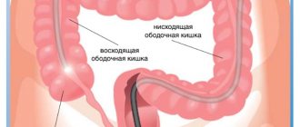
Here you see what the cecum area looks like when following these recommendations. Look how clean the mucous membrane is and how it is accessible not only to traditional examination, but also to examination using the technologies I spoke about. Ascending colon, transverse colon, descending colon, sigmoid colon and rectum. Thus, observing the criteria and principles of colonoscopy that I spoke about, colonoscopy is a tool that will help a gastroenterologist or clinician make the correct diagnosis and, accordingly, begin the correct treatment of patients with irritable bowel syndrome. Thank you for your attention.
How is a colonoscopy performed?
During the examination, the doctor uses a special device - a colonoscope, which is a long flexible tube with a diameter of about 13 mm. The colonoscope is inserted through the anus and gradually moves throughout the colon, allowing the doctor to visualize the entire mucous membrane.
During a colonoscopy, the doctor can take a small piece of the mucous membrane (which is completely safe) of the intestine for a clarifying study - histology or cytology. Also during the procedure, in agreement with the patient, the doctor can remove polyps - precursors of cancer, if they are diagnosed in the patient.
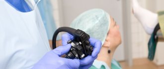
- Colonoscopy, at the request of the patient, can be performed either without anesthesia or with anesthesia - sedation or “sleep colonoscopy”.
- Duration of a colonoscopy: The procedure to examine the entire colon takes about 20 minutes. In the case of polyp removal, the procedure may take 40-45 minutes - the duration directly depends on the number and size of tumors.
- If a colonoscopy is performed under medicated sleep conditions, the patient needs an additional 30 minutes until normal health is completely restored.
- After the colonoscopy, the patient can return to their normal daily activities.
Diet before colonoscopy
An important part of preparing for a colonoscopy is proper diet in the preceding days and on the day of the examination. In general terms, the recommendations are as follows:
- A few days before the procedure, you need to switch to a low-fiber diet, reduce the consumption of fresh vegetables and fruits, dried fruits, nuts, and whole grains.
- 1–3 days before your colonoscopy and on the day of the procedure, you should avoid eating solid foods. Broths, clear fruit juices (for example, clarified juice from apples, white grapes), tea and coffee, and jelly are allowed. It is recommended to drink more fluid the day before.
- You should not eat or drink anything 2–4 hours before the procedure The procedure can only be carried out under medicated sleep conditions on an empty stomach.
In parallel with the diet, in the afternoon before, the intestines are prepared for the procedure using laxatives. Recommendations regarding types of drugs and dosage regimens may vary from doctor to doctor.
What should you watch out for during a colonoscopy?
Colonoscopy has become a fairly popular procedure, which is now performed in various clinics. Despite its widespread use, colonoscopy is an invasive procedure that should only be performed by an experienced and qualified specialist using modern high-tech equipment.
Side effects of a “poor” colonoscopy:
- Damage (perforation) of the colon is an extremely rare, but very dangerous complication that requires immediate surgical correction and most often occurs when the medical staff is extremely poorly prepared, excessive haste, and the desire to perform the study “fastest in Moscow.”
- Bleeding after colonoscopy is also an extremely rare complication and has the same causes as perforation.
- Improper removal of the polyp means the persistence of residual tumor, which entails progression of the disease, while the patient is completely confident in his recovery.
- Incorrectly selected anesthesia causes difficult awakening, nausea, dizziness and even vomiting, headaches, general weakness, and temporary loss of performance.
Be careful when choosing a clinic for a colonoscopy, beware of “counterfeits”. The study must be carried out by an experienced and qualified specialist.
Specialists
The colonoscopy procedure in our Clinic is performed by the head of the department of diagnostic and therapeutic endoscopy, Ph.D. Pavel Vladimirovich Pavlov is a specialist who has been performing this research since 2003, and also performs all known endoscopic intraluminal operations on the intestine: polypectomies, mucosectomies, dissections in the submucosal layer, bougienage, balloon dilatations, stenting and others. Pavel Vladimirovich has extensive experience in the diagnosis and treatment of early cancer not only of the colon, but also of the entire digestive tract.
Candidate of Medical Sciences also deals with diagnostics and therapeutic interventions on the intestine. Afanasyeva Aigul Fanzirovna is a doctor who has devoted herself to endoscopy since 2003, performing most endoscopic intraluminal interventions on the digestive tract and has deservedly established herself as a brilliant specialist.
results
After the examination, the radiologist provides a detailed description of the images obtained and issues a general conclusion on them within about one hour. If this is a complex case, consultation with experts is necessary, the conclusion will be ready the next day.
Modern CT scanners at SM-Clinic and experienced radiologists will help your child undergo a CT examination comfortably and safely with minimal discomfort. At the same time, the results will be ready quickly, they will be as accurate as possible, helping doctors clarify the diagnosis and select treatment.
Anesthesia
A special type of intravenous sedation used during colonoscopy is anesthesia with the drug “propofol”, the most gentle way of temporarily switching off consciousness, after which the patient wakes up alert and rested at the end of the procedure. This anesthesia is used in the most advanced clinics in the world.
Our Clinic performs about 2,000 diagnostic and therapeutic colonoscopies annually. An objective indicator of the quality of diagnosis is the highest rate of finding “polyps” during colonoscopy in Russia. Repeated visits by patients to the same specialists for follow-up studies also indicate a high level of trust and quality of medical services provided!
Multislice computed tomography
MSCT is an improved version of tomographic diagnostics, which has important distinctive characteristics. The information content of the method is ensured by the phenomenal clarity of a series of images obtained in just one revolution of the X-ray tube. The method allows not only to assess the condition of the organ under study, but also to monitor its physiological functioning in real time.
Main advantages of MSCT:
- high scanning speed;
- reduction of radiation dose;
- improved spatial and temporal resolution;
- increasing image contrast;
- increasing the area of anatomical coverage;
- efficiency of using an x-ray tube.