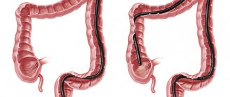
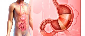
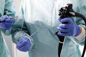
Colonoscopy is a diagnostic method that is used to assess the condition of the colon mucosa. The examination is carried out using a special instrument - a colonoscope. It is a thin flexible tube with a video camera at the end. The colonoscope is inserted into the intestines through the anus.
- Goals of performing a colonoscopy
- Preparing for a colonoscopy
- Diet before colonoscopy
- Indications and contraindications
- Diagnostic colonoscopy
- Therapeutic manipulations during colonoscopy
- Colonoscopy in a state of medicated sleep
- Colonoscopy technique
- Recording the procedure on video
- Recovery after colonoscopy
- Possible complications
- Alternatives to Colonoscopy
- Colonoscopy results
Goals of performing a colonoscopy
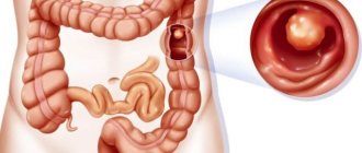
Using a colonoscope, the doctor can detect ulcers, polyps, foci of inflammation, tumors, and sources of intestinal bleeding. In addition, during a colonoscopy, tissue samples can be obtained from the intestines for histological and cytological examination, and polyps or other formations can be removed.
It is recommended to have your first colonoscopy at age 50. Further recommendations are given individually, depending on the results of the study. Unfortunately, most colon tumors are detected at late stages. There is only one reason here - late diagnosis of the tumor. The only way to detect a tumor at an early stage is regular examination of the colon, which allows us to identify pre-tumor pathology and remove it, or carry out other necessary treatment to prevent it from degenerating into cancer. The key to curing cancer is early diagnosis. Early diagnosis can be done using colonoscopy.
Colonoscopy under anesthesia in Moscow
At the Central Clinical Hospital of the Russian Academy of Sciences, colonoscopy is performed using modern equipment. Thin endoscopes and high-quality optical equipment allow you to obtain accurate data with a minimum of discomfort for the patient. For anesthesia, harmless drugs are used, which are completely eliminated from the body within a few hours after the examination.
High-quality latest equipment and the work of experienced doctors allow:
- carry out the procedure as carefully and carefully as possible;
- perform tissue sampling or endoscopic treatment as necessary;
- correctly interpret the research results.
The high quality of the service provided allows the attending physician to obtain correct information about the patient’s condition, make an accurate diagnosis at an early stage of the pathology and begin treatment immediately.
Advantages of visiting the RAS Medical Center:
- attentive and sensitive attitude towards each patient;
- use of modern equipment;
- the possibility of performing the procedure under general anesthesia or local anesthesia;
- the ability to study hard-to-reach parts of the intestine;
- correct and accurate interpretation of research results by experienced, highly qualified specialists.
Contraindications to colonoscopy under anesthesia:
- hypertension 3 degrees;
- condition after a stroke;
- severe pulmonary and heart failure;
- severe forms of Crohn's disease and ulcerative colitis (increased risk of intestinal perforation);
- adhesive disease;
- peritonitis.
Be sure to tell your doctor about the following risk factors:
- pregnancy;
- history of epilepsy;
- allergies to anesthetic drugs;
- psychiatric or neurological diseases;
- blood clotting disorder.
Diet before colonoscopy
An important part of preparing for a colonoscopy is proper diet in the preceding days and on the day of the examination. In general terms, the recommendations are as follows:
- A few days before the procedure, you need to switch to a low-fiber diet, reduce the consumption of fresh vegetables and fruits, dried fruits, nuts, and whole grains.
- 1–3 days before your colonoscopy and on the day of the procedure, you should avoid eating solid foods. Broths, clear fruit juices (for example, clarified juice from apples, white grapes), tea and coffee, and jelly are allowed. It is recommended to drink more fluid the day before.
- You should not eat or drink anything 2–4 hours before the procedure The procedure can only be carried out under medicated sleep conditions on an empty stomach.
In parallel with the diet, in the afternoon before, the intestines are prepared for the procedure using laxatives. Recommendations regarding types of drugs and dosage regimens may vary from doctor to doctor.
Indications and contraindications
Indications for colonoscopy are:
- blood and mucus in the stool;
- presence of relatives with colon cancer;
- previous colon surgery;
- chronic abdominal pain of unknown etiology;
- suspicions of cancer, ulcerative colitis, Crohn's disease;
- elevated temperature for a long period of time, accompanied by anemia and weight loss.
Regular colonoscopy is also recommended for all people over 50 years of age for the early detection of tumors, polyps and other diseases of the colon.
Contraindications to colonoscopy are the active stage of Crohn's disease or ulcerative colitis.
Indications for colonoscopy under anesthesia
This type of examination is prescribed if pathological changes in the colon are suspected. The procedure can also be recommended for chronic diseases of the upper gastrointestinal tract (stomach ulcers, gastritis, cholecystitis), which can cause complications associated with the condition of the intestines.
The doctor may decide on the need for this type of examination if the patient has complaints and symptoms indicating diseases of the colon:
- blood or mucus found in the stool by the patient himself or during a laboratory test;
- pain during defecation;
- frequent feeling of incomplete bowel movement;
- severe cramps, pain, cramping in the abdomen that occur before or after bowel movements;
- frequent constipation;
- frequent feeling of fullness in the intestines;
- frequent diarrhea;
- the presence of undigested pieces of food in the stool;
- pain on palpation;
- hard belly;
- bloating;
- increase in the number of tumor markers in the blood;
- anemia;
- allergic reactions or intoxication of unknown origin;
- elevated temperature, combined with pain in the intestinal area.
Colonoscopy may be necessary in preparation for gynecological operations if the patient has unremoved polyps or tumors or is diagnosed with endometriosis.
Regular diagnostic procedures are required for patients:
- over 45 years old;
- with a genetic predisposition to intestinal cancer;
- with chronic gastrointestinal diseases that negatively affect the intestines;
- leading a predominantly sedentary lifestyle and allowing eating disorders;
- having a history of surgery in the abdominal region;
- with diagnosed Crohn's disease.
Diagnostic colonoscopy
A diagnostic colonoscopy is performed to identify certain pathological formations in the intestines. There are two special types of diagnostic colonoscopy:
- A screening colonoscopy is recommended for all people over 50, even if they have no symptoms. For some bowel diseases, screening may need to start at a younger age. This type of screening is used for early diagnosis of polyps that can develop into a malignant tumor and intestinal cancer.
- Control colonoscopies are periodically performed in people with a history of polyps, colon cancer, and inflammatory bowel diseases.
Having discovered a pathological neoplasm in the intestine, the doctor removes it using a special instrument inserted through a colonoscope and sends it to the laboratory for histological and cytological examination. This procedure is called a biopsy .
The Euroonko clinics in Moscow and St. Petersburg are running a promotion for “Gastro- and colonoscopy under intravenous sedation.” The cost of the complex service includes endoscopic examinations of the stomach and colon, light anesthesia (sedation, “medicated sleep”), a preliminary appointment with a specialist (candidate of medical sciences or doctor of medical sciences), a three-hour stay in a comfortable room. More details about the program.
Therapeutic manipulations during colonoscopy
During an endoscopic examination of the colon, the doctor may perform some therapeutic procedures:
- Remove polyp.
- Stop intestinal bleeding, for example, using electrocoagulation or clipping.
- Eliminate stenosis (narrowing of the intestinal lumen) caused by a malignant tumor or other causes. A special balloon is inserted into the narrowed area and inflated to expand the lumen, and then a stent is installed - a tube with a mesh wall made of metal or plastic (this procedure is called stenting ).
During the work of the endoscopy department at Euroonko, more than a hundred endoscopic stentings of the colon were performed under the guidance of an endoscopist, Doctor of Medical Sciences Mikhail Sergeevich Burdyukov. This minimally invasive procedure helps restore intestinal patency in inoperable cancer.
Often, a colonoscopy is initially performed as a diagnostic procedure and, if pathological changes are detected, it becomes a therapeutic procedure.
Colonoscopy in a state of medicated sleep
Colonoscopy is usually painless, but the patient may experience discomfort from a feeling of bloating (this goes away immediately after the procedure) and the probe moving through the loops of the intestine. In Euroonco clinics it is possible to carry out the procedure in a state of medicated sleep. In this case, the patient is given a special sedative, under the influence of which he is plunged into deep sleep. After approximately 40 minutes, the effect of the drug ends, and within 5-10 minutes after waking up the patient can walk and talk, and after an hour he can go home.
Benefits of colonoscopy under general anesthesia
Despite the informative nature of the method, the procedure is quite painful for the patient. Because of this, during the examination it can move, preventing the passage of the probe, which is extremely undesirable. Therefore, colonoscopy is now usually performed under anesthesia. This allows:
- relieve the patient from psychological discomfort and pain;
- speed up the procedure;
- increase the accuracy of the study.
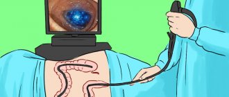
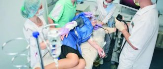
During the procedure, the patient lies on his side with his knees drawn to his chest. With the help of intravenous anesthesia, he falls into a shallow sleep. The anesthesiologist monitors the patient's condition throughout the entire procedure. The coloproctologist inserts the endoscope into the intestines only after the patient falls asleep. Since light anesthesia is used for pain relief, you can go home immediately after its effect ends; a hospital stay is not required. Upon completion of the procedure, you can consult a proctologist who will interpret the results of the study.
In some cases, after a colonoscopy under anesthesia, the patient may experience slight discomfort due to the fact that during the examination a small amount of air is sometimes introduced into the intestines to improve visibility. However, the work of an experienced doctor and the use of modern equipment make it possible to minimize all unpleasant sensations.
Colonoscopy technique
A diagnostic colonoscopy lasts on average 30 minutes. If therapeutic manipulations are required, the procedure time increases.
In order for the patient to undergo the procedure without pain and discomfort, sedation is used - the patient is immersed in a state of “medicated sleep”.
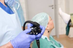
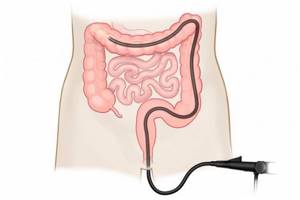
The patient must be completely undressed from the waist down. He is placed on his left side, with his legs bent and knees brought to his stomach. The doctor lubricates the colonoscope with Vaseline, carefully inserts it through the anus and slowly advances it, examining the intestinal mucosa. In this case, the intestines are inflated with gas to provide a better view. At Euroonko, carbon dioxide is used for this, because it acts as an antispasmodic, is absorbed through the intestinal walls faster than oxygen and nitrogen and is excreted from the body.
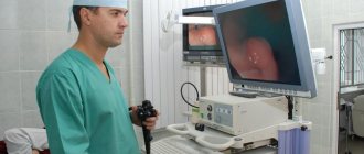
The image from the colonoscope camera is transmitted to the device screen. Videos of the study are recorded and saved on a computer.
After the intestines are examined, the doctor carefully removes the colonoscope. The study is completed.
Recovery after colonoscopy
Hospitalization is not necessary, and colonoscopy can be performed on an outpatient basis. The patient can leave the clinic as soon as he recovers from anesthesia. But you are not allowed to drive on your own on this day. You need to take one of your relatives to the clinic so that you can be escorted home.
Notes and recommendations regarding the recovery period after colonoscopy:
- When the effect of sedation wears off, a feeling of cramping and fullness of the intestines may appear. It will soon pass. In order for the gas to leave the intestines faster, it is recommended to walk.
- For 24 hours after the procedure, you should avoid drinking alcohol, driving vehicles, and doing work that requires concentration.
- Unless otherwise advised by your doctor, you can begin eating as usual immediately after the sedation ends. A special diet is prescribed after removal of polyps and other surgical procedures.
- It is recommended to rest for one day after the study, then you can do all your usual activities without restrictions, and go to work.
- If a biopsy was performed during a colonoscopy, you may notice some blood in your stool for 1 to 2 days afterward.
- If you need to take blood clot preventative or other medications continuously, ask your doctor how long after your colonoscopy you can start taking them again.
- Because your bowels were cleaned before the colonoscopy, another bowel movement may take a few days. It depends on your diet. If you do not have stool for a very long time after the procedure, consult your doctor.
You should immediately seek medical help if symptoms such as high fever, bleeding from the anus, or abdominal pain appear.
Colonoscopy of the intestine
Colonoscopy is a method of diagnosing the intestines using a flexible probe with a high-resolution video camera. The image with a magnification of up to 10 times visualizes the condition of the tissues, the presence of formations, inflammation, and sources of bleeding. During the examination of the intestines, the doctor can take biomaterial for histological examination, stop bleeding, and remove polyps.
Indications for examination
- Age over 55 years. According to statistics, colorectal oncology accounts for 10-11% of the total number of diagnosed cancer cases. The largest number of cases are in the age group over 55 years. The danger of localizing tumors in the intestines is that in the first stages there may be no symptoms, or the manifestations may be uncharacteristic. To diagnose pathology at an early stage, it is recommended to do a colonoscopy of the intestines annually.
- Pain in the intestines. If there is pain, the causes of which have not been established during other examinations, the patient is indicated for a colonoscopy. Pain sensations can be pulling, sharp or spasmodic.
- Regular constipation and bowel irregularities. Excessive pushing and hard stools can lead to intestinal injury and the formation of foci of inflammation. In addition, constipation can be a consequence of other pathologies that also require diagnosis and treatment.
- Presence of blood in stool. With intense bleeding, clearly visible veins appear. This may be a symptom of bleeding in the sigmoid or rectum. The presence of blood in the stool can also be detected by analysis.
- The presence of pus and mucus in the stool. This sign indicates an infectious process - an abscess, a disintegrating tumor, tuberculosis or acute inflammation of an ulcer. Such symptoms require immediate medical attention.
- Regular stool disorders of a non-infectious nature. This is one of the manifestations of disorders of the small intestine. In this case, beneficial substances from food are not absorbed, malabsorption develops, and metabolism is disrupted.
- Weight loss with normal nutrition and constant physical activity. If body weight decreases for no apparent reason, it is necessary to examine the digestive system, including a colonoscopy.
- Anemia and increased erythrocyte sedimentation rate (ESR) that do not go away with drug therapy. These are signs of constant blood loss or pathologies of the hematopoietic system. Blood loss may be a sign of hidden bleeding, including in the intestines.
In addition, intestinal colonoscopy is indicated for those who have a history of polyps or the appearance of neoplasms of different locations. If among your relatives there are those who have suffered from intestinal oncology, it is recommended to conduct an examination once a year, regardless of age. In addition to routine research, the procedure can be prescribed for emergency indications:
- The presence of a foreign object in the intestine.
- Bleeding due to the formation of protrusions on the intestinal walls with their further rupture.
- Impaired patency, narrowing of the intestinal lumen.
During emergency colonoscopy, the examination is combined with treatment procedures. The doctor can provide first aid and plan further therapy.
Contraindications to colonoscopy
Despite the sensitivity of the procedure, colonoscopy is usually well tolerated by patients. But there are a number of conditions when the study is postponed or impossible to carry out:
- For acute respiratory diseases, infectious intestinal diseases, peritonitis, severe poor health.
- With exacerbation of chronic diseases of the cardiovascular system, decompensated endocrine diseases.
- For blood clotting disorders. If you are taking blood thinning medications, you must inform your doctor about this to assess the risks of the procedure.
- For chronic or acute diverticulitis, large hernias.
- For colitis, ulcerative lesions of the intestine in the acute stage, granulomatous inflammation of the gastrointestinal tract.
- With heavy bleeding, anal fissures, exacerbation of hemorrhoids.
- After intestinal surgery.
During pregnancy, the advisability of a colonoscopy is assessed individually based on the patient’s condition.
Important:
If the intestines are poorly cleansed, research becomes difficult. In this case, the doctor may interrupt the procedure and reschedule it for another day.
How is a colonoscopy performed?
For the examination, an endoscopic device is used, which allows the doctor to see the condition of the intestines and perform manipulations.
The endoscope consists of a flexible tube about 2 meters long. The tube is made of soft hypoallergenic material, which increases the safety of intestinal colonoscopy.
At the end of the tube there is a video camera, openings for the irrigation and instrumental channels, and illumination.
- The video camera has high resolution and light sensitivity, which allows you to get a clear image in low light conditions.
- Backlight improves video quality and helps you work with tools.
- An irrigation channel is needed to supply air. This facilitates the passage of the probe and, in some cases, allows the stenosis to be relieved.
- The instrumental channel is needed for inserting biopsy syringes, injection needles, wire loops, clips and other devices into the intestine.
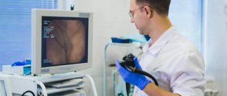
During endoscopy, the doctor controls the probe using a control unit; the same unit allows manipulation of the intestine using instruments.
The image from the camera is sent to the monitor on an enlarged scale, which allows you to see even small formations (about 1 mm), foci of inflammation and other pathological processes.
During the procedure, the patient lies on the couch on his left side with his knees tucked. In this position, intestinal colonoscopy is performed as comfortably and informatively as possible.
The doctor treats the endoscope and the area around the anus with a disinfectant. To facilitate sliding, lubricant is applied to the distal end of the probe.
When the patient is ready, the doctor inserts the endoscope into the anus and begins to move along the intestines. Air is supplied from the irrigation channel to straighten the folds and facilitate the passage of the probe. In this case, the patient may feel bloating or a feeling of fullness. This is a normal reaction that accompanies the procedure.
The examination lasts about 30 minutes, which allows you to assess the condition of the rectum and large intestinal cavity. If medical procedures are required, a colonoscopy will take longer.
After completing the examination and treatment operations, the doctor carefully removes the endoscope. If general anesthesia was not used during the procedure, the patient can stand up immediately, but sudden movements should be avoided. A feeling of bloating and fullness in the intestines is acceptable.
If the colonoscopy was performed in a medicated sleep, the patient is woken up, his well-being is assessed and he is also allowed to get up.
No hospital stay is required; you can leave the clinic after the colonoscopy. The examination results can be provided in the form of a form filled out by a doctor and on electronic media with a video recording.
Treatment during colonoscopy
If pathologies are found during an examination of the intestines, some of them can be eliminated using the endoscopic method:
- Intestinal adenomas and polyps. Neoplasms that can develop into a malignant tumor. Usually their size does not exceed 1 cm. They can occur singly or form conglomerates. In addition to the likelihood of progression to oncology, the formations make it difficult to pass stool. If adenomas are injured when passing strong stools, then there is a risk of an inflammatory process. During colonoscopy, it is possible to remove small lesions using a loop clamp and a coagulation electrode.
- Bleeding. Injuries to intestinal tissue can become inflamed without treatment. During colonoscopy, bleeding can be controlled using electrocoagulation or the application of clips. It is also possible to treat angiodysplasia (vascular pathology) of the mucous membrane.
- Neoplasms larger than 1 cm. When a tumor of this size is detected, colonoscopy is combined with sampling for histological examination.
- Removing foreign objects. The manipulation clamp allows you to remove solid objects from the intestines.
Biopsy, removal of polyps and adenomas, coagulation of blood vessels are performed using local anesthesia or general anesthesia.
Use of anesthesia during colonoscopy
Typically, anesthesia or sedation is used during medical procedures, but in some cases they are also needed for colonoscopy:
- When the patient has a low pain threshold. To relieve discomfort, a topical anesthetic may be applied.
- If there is severe inflammation or adhesions, sharp pain may occur during the procedure. Depending on the situation, the doctor assesses the possibility of continuing the study under anesthesia.
- If the patient has mental disorders or uncontrolled movements, then clonoscopy is performed under general anesthesia.
- If a person feels uncomfortable, the procedure can also be performed in a dream.
Possible complications of colonoscopy
The procedure is considered safe and atraumatic. The study is well tolerated by most patients. After a colonoscopy you may experience:
- Bloating that goes away within a day.
- Minor discomfort in the anal area goes away after a few hours.
- If polyp resection or biopsy was performed, abdominal pain and fever within 38.0°C are acceptable. Symptoms should subside within 1-3 days.
Important:
Successful colonoscopy depends on the experience of the doctor. Incorrect technique or careless use of instruments may result in intestinal damage and bleeding.
Also, during the study, allergic reactions to anesthesia and other drugs may develop. To prevent such complications, the doctor collects anamnesis before performing a colonoscopy. If the patient has an allergy, this must be reported.
If after the examination the patient develops weakness, dizziness, decreased ability to concentrate, vomiting with blood or diarrhea, then you should seek help immediately. This may be a sign that the colonoscopy caused internal bleeding.
What does the examination show?
During a colonoscopy, the following intestinal pathologies can be diagnosed:
- Neoplasms. In the video you can differentiate polyps, adenomas, tumors at an early stage.
- Colitis. With this disease, the walls are ulcerated, blood may ooze from them, and pus may be released. Outside of exacerbation, the mucous membranes may be hyperemic, without a pronounced vascular pattern.
- Diverticulosis. This is a protrusion of the intestinal wall to the side with the formation of a cavity inside. Feces accumulate and stagnate in the cavity, and the tissue becomes thinner, even to the point of rupture. This pathology is not accurately determined by ultrasound, but colonoscopy makes it possible to diagnose and treat hernial elements.
- Adhesions and foreign bodies. The formation of adhesions and the entry of foreign bodies often provoke obstruction. Colonoscopy accurately determines the cause of the disorders.
- Tuberculosis. The disease can develop as a result of pulmonary tuberculosis. During a colonoscopy, the doctor will see tissue damage by specific granulomas.
- Ischemia. Circulatory disorders can occur due to vascular pathologies. In this case, the tissues do not receive nutrition, as a result of which necrotic processes develop and ulcers appear.
During the examination, other diseases may be detected. In some cases, after a colonoscopy, the patient is sent for additional diagnostics: ultrasound, CT, MRI, irrigoscopy and other studies.
Preparation for the procedure
The possibility of performing a colonoscopy and its informative content depends on the quality of preparation.
The main task is to clean the intestines so that solid contents do not interfere with the advancement of the probe and do not complicate the visualization of tissues. General preparation scheme:
- For 7-10 days, stop taking blood thinning drugs (unless otherwise agreed with the attending physician), drugs to increase hemoglobin levels, and activated charcoal.
- If a colonoscopy is performed under anesthesia, then 10 days before you need to take tests for syphilis, HIV, hepatitis B and C, and general blood and urine tests.
- 2-3 days before the colonoscopy, you need to switch to a diet that prevents the formation of toxins and gases. Legumes, nuts, fruits, raw vegetables, flour, carbonated drinks, and sweets are excluded. Also, you should not eat or drink foods and liquids with a red and purple color (beets, cherry nectar, etc.). The basis of nutrition in preparation for colonoscopy can be clear broths, jelly, light juices without pulp.
- During the entire preparation period, it is recommended to drink as much plain water as possible.
- The last meal before a colonoscopy is acceptable 20 hours before the examination.
Preparing for a colonoscopy includes taking laxatives. The night before (3-4 hours before bedtime) and in the morning 5 hours before the examination, it is necessary to cleanse the intestines using the drug Fortrans or analogues.
- 2 sachets of powder are dissolved in 2 liters of water, after which 200 ml of the drug is taken every 7-10 minutes.
- The effect of the drug will last for the next three hours. During this period, it is recommended to stay at home as frequent bowel movements will occur.
To improve the taste of the prepared solution, you can cool it for several hours and replace 100 ml of water with 100 ml of freshly squeezed lemon or orange juice.
After squeezing, the juice must be filtered so that the pulp does not get into the solution. During cleaning, it is recommended to drink plenty of fluids (water, water with lemon), the last intake of liquid is 2 hours before the examination.
You should choose loose, comfortable clothing that does not compress the abdominal area. It is not recommended to smoke on the eve and on the day of a colonoscopy, as nicotine causes spasms of blood vessels and makes them fragile, increasing the risk of complications such as bleeding.
If you have a history of chronic diseases, food allergies, preparation for a colonoscopy is agreed with your doctor. The discontinuation of medications taken, the use of microenemas, and replacement of the active ingredient in a laxative should also be discussed with a specialist.
Important:
proper preparation for colonoscopy will allow the endoscopist to examine the condition of the mucous membranes as thoroughly as possible and detect formations 1 mm in size. If cleaning is not done well enough, diagnosis becomes difficult.
Capsule examination or colonoscopy: what is the difference?
An alternative to colonoscopy is capsule diagnostics.
The mini-camera is built into a capsule that the patient swallows. As the capsule moves, it takes pictures and the data is transmitted to a receiver attached to the patient’s body. The capsule comes out naturally. The method does not require preliminary measures, is more comfortable from a psychological point of view and allows you to examine the entire gastrointestinal tract.
But a significant advantage of colonoscopy is the ability to linger on the desired areas for a more detailed study and carry out treatment. In addition, the cost of endoscopic diagnostics is significantly lower.
The main thing about colonoscopy
- Unlike radiation diagnostic methods, with colonoscopy it is possible to visually examine the condition of the mucous membranes and combine examination with treatment.
- The correctness of the results depends on the thoroughness of preparation. You need to take this stage seriously. Colonoscopy may be delayed due to stool accumulation in the intestines.
- The patient should be in a calm state. If psychological difficulties arise and the nervous condition does not go away, it is better to perform a colonoscopy under sedation.
The qualifications and experience of the endoscopist are of great importance. You need to undergo a colonoscopy in a clinic with a good reputation, high-quality modern equipment and a proven staff of doctors.
Possible complications
Colonoscopy is a safe test. The risk of complications during a diagnostic examination of the colon is minimal. The risks of surgical endoscopy of the colon are less than one percent, which allows us to speak with confidence about its safety. Complications during endoscopic examination of the colon are very rare, including:
- Allergic reactions to sedative medications.
- Bleeding after biopsy, polyp removal.
- Perforation (rupture of the wall) of the intestine.
Contraindications
Regardless of what examination is planned to be performed - an X-ray examination of the intestines or a colonoscopy - both methods have similar contraindications, and they are not associated with barium, since this contrast agent is well tolerated by almost all patients. The most serious contraindications include:
- Pregnancy.
- Severe diseases of the cardiovascular system, blood clotting disorders.
- Deep intestinal ulcers accompanied by heavy bleeding.
Important
Keep in mind that during irrigoscopy there is a certain radiation exposure to the body, so the procedure cannot be prescribed too often.
Alternatives to Colonoscopy
In addition to colonoscopy, other tests may also be used for the early diagnosis of malignant tumors of the colon:
- Fecal occult blood test (annually). The accuracy of the study is 62–79%. Out of 10 people with colon cancer, only 6-8 will test positive. False-positive results are possible if the patient has recently eaten red meat, foods rich in vitamin C, or is taking drugs from the group of non-steroidal anti-inflammatory drugs. If the test shows a positive result, a colonoscopy is required to clarify the diagnosis.
- Flexible sigmoidoscopy (every 5 years) is an endoscopic examination of the rectum and lower third of the colon. It is performed in the same way as a colonoscopy, but during it, a smaller part of the intestine is examined. Helps identify 70–80% of polyps and malignant tumors in the rectum and lower colon.
- Fecal DNA testing combined with stool occult blood testing (every 3 years). Can detect up to 92% of malignant tumors and up to 42% of precancerous conditions.
- Virtual colonoscopy (CT colonography) (every 5 years) - multislice computed tomography (MSCT), during which the colon is filled with air. The detection rate of tumors larger than 1 cm using this study is 94%, polyps measuring 6–9 mm is 65%. Polyps smaller than 6 mm are not detected. If a polyp is found during CT colonography, a colonoscopy will still have to be performed to remove it.
Thus, colonoscopy is the most accurate and informative method of screening for colon cancer, while it makes it possible to immediately carry out a biopsy and some therapeutic manipulations.